|
|
ascospores and dimorphic conidiophores (Schroers, 2001). This fungus is commonly found on the recently dead trees in the pantropical areas (Samuels, 1988a; Samuels et al.,
1990).
Bionectria ochroleuca (Schwein.) Schroers & Samuels, Z.
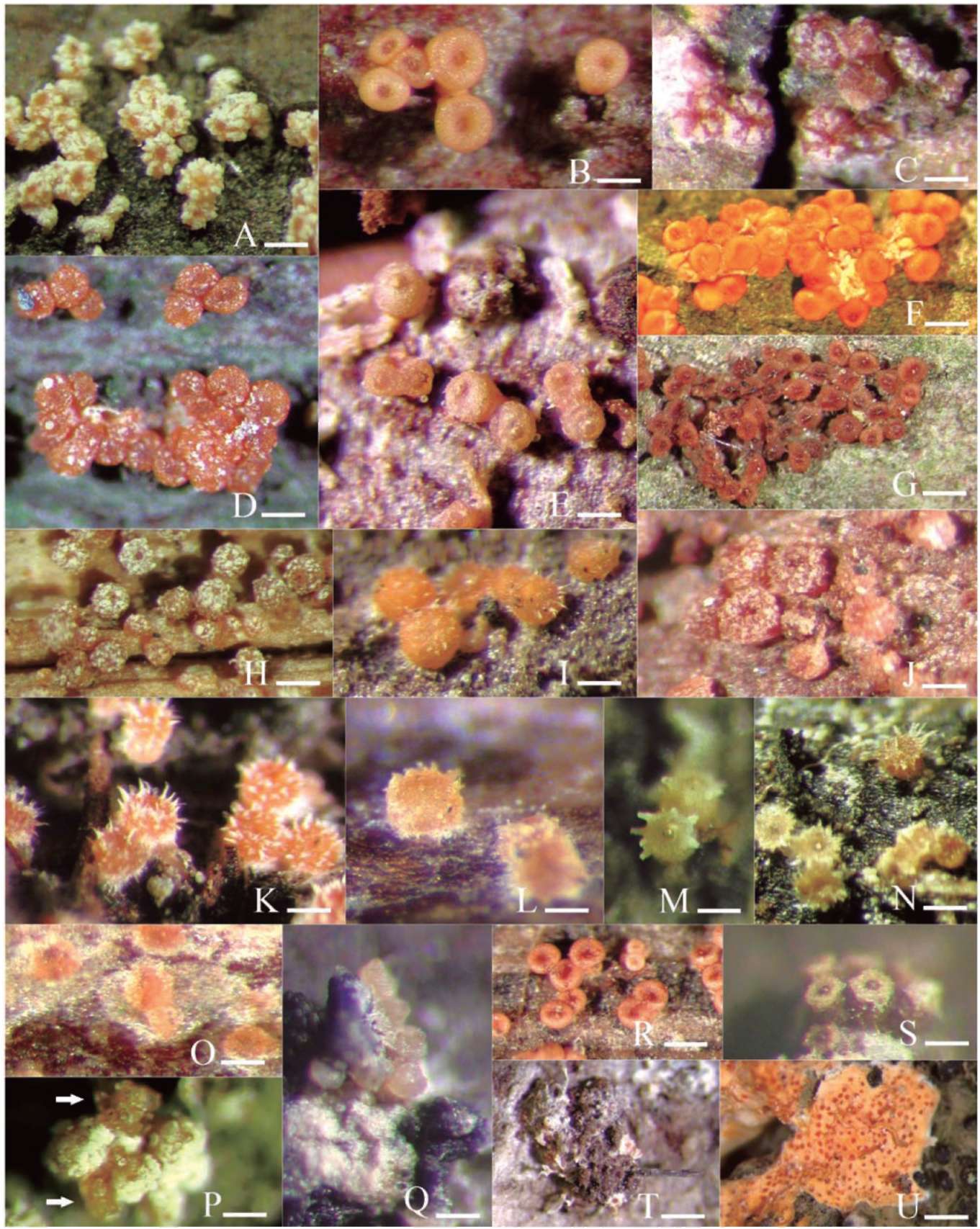
Mykol. 63: 151. 1997. Figure 1D, 2I
Anamorph. Clonostachys rosea (Link: Fr.) Schroers, Samuels, Seifert & W. Gams, Mycologia 91: 369. 1999.
Figure 2C, D, Q
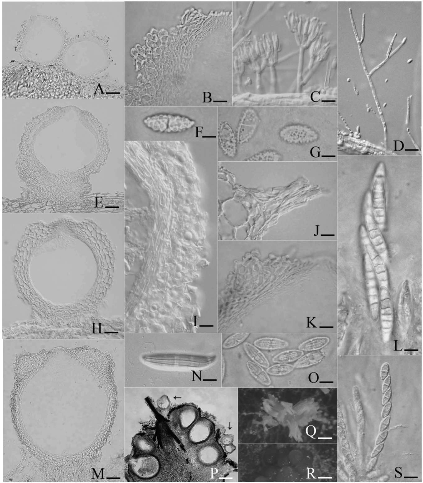
Stromata flattened to pulvinate. Perithecia aggregated in groups of 5-20, yellowish or pale orange, globose to subglobose, 200-350 fm diam, laterally pinched or not when dry, smooth or roughened; perithecial wall 20-55 fm thick, three-layered, orange droplets in the cells of the outermost wall layer; ostiolar openings nonpapillate. Asci clavate, 40-70 x 5-8.5 fm, with an apical ring. Ascospores
hyaline, ellipsoidal, 1-septate, (8.5-)9-12(-14.5) x
(2.5-)3-4(-4.5) fim, finely roughened.
Cultures and anamorph. Colonies 3-4 cm diam in 7 days at 20°C, with aerial hyphae forming strands, diffusing a yellow pigment into the medium, not forming sporodochia. Conidiophores dimorphic, verticillate and penicillate. Verticillate conidiophores frequently observed, up to 200 fm high, solitary, bearing 2 or 3 divergent phialides; phialides cylindrical, 15-30 x 1.5-2 fm. Penicillate conidiophores solitary or aggregated into sporodochia, bi- to terverticillate, bearing 2-4 divergent phialides; phialides flask-shaped to cylindrical, 9-12 x 1.5-2.5 fm. Intercalary phialides absent. Conidia borne on verticillate conidiophores hyaline, ellipsoidal, slightly
curved, aseptate, (5.5-)6-9(-10.5) x (2-)2.5-4 (-4.5) fm.
Conidia borne on penicillate conidiophores hyaline, ovoidal to subglobose, with a lateral hilum, aseptate, 4-6 x 2.5-3 fm, with imbricate conidial chains adhered laterally to form yellowish or white conidial columns.
Specimens examined. Taipei County, Sunshai, Man-
yue-yuan, on bark, 27 Sep 2000, JYM 89092712. Tainan County, Nansi, on bark, 14 Apr 2001, JYM & HHM
90041411. Taipei City, Nankang, Hu-shih park, on bark, 6 Apr 2003, JYM & HHM 92040604. Taipei City, Shihlmn,
Ping-ding-gu-jyun, on bark, 9 Aug 2003, GJR 92080905.
Taipei City, Shihlinn, Ping-ding-gu-jyun, on bark, 25 Sep
2003, GJR 92092502 (cultured). Taipei County, Hsintien,
Jhih-tan-shan, on bark with Albonectria rigidiuscula and
Gibberella sp., 9 Oct 2003, GJR 92100901 (cultured).
Taipei County, Hsintien, Yinhodong, on bark, 16 Oct
2003, GJR 92101602 (cultured). Taipei County, Jingtung, Shih-shun-chien, on bark, 5 Jan 2004, GJR 93010505
(cultured). Taipei County, Wulai, Fusan, Ha-pen, on bark,
6 Mar 2004, GJR 93030601. Taipei County, Jingtung,
Jing-tung historical trail, on bark, 18 Mar 2004, GJR 93031806. Taipei County, Hsintien, Shih-zih-tou-shan, on
bark, 21 Mar 2004, GJR 93032104 (cultured). Taipei City,
Kungguan, National Taiwan Univ., on Cycas taitungensis Shen, Hill, Tsou & Chen, 26 Apr 2004, WJH & YRL 93042601 (cultured). Taipei County, Jingtung, Jing-
|
tung historical trail, on bark, 4 Sep 2004, GJR & KHP 93090405. Taipei City, Muja, on bark, 25 Sep 2004, SSH
93092509. Taipei County, Pingshi, Chou-tou-shan, on Sphaeropteris lepifera (J. Sm. ex Hook.) Copel., 31 Oct
2004, GJR 93103108. Taipei County, Hsintien, Yinhodong, on bark, 25 Nov 2004, GJR 93112504. Taipei County,
Hsintien, Shih-zih-tou-shan, on bark, 11 Dec 2004, GJR 93121110. Taipei County, Wanlee, Linnshih historical
trail, on bark, 15 Jan 2005, GJR 94011506 (cultured).
Taipei County, Wanlee, Linnshih historical trail, on bark, 15 Jan 2005, GJR 94011509. Taipei City, Peitou, Chintien
Temple, on bark, 22 Jan 2005, SSH & CTH 94012209
(cultured). Kaohsiung County, Liouguei, Shanping, on
bark, 10 Mar 2005, GJR 94031013 (cultured). Ilan County, Yuanshan, Fushan, on bark, 29 Apr 2005, HHM 94042906 (cultured); 94042909. Taipei City, Kungguan, National
Taiwan Univ., on bark, 12 Jun 2005, GJR 94061201 (cultured). Taipei County, Wanlee, Rueichuan historical
trail, on bark, 23 Jul 2005, GJR 94072305 (cultured).
Taipei County, Wanlee, Rueichuan historical trail, on Sphaeropteris lepifera, 23 Jul 2005, GJR 9407231 3 (cultured). Kaohsiung County, Maolin, on bark, 14 Sep
2005, GJR 94091404 (cultured). Chiayi County, Shihjhuo, on bark, 20 Nov 2005, GJR 94112008. Taipei City, Chi-shinn-shan, on bark, 26 Nov 2005, SSH & CTH 94112603.
Ilan County, Yuanshan, Fushan, on bark, 6 Dec 2005, GJR
94120605.
Notes. Bionectria ochroleuca is frequently encountered in tropical and neotropical regions (Samuels, 1976a; Schroers and Samuels, 1997; Schroers, 2001) and is well represented among our Taiwan collections. Its perithecial wall possesses unevenly thickened cells as in B. byssicola; however, B. byssicola has coarse warts on the perithecial surface. The orange droplets are located in the cells of the outermost wall layer, which distinguishes B. ochroleuca from other members of Bionectria. Droplets present in other species of Bionectria are essentially hyaline. While most of our collections were made from the bark of broad-leaf trees, some were from tree ferns: specimens 93103108 and 94072313 were from Sphaeropteris lepifera. Like Samuels (1976a) and Schroers (2001), we were also able to obtain the teleomorph in cultures from specimens
92101602, 93010505, 93032104, and 94061201.
Bionectria parviphialis (Samuels) Schroers, Stud. Mycol.
46: 182. 2001. Figures 1E, 2E, F
Anamorph. Clonostachys pseudosetosa (Samuels) Schro-
ers, Stud. Mycol. 46: 182. 2001.
Stromata lacking. Perithecia aggregated, superficial, pale orange, globose, 250-350 [m diam, not collapsed when dry, roughened; perithecial wall 40-50 [m thick, two-layered; ostiolar openings nonpapillate. Asci clavate, 55-70 x 10-15 [m, with an apical ring. Ascospores hyaline,
ellipsoidal, 1-septate, (14.5-)15-17.5 x (4.5-)5-6 [m, with
coarse warts up to 1 [m diam.
Specimen examined. Taipei County, Pingshi, Chung-
yang-chien-shan, on bark, 20 Nov 2003, GJR 92112008.
|
|