Botanical Studies (2010) 51: 343-350.
MICROBIOLOGY

Aquaperonospora taiwanensis gen. et sp. nov. in
Peronophythoraceae of Peronosporales
Wen-Hsiung KO1*, Mei-Ju LIN1, Chung-Yue HU2, and Pao-Jen ANN2
1Department of Plant Pathology, National Chung Hsing University, Taichung, Taiwan
2Division of Plant Pathology, Taiwan Agricultural Research Institute, Wufeng, Taichung, Taiwan


(Received June 16, 2009; Accepted November 25, 2009)
ABSTRACT. Twelve isolates of a Pythium-like organism capable of producing Peronospora-like sporangiophores were isolated by baiting from an irrigation ditch in central Taiwan. This organism is described herein as a new genus and species, Aquaperonospora taiwanensis, in Peronophythoraceae of Peronosporales. Low sequence identities in both the ITS and 28S rDNA sequences between A. taiwanemis and representative species of other genera in Peronosporales supported the validity of the establishment of Aquaperonspora as a new genus. The groupings of A. taiwanensis and Pythium ostracodes, and Peronophythora litchii and Phytophthora infestans in both ITS and 28S phytogenetic trees were consistent with the suggestions that Aquasponospora and Peronophythora are transitional genera between Pythium and Peronospora, and Phythophthora and Peronospora, respectively. Based on this study and those reported by others, the determinate growth of sporangiophores is no longer a tenable distinguishing characteristic of Peronosporaceae or Peronophythoraceae. A new key to the families of Peronosporales is, therefore, presented.
Keywords: Albuginaceae; Determinate growth; Irrigation ditch; Peronophythoraceae; Peronosporaceae; Peronosporales; Pythiaceae.
INTRODUCTION
During our survey of the distribution of Phytophthora and Pythium in Taiwan (Ko et al., 2004; 2006), ten isolates of a Pythium-like organism capable of producing rigid, erect, and branched Peronospora-like sporangiophores were obtained by baitings performed in 2003 at different sites in an irrigation ditch at the experimental farm of Taiwan Agricultural Research Institute, Wufeng, Taichung. Two more isolates with such morphological characteristics were obtained from the same ditch in 2008. These unusual characteristics do not fit the descriptions of any members of known genera in Peronosporales. A new genus Aquaperonospora, therefore, is erected for this organism.
MATERIALS AND METHODS
Isolation and culture of organisms
To isolate the organisms, 10 leaf disks (9 mm diam) of citrus (Grimm and Alexander, 1973), azalea, or camellia (Zhou et al., 1992), depending on availability of baiting materials at the time of baiting, were wrapped in a single layer of Miracloth, tied with string and placed in the
*Corresponding author: E-mail: kowh@dragon.nchu.edu.tw; Tel: +886-4-2284-0780 ext 371; Fax: +886-4-2287-7585.
ditch at the experimental farm of the Taiwan Agricultural Research Institute, Wufeng, Taichung. Water in the ditch originated from runoff water from the forests on the mountain. The baiting bags were suspended with the string in the slowly moving water at about 10- to 15-m intervals. After 2 days, bags were retrieved from the ditch, and baits were blotted dry and placed on a selective medium consisting of 5% V-8 juice, 0.02% CaCO3, 100 ppm ampicillin, 50 ppm nystatin and 10 ppm pentachloronitrobenzene, and 2% agar (Ko et al., 1978a). Plates were incubated on the laboratory bench at 24°C. The hyphae emerging from a colonized leaf disk after 2 days were transferred to a plate of 10% V-8 agar (10 ml V-8 juice, 0.1 g CaCO3, 15 g agar per L). A culture block (5 x 5 mm) cut from an isolated culture was immersed in 8 ml sterile distilled water in a small plate (6 cm diam), and incubated at 24°C under light for production of sporangia and zoospores. Plates were observed under 10 X objective after 24 h. Single-zoospore isolates were obtained from those cultures producing rigid, erect, and branched Peronospora-like sporangiophores by spreading a loopful of zoospore suspension on 2% water agar and transferring the colony originating from a zoospore to a V-8 agar plate. A single-zoospore isolate from a different leaf disk was stored in sterile distilled water in a test tube at 24°C (Boeswinkel, 1976).
344
Botanical Studies, Vol. 51, 2010

DNA extraction, sequencing and phylogenetic analysis
Using a plant genomic DNA extraction kit (GenMark Technology Co., Taichung, Taiwan), DNA of A. taiwanensis was extracted from 0.1 g of three-day-old mycelia growing on cellophane placed on V-8 agar The nucleic acid sequence of its ITS was amplified with primer pair ITS1 and ITS4 (White et al., 1990). A PCR reaction was performed in a 25 fil volume reaction containing 50 ng DNA, 0.5 pmole each of ITS1 and ITS4 primers and a 1.5 unit of SuperTaq polymerase (Protech Technology Enterprise Co., Ltd., Taiwan) with a buffer system recommended by the manufacturer. The PCR cycling conditions included an initial denaturation of 94¢XC for 2 mm, 30 cycles of 94¢XC for 30 s, 55¢XC for 30 s, 72¢XC for 1 min, and a final elongation at 72¢XC for 6 min.
Amplified PCR product was analyzed by electrophoresis in a 1.2% agarose gel and cloned into pCR2.1-TOPO vector (Invitrogen, Carlsbad, California)
according to the manufacture's instructions. Plasmid clones with expected size DNA inserts were screened and used for sequencing. Sequencing of the target DNA insert was done by an automatic DNA sequencer (ABI PRISM 377, Perkin-Elmer, California) with the BigDye Terminator Cycle Sequencing Kit (Perkin-Elmer Applied Biosystem, California). The large ribosomal subunit, 28S, was analyzed in the same manner with primer pair LROR and LR7 (Vilgalys and Hester, 1990). The annealing temperature was changed to 50¢XC. To examine phylogenetic relationships, the new sequence and those of representative taxa in the Peronosporales retrieved from GenBank (Table 1) were aligned using the ClustalX, Version 1.81 (Thompson et al., 1997).
The multiple alignments were adjusted after visual examination with Sequence Alignment Editor (Se-A1), Version 1.0 alpha 1 (Rambaut, 1996). Phylogenetic tree building based on the neighbor-joining (NJ) method (Saitou and Nei, 1987) and distance matrix for the aligned
Table 1. Collection and GenBank accession numbers of ITS and 28S sequences of representative members in Peronosporales retrieved from GenBank for phylogenetic analysis.
Species (Isolate)
Associated habitat
Location
Source
GenBank accession no.
ITS
Albugo candida (BPI184865)
Diplotaxis tenuifolia
Korea
Choi et al.
DQ418497
Bremia lactucae (SMK18913)
Lactuca indica var. laciniata Korea
Choi et al.
DQ235797
Peronospora parasitica (05NF)
Raphanus raphanistrum
Australia
Maxwell and Scott
DQ447120
Peronophythora litchii (CBS 100.81)
Unknown
Austria
Voglmayr
AY198308
Phytophthora infestans (3015)
Solonacea
Colombia
Restrepo et al.
EU200300
Plasmopara viticola (unknown)
Vitis vinifera
Germany
Thines
DQ665668
Pseudoperonospora cubensis (HV 222)
Cucumis sativa
Austria
Voglmayr
AY198306
Pythium monospermum (CBS 158.73)
Unknown
Canada
Levesque and Cock
AY598621
Pythium ostracodes (CBS 768.73)
Unknown
Japan
Kageyama et al.
AB108022
Pythium vexans (MAFF305905)
Unknown
Japan
Matsumoto et al.
AJ233462
28S
Albugo candida (MG 15-7)
Capsella bursa-pastoris
Germany
Riethmueller et al.
AY035538
Basidiophora entospora (HV 123)
Conyza canadensis
Germany
Riethmueller et al.
AY035513
Bremia lactucae (HV 656)
Picris hieracioides
Germany
Riethmueller et al.
AY035512
Peronospora parasitica (GG92(MA))
Raphanus raphanistrum
Spain
Goeker et al.
EU054927
Paraperonospora leptosperma (HV 383)
Tripleurospermum inodorum Germany
Riethmueller et al.
AY035515
Peronophythora litchii (AR 178)
Unknown
Germany
Riethmueller et al.
AY035531
Phytophthora infestans (CBS 560.95)
Unknown
Germany
Riethmueller et al.
AF119602
Plasmopara viticola (AR 160)
Vitis vinifera
Germany
Riethmueller et al.
AY035524
Pseudoperonospora cubensis (HV 221h)
Cucumis sativus
Germany
Riethmueller et al.
AY035496
Pythium monospermum (AR 213)
Unknown
Germany
Riethmueller et al.
AY035535
Pythium ostracodes (CBS 768.73)
Unknown
Switzerland
Belbahri et al.
EF426542
Pythium vexans (CBS 119.80)
Unknown
Switzerland
Belbahri et al.
EF426541
Sclerospora graminicola (HV 532)
Setaria viridis
Germany
Riethmueller et al.
AY035514
KO et al. ― Aquaperonospora taiwanesis gen. et sp. nov.
345
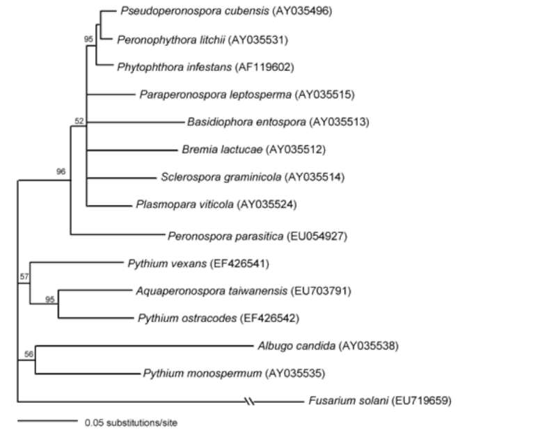
sequences was done using Kimura's two-parameter method (Kimura, 1980) of the program PAUP* 4.0 (Swofford, 1998). Bootstrap values were generated with 1000 replicate heuristic searches to estimate support for the clade stability of the consensus tree (Felsenstein, 1985).
TAXONOMY
Aquaperonospora Ko gen. nov.
Sporangiophora e myceliis, rigida, erecta, dichotome ramosa. Sporangia super ramunculis sporangiophorum simul formata, sporangiis additis super sporangiophoris e basi sporangiorum priorum interne vel externe prolificantibus factis successive. Zoosporae intra vesiculam externam sporangiorum formatae. Periplasma oogonii inconspicuum.
Etymology. Refers to its water habitat and sporangiophore morphology similar to that of the genus Peronospora.
Type species. A. taiwanensis Ko.
Aquaperonospora taiwanensis Ko sp. nov.
Hyphae coenocyticae, 3.0-4.4 £gm latae. Sporangiophora e myceliis, rigida, erecta, dichotome ramosa, 475-1540 £gm longa, 4.0-6.0 £gm lata, unoquoque sporangia 1-30 ferenti. Sporangia citriformia, 33.0-41.0 x 22.0-29.6 £gm, super ramunculis sporangiophororum simul formata, sporangiis additis super sporangiophoris e basi sporangiorum priorum interne vel externe prolificantibus factis successive. Zoosporae reniformes, 14-18 x 12-15 £gm, intra vesiculam externam sporangiorum formatae. Periplasma oogonii inconspicuum. Oogonia globosa, terminalia vel intercalaria, laevia, 15-32 £gm diam. Antheridia elongata, 12-21 x 3-5 £gm, ad oogonium longitudine tota maximam partem singulariter adhaerentia. Oosporae apleroticae, 13-28 £gm diam, pariete usque ad 3 £gm crasso.
Etymology. Taiwanensis refers to the country of origin.
Holotype. HAST 109384 (dried culture), Herbarium,
Biodiversity Research Center, Academia Sinica, Taipei, Taiwan.
A culture from the holotype has been deposited at the Bioresource Collection and Research Center, Food Industry Research and Development Institute, Hsinchu, Taiwan (BCRC 34009).
Aquaperonospora taiwanensis was isolated by baiting from an irrigation ditch, with ten isolates collected in 2003 and two isolates in 2008. It is a fast growing organism compared to other fungi, with mycelia extending 23 mm/ day at 24°C on V-8 agar. The optimum temperature for growth was 32°C while the maximum and minimum temperatures were 40 and 12°C, respectively. The growth rate at 32°C was 32.3 mm for 24 h. Aquaperonospora taiwanensis did not produce asexual propagules on V-8 agar at 24°C under light. However, when a culture block was immersed in water and incubated at 24°C under light
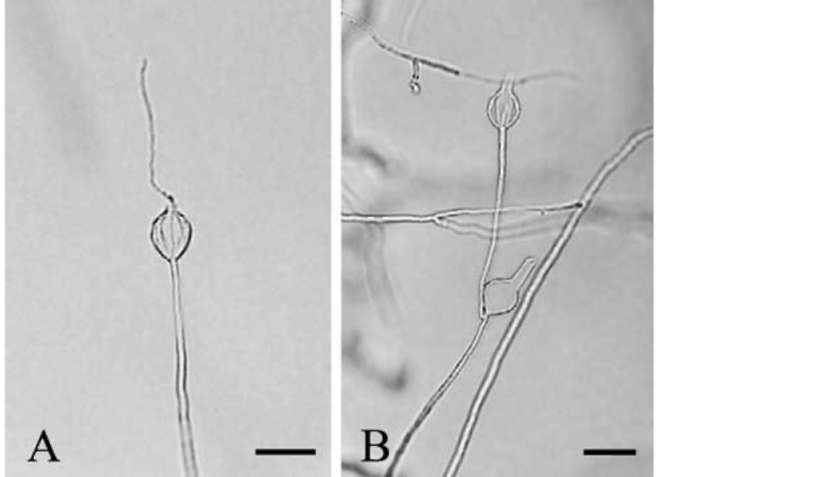
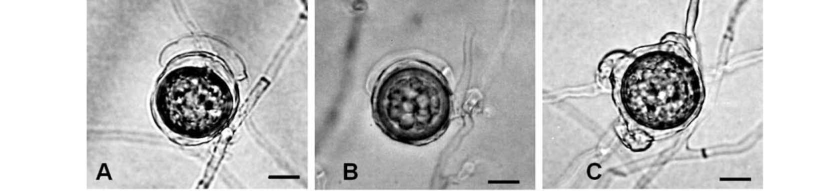
as described above, the organism produced numerous well differentiated Peronospora-like sporangiophores (Figure 1) within 24 h. Sporangiophores (475-1540 £gm long, 4-6 £gm wide) were rigid, erect, and dichotomously branched, bearing 1-30 sporangia each. Sporangia (33.0-41.0 x 22.0-29.6 £gm) were lemon-shaped and formed synchronously on terminal branchlets of sporangiophores (Figure 2). Additional sporangia were mostly formed
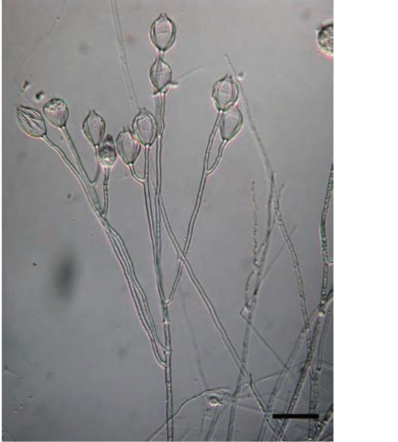
Figure 1. Differentiated sporangiophore of Aquaperonospora taiwanensis. Bar=50 [im.
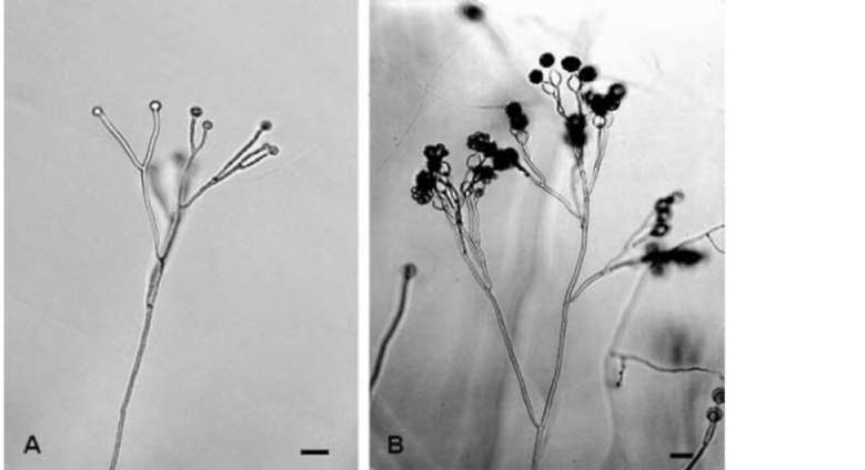
Figure 2. Sporangiophores of Aquaperonospora taiwanensis. A, synchronous formation of sporangia on branchlets of a sporangiophore. Bar= 100 £gm; B, concomitant discharge of cytoplasmic material from sporangia on a sporangiophore into vesicles during zoospore formation. Bar= 100 £gm.
346
Botanical Studies, Vol. 51, 2010

within the empty sporangia (Figure 3A). Occasionally, a sporangium was also formed on the tip of a secondary sporangiophore originating from the base of a primary sporangium (Figure 3B). Before releasing zoospores, each sporangium developed a discharge tube of various lengths and moved its undifferentiated content through this tube to form a vesicle at its end. When the vesicle ceased to enlarge, zoospores were delimited and started moving. Subsequently, the membranes of the vesicle disappeared and zoospores swam away. Zoospores were reniform, measuring 14-18 x 12-15 £gm. Aquaperonospora taiwanensis is homothallic and produced abundant oospores in V-8 agar after incubation at 24°C in darkness for 2 weeks. Oogonial periplasm was inconspicuous, and oogonia (15-32 £gm diam) were globose, terminal or intercalary, and smooth (Figure 4). Antheridia were paragynous, elongated, measured 12-21 x 3-5 £gm, and mostly singly adhered along their lengths to oogonium. Oospores (13-28 £gm diam) were aplerotic with walls up to 3 £gm thick.
PHYLOGENETICS
Rigid, erect, and branched sporangiophores and synchronous formation of spoangia on branchlet tips of A. taiwanensis are similar to species of Peronospora. However, the mode of zoospore release and morphology of sexual organs resemble those of Pythium species. Therefore, three species of Pythium were used in the phylogenetic study while only one representative species from each of other genera in Peronosporales was included. Comparison of ITS sequence similarity showed low sequence identity between A. taiwanensis and other representative taxa in Peronosporales. The
highest sequence identity was between A. taiwanensis and Phytophthora infestans with a 63.7% identity, followed by a 56.4% identity between A. taiwanensis and Pseudoperonospora cubensis (Table 2). The lowest sequence identity was between A. taiwanensis and Abugo candida with a 22.3% identity follow by a 35.8% identity between A. taiwanensis and Pythium monospermum. An analysis of phylogenetic relationships among representative taxa in Peronosporales based on ITS sequences revealed that A. taiwanensis and Pythium ostracodes belonged to a well supported clade (82%) and that the other two Pythium species, Py. monospermum and Pythium vexans shared the same clade with Al. candida (67%) (Figure 5). Moreover, Ph. infestans, Ps. cubensis,
Figure 3. Growth renewal of sporangiophores of Aquaperonospora taiwanensis. A, sporangiophore proliferating internally. Bar= 40 £gm; B, sporangiophore proliferating externally. Bar= 40 £gm.
Table 2. Sequence identity of ITS and 28S regions between Aquaperonospora taiwanensis and representative taxa in Peronosporales retrieved from GenBank.
ITS
28S
Species (isolate)
Identity
Species (isolate)
Identity
Albugo candida (BPI184865)
22.3%
Albugo candida (MG 15-7)
71.6%
Bremia lactucae (SMK18913)
41.1%
Basidiophora entospora (HV 123)
76.4%
Peronospora parasitica (05NF)
38.6%
Bremia lactucae (HV 656)
76.7%
Peronophythora litchii (CBS 100.81)
57.5%
Peronospora parasitica (GG92(MA))
76.8%
Phytophthora infestans (3015)
63.7%
Paraperonospora leptosperma (HV 383)
79.8%
Plasmopara viticola (unknown)
48.4%
Peronophythora litchii (AR 178)
83.6%
Pseudoperonospora cubensis (HV 222)
56.4%
Phytophthora infestans (CBS 560.95)
84.5%
Pythium monospermum (CBS 158.73)
35.8%
Plasmopara viticola (AR 160)
81.9%
Pythium ostracodes (CBS 768.73)
54.6%
Pseudoperonospora cubensis (HV 221h)
83.2%
Pythium vexans (MAFF305905)
41.5%
Pythium monospermum (AR 213)
79.6%
Fusarium solani (LW-1, out group)
4.9%
Pythium ostracodes (CBS 768.73)
87.6%
Pythium vexans (CBS 119.80)
82.7%
Sclerospora graminicola (HV 532)
78.6%
Fusarium solani (LW-1, out group)
35.5%
KO et al. ― Aquaperonospora taiwanesis gen. et sp. nov.
347
Figure 4. Sex organs (A, B, C) of Aquaperonospora taiwanensis. Bar=10 [im.
Figure 5. Phylogenetic relationship among representative taxa in the Peronosporales based on rDNA sequence of the ITS1-5.8S-ITS2 region using neighbor-joining method (Saitou and Nei, 1987). The distance matrix for the aligned sequences was calculated using Kimura's two-parameter method (Kimura, 1980) of the program PAUP* 4.0 (Swofferd, 1998). Bootstrap values were generated with 1000 replicate heuristic searches to estimate support for clade stability of the consensus tree (Felsenstein, 1985).
and Peronophythora litchii were grouped in a highly supported clade (97%) while the other clade consisted of Bremia lactucae, Plasmopara viticola, and Peronospora parasitica with 79% bootstrap values.
Comparison of 28S sequence similarity also showed relatively low sequence identity between A. taiwanensis and other representative taxa in Peronosporales. The highest sequence identity was between A. taiwanensis and Py. ostracodes with an 87.6% identity, followed by an 84.5% identity between A. taiwanensis and Ph. infestans (Table 2). Aquaperonospora taiwanensis also shared a 83.6 and 83.2% sequence identity with Pp. litchii and Ps. cubensis, respectively. The lowest sequence identity was between A. taiwanensis and Al. candida with 71.6% followed by 76.4% between A. taiwanensis and Basidiophora entospora. Analysis of phylogenetic relationship among representative taxa in Peronosporales based on 28S sequences showed the formation of a relatively large clade supported by a bootstrap value of 96% among Ps. cubensis, Pp, litchii, Ph. infestans, Paraperonospora leptosperma, Ba. entospora, Br.
Figure 6. Phylogenetic relationship among representative taxa in the Peronosporales based on rDNA sequence of the 28S region using neighbor-joining method (Saitou and Nei, 1987). The distance matrix for the aligned sequences was calculated using Kimura's two-parameter method (Kimura, 1980) of the program PAUP* 4.0 (Swofferd, 1998). Bootstrap values were generated with 1000 replicate heuristic searches to estimate support for clade stability of the consensus tree (Felsenstein, 1985).
lactucae, Sclerospora graminicola, Pl. viticola, and Pe. parasitica (Figure 6). The first three species formed a distinct group within this clade with a high bootstrap support (96%). Aquaperonospora taiwanensis, Py. Ostracodes, and Py. vexans were placed in the same clade supported by a 67% bootstrap value with the first two species forming a well supported distinct group (96%) within the clade. Al. candida and Py. monospermum shared a moderately supported clade (56%).
DISCUSSION
Comparison of partial rDNA sequences in this study reveals a low sequence identity between A. taiwanensis
348
Botanical Studies, Vol. 51, 2010

and species of other genera in Peronosporales and supports the validity of the establishment of Aquaperonospora as a new genus. The highest ITS sequence identity between A. taiwanensis and representative species of other genera in Peronosporales was 63.7% (Table 2) while the reported ITS sequence identity between species of different genera in the same order ranged from 88.2 to 92.4% (Voglmayr, 2003; Zhang et al., 2007). Moreover, the highest 28S sequence identity between A. taiwanensis and representative species of other genera in Peronosporales was 87.6% (Table 2) while the reported 28S sequence identity between species of different genera in the same order ranged from 87.0 to 98.4% (Riethmuller et al., 2002; Zhang et al., 2007).
Aquaperonospora taiwanensis resembles Pp. litchii (Ko et al., 1978b; Zhang et al., 2007) in its ability to produce Peronospora-like sporangiophores on artificial medium. However, zoospore release from sporangia of A. taiwanensis has the characteristics of Pythium while that of Pp. litchii has the characteristics of Phytophthora (Ko et al., 1978b). The sex organs of A. taiwanensis and Pp. litchii (Ko et al., 1978b) resemble those in Pythiaceae in having inconspicuous oogonial periplasm. These data show that A. taiwanensis has the characteristics of both Peronosporaceae and Pythiaceae, just like Pp. litchii. It is, therefore, also placed under Peronophythoraceae.
The transfer of Pp. litchii to Phytophthora has been suggested because of the close sequence relationship of some of its DNA fragments including ITS and 28S with those of certain Phytophthora species (Riethmuller et al., 2002; Voglmaryr, 2003; Goker et al., 2007). The validity of the suggestion is dependent on the accuracy of the hypothesis of the existence of a negative correlation between the order of the taxonomic ranks and the level of the sequence similarity of these DNA fragments. The hypothesis was refuted by Zhang et al. (2007) who showed that the ITS sequence similarity level between Phytophthora sojae and Pl. viticola (99.3%) was higher than that between Ph. sojae and Phytophthora tropicalis (89.5%), and that the 28S sequence similarity level between Pythium undulatum and Pythium monospermum (82.6%) was lower than that between Peronospora potertillae-sterilis and Phytophthora nicotianae (95.8%). The DNA fragments currently used in phylogenetic studies are not valid in the determination of the taxonomic status of Oomycota because they are probably not related to morphological and physiological traits (Zhang et al., 2007). When genes related to taxonomical characters are discovered in the future, they should be useful in the determination of taxonomic ranks and phylogenetic relationship in Oomycota and in other groups of organisms as well.
The determinate growth of sporangiophores was originally considered one of the pivotal characteristics of Peronosporaceae and Peronophythoraceae (Ko et al., 1978b; Alexopoulos and Mims, 1979). Subsequently, the occasional growth renewal of sporangiophores of Pp.
litchii on artificial media away from the litchi host (Litchi sinensis Sonn.) was observed by several researchers (Chi et al., 1982; Huang et al., 1983; Ho et al., 1984). Sporangiophores of A. taiwanensis were also capable of new growth on agar medium under submerged conditions. In 1969, Tiwari and Arya reported the growth of Sc. graminicola from infected host callus onto modified White's medium (Tiwari and Arya, 1969). The organism maintained this saprophytic growth independent of the host callus during two subsequent subcultures made at intervals of 20 days on the same fresh medium. Sporangiophores produced under such conditions sometimes gave rise to secondary and tertiary sporangiophores. These results show that determinate growth of sporangiophores is not a stable distinguishing character for either Peronosporaceae or Peronophythoraceae. A new key to the families of Peronosporales is, therefore, presented.
Key to the families of Peronosporales
1. Sporangia borne in chain, periplasm conspicuous, obligate plant parasites.............................. Albuginaceae
1. Sporangia borne singly ................................................. 2
2. Sporangiophores undifferentiated, periplasm inconspicuous, nonobligate plant parasites or saprophytes ................................................... Pythiaceae
2. Sporangiophores differentiated .................................... 3
3. Periplasm conspicuous, obligate plant parasites ............
.............................................................. Peronosporaceae
3. Periplasm inconspicuous, nonobligate plant parasites or saprophytes ................................... Peronophythoraceae
Acknowledgements. This work was supported in part by a grant from the National Science Council of Taiwan (NSC 97-2811-B-005-001). We thank Dr. Yu-Ming Ju for preparing the Latin diagnosis.
LITERATURE CITED
Alexopoulos, C.J. and C.W. Mims. 1979. Introductory Mycology, 3rd ed. John Wiley & Sons, New York.
Boeswinkel, H.J. 1976. Storage of fungal cultures in water.
Trans. Br. Mycol. Soc. 66: 183-185. Chi, P.K., X.P. Pang, and R. Liu. 1982. Studies on Phytophthora
disease of litchi. I. Identification of causal agent. Proceedings of the 1981 Annual Meeting of Chinese Phytopathological Society, pp. 72-73.
Felsenstein, J. 1985. Confidence limits on phylogenies: An approach using the bootstrap. Evolution 39: 783-791.
Goker, M., H. Voglmayr, A. Riethmuller, and F. Obervinkler. 2007. How do obligate parasites evolve? A mutli-gene phylogenetic analysis of downy mildews. Fungal Genet.
Biol. 44: 105-122.
Grimm, G.R. and A.F. Alexander. 1973. Citrus leaf pieces as traps for Phytophthora parasitica from soil slurries.
Phytopathology 63: 540-541.
KO et al. ― Aquaperonospora taiwanesis gen. et sp. nov.
349
Ho, H.H., J.Y. Lu, and L.Y. Gong. 1984. Observations on
asexual reproduction by Peronophythora litchii. Mycologia 76: 745-747.
Huang, H., C.P. Wang, and D.Y. Xu. 1983. On Peronophythora
litchii. Acta. Mycol. Sin. 2: 201-206.
Kimura, M. 1980. A simple method for estimating evolutionary rate of base substitutions through comparative studies of nucleotide sequences. J. Mol. Evol. 16: 111-120.
Ko, W.H., H.S. Chang, and H.J. Su. 1978a. Isolates of
Phytophthora cinnamomi from Taiwan as evidence for an Asian origin of the species. Trans. Br. Mycol. Soc. 71:
496-499.
Ko, W.H., H.S. Chang, H.J. Su., C.C. Chen, and L.S. Leu. 1978b. Peronophythoraceae, a new family of Peronosporales. Mycologia 70: 380-384.
Ko, W.H., S.Y. Wang, and P.J. Ann. 2004. Pythium sukuiense, a new species from undisturbed natural forest in Taiwan.
Mycologia 96: 647-649. Ko, W.H., S.Y. Wang, and P.J. Ann. 2006. The possible origin
and relation of Phytophthora katsurae and P. heveae, discovered in a protected natural forest in Taiwan. Bot.
Stud. 47: 273-277.
Rambaut, A. 1996. Se-Al version 1.0 alpha 1. Sequence Alignment Editor. Department of Zoology, University of
Oxford, South Parks Road, Oxford OX1 4JD, UK.
Riethmuller, A., H. Voglmayr, M. Goker, M. Weib, and F. Oberwinkler. 2002. Phylogenetic relationship of the downy mildews (Peronosporales) and related groups based on nuclear large subunit ribosomal DNA sequences. Mycologia
94: 834-849.
Saitou, N. and M. Nei. 1987. The neighbor-joining method: A new method for reconstructing phylogenetic trees. Mol.
Biol. Evol. 4: 406-425. Swofford, D.L. 1998. PAUP* 4.0: Phylogenetic Analysis Using
Parsimony (*and Other Methods). Sinauer Associates, Sunderland, Massachusetts.
Thompson, J., T. Gibson, F. Plewniak, F. Jeanmougin, and D. Higgins. 1997. The CLUSTAL¡XX windows interface: flexible strategies for multiple sequence alignment aided by quality analysis tools. Nucleic Acids Res. 25: 4876-4882.
Tiwari, M.M. and H.C. Arya. 1969. Sclerospora graminicola axenic culture. Science 163: 291-293.
Vilgalys, R. and M. Hester. 1990. Rapid genetic identification and mapping of enzymatically amplified DNA from several Cryptococcus species. Bacteriology 172: 4328-4243.
Voglmayr, H. 2003. Phylogenetic relationships of Peronospora and related genera based on nuclear ribosomal ITS
sequences. Mycol. Res. 107: 1132-1142. White, T.J., T. Bruns, S. Lee, and J.W. Taylor. 1990.
Amplification and direct sequencing of fungal ribosomal RNA genes for phylogenetics. In M.A. Innis, D. H. Gelfand,
J. J. Sninsky, and T. J. White (eds.), PCR Protocols: a Guide
to Methods and Applications. Academic Press. New York,
pp. 315-322.
Zhang, Z.G., X.B. Zheng, Y.C. Wang, and W.H. Ko. 2007.
Evaluation of the rearrangement of taxonomic position of Peronophythora litchii based on partial DNA sequences.
Bot. Stud. 48: 79-89. Zhou, X.G., Z.Y. Zhu, C.P. Lu, S.J. Wang, and W.H. Ko. 1992.
Phytophthora cinnamomi in Shanghai and its possible origin. Mycopathologia 120: 29-32.
350
Botanical Studies, Vol. 51, 2010

ÅSµß¥Ø¤§ÅS¬Ìµß¬ì¤¤·sÄÝ·sºØ¡G¥xÆW¤ô¥ÍÅSµß
¬_¤å¶¯1 ªLª´¯]1 ­Jã¤ë2 ¦wÄ_­s2
1°ê¥ß¤¤¿³¤j¾Ç´Óª«¯f²z¾Ç¨t
2¦æ¬F°|¹A·~©e­û·|¹A·~¸ÕÅç©Ò´Óª«¯f²z²Õ
¥Ñ¥xÆW¤¤³¡Äéµ@¤ô·¾¸Ì»¤³¨¨ì12®è¯à²£¥Í¹³ÅSµß¤§ÌU¤lÅn±ð¡AÃþ¦ü»GÅðµßªºµß®è¡C¥»¤å§â³o¨Çµß
©R¦W¬°·sÄݤηsºØªº¥xÆW¤ô¥ÍÅSµß¡AÄÝ©óÅSµß¥Ø¤§ÅS¬Ìµß¬ì¡C¤ô¥ÍÅSµß¤§ITS¤Î28Sªº®Ö»Ä§Ç¦C»P¦UÅS
µß¥Ø¤¤¨ä¥LÄݪº¥NªíºØªº§Ç¦C¬Û¦ü«×§C¡Aªí¥Ü¨ä¬°¤@·sÄݬO¥¿½Tªº¡C¥xÆW¤ô¥ÍÅSµß¦P½¼§Î»GÅðµß¥H¤Î¯ï
ªKÅS¬Ìµß¦P±ß¬Ì¯fµß¦bITS»P28Sªº¿Ë½t¾ð¡A¦UÄÝ©ó¦P¤@±Ú¸s¡A³o»P¤ô¥ÍÅSµßÄݬ°»GÅðµßÄÝ»PÅSµßÄݤ§
¹L´çÄÝ¡A¥H¤ÎÅS¬ÌÄݬ°¬ÌµßÄÝ»PÅSµßÄݤ§¹L´çÄݤ§«Øij§k¦X¡C¨Ì¾Ú¦¹¬ã¨s¤Î§O¤H³ø§i¡AÌU¤lÅn±ð¤§¤£¦A
¥Í,¤£¦A¬OÅSµß¬ì»PÅS¬Ìµß¬ì¤§¯S¦³©Êª¬¡C¦]¦¹¦b¦¹³ø§i¤º¥ç«Ø¥ß¤@·sªºÅSµß¥Ø¤U©Ò¦³¬ìªºÀ˯Áªí¡C
ÃöÁäµü¡G¥Õù׵߬ì¡F¤£¦A¥Í¡FÄéµ@¤ô·¾¡FÅS¬Ìµß¬ì¡FÅSµß¬ì¡FÅSµß¥Ø¡F»GÅðµß¬ì¡C