Botanical Studies (2011) 52: 23-34.
Biochemical characterization of a β-N-acetylhex-osaminidase from fig latex
Ya-Min CHANG1, Yun-Chin CHUNG1, Chia-Chen HSU1, Li-Chun CHEN1, Chui-Liang CHIANG2, Chen-Tien CHANG1*, and Hsien-Yi SUNG3'*
1Department of Food and Nutrition, Providence University, Shalu, Taichung 433, Taiwan
2Department of Food Science, Central Taiwan University of SScience and Technology, Taichung 406, Taiwan
3Department of Biochemical Science and Technology, National Taiwan University, Taipei 106, Taiwan
(Received April 16, 2010; Accepted July 8, 2010)
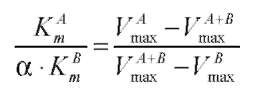
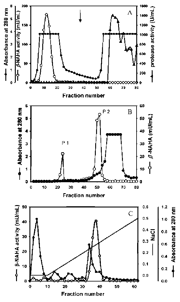
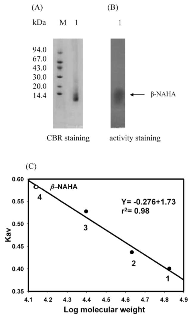
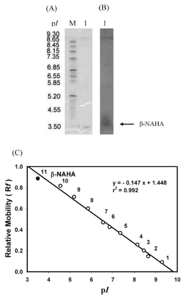
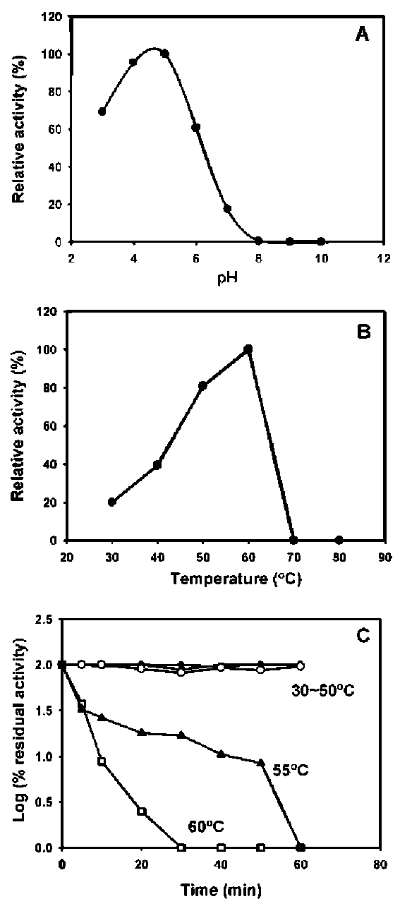
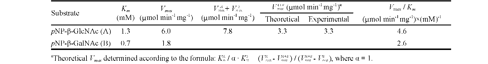
ABSTRACT. A major isoform of β-N-acetylhexosaminidase (β-NAHA) (EC 3.2.1.52) was purified from fig latex in three chromatography steps, affinity chromatography on pAPMA-Sepharose CL-4B to remove cysteine proteases, Sephacryl S-100 HR gel filtration, and DEAE-Sephacel ion-exchange chromatography. The purified β-NAHA appeared almost homogeneous on SDS-polyacrylamide gel electrophoresis and enzyme-ac­tivity staining. The purified enzyme catalyzed the hydrolysis of both p-nitrophenyl-N-acetyl-p-D-glucosaminide (pNP-β-GlcNAc) and β-nitrophenyl-N-acetyl-β-D-galactosaminide pNP-β-GalNAc). The optimum pH for the pNP-β-GlcNAc hydrolysis was 4.5, the optimum temperature was 60°C, the Km was 1.3 mM, the Vmax was 6.0 fimol min-1 mg-1 and the activation energy was 8.93 kcal/mol. The molecular mass of the enzyme was 13.7 kDa, as estimated by gel filtration. The isoelectric point of the enzyme was 3.5, as estimated by isoelectric fo­cusing electrophoresis and activity staining. The enzyme was thermally stable after 60 min at 30-50°C, but its activity decreased significantly at temperatures greater than 55°C. Both the heavy metal ion Hg2+ (0.25 mM) and the chemical modification reagent diethyl pyrocarbonate (2.5 mM) significantly inhibited enzyme activity. Substrate specificity and competition kinetics analysis indicated that the activity of the purified β-NAHA was specific for the p-glycosidic linkage, and the enzyme had only one active site for substrates, pNP-β-GlcNAc and pNP-β-GalNAc.
Keywords: Characterization; Fig (Ficus carica) latex; β-N-acetylhexosaminidase; Purification.
INTRODUCTION
also seen in the whole tissues of individual seeds and seed­lings (Hodge et al., 1996) and in rubber tree latex (Martin, 1991; Giordani et al., 1992). Some plant β-NAHAs also degrade chitin and chitin oligomers (Li and Li, 1970; Yi, 1981; Barber and Ride, 1989), suggesting that the enzyme plays a role in the defense system in plants against chitin-ous pathogens. However, the physiological signification of this enzyme in plants has yet to be fully elucidated.
Latex is the cytoplasmic fluid, containing the usual plant organelles in laticifer cells. Laticifers are anasto­mosed as a result of partial hydrolysis of adjacent walls, and thus form a tube-like network, or paracirculatory sys­tem, through-out the plant (Esau, 1967). When laticifers are injured, latex flows from the wound site. The latex exuded from laticifers is known to contain a variety of de­fense-related proteins such as chitinase, p-1,3-glucanase, hevamine and hevein (Broekaert et al., 1990; Rozeboom et al, 1990; Van Parijs et al., 1991). It has been suggested that latex secretion provides a defense against wounds and/or predators such as insects and microorganisms. Fig tree latex contains proteolytic enzymes (ficin) (Robbins and Lamson, 1934; William, 1958; Whitaker, 1959), chi-tinolytic enzymes such as plant lysozyme (Glazer et al.,
β-N-acetylglucosaminidase catalyzes the release of N-acetylglucosaminyl residues from the non-reducing terminus of oligosaccharides. It is also referred to as p-N-acetylhexosaminidase (β-NAHA) because it is capable of cleaving terminal N-acetyl-β-D-galactosaminyl residues from oligosaccharides as well (Dey and Campillo, 1984; Conzelmann and Sandhoff, 1987; Webb, 1992). This en­zyme is widely distributed in nature and has been detected in animal tissues, microorganisms, and plants (Conzel-mann and Sandhoff, 1987). In plants, β-NAHA is thought to participate in the processing and turnover of glycopro-tein during germination (Neely and Beevers, 1980; Yi, 1981; Vitale and Chrispeels, 1984) and the metabolism of N-glycans during ripening in apples (Choi and Gross, 1994). However, β-NAHA and chitinase activites were

*Corresponding authors: E-mail: sunghy@ntu.edu.tw; Fax: 886-2-23634729; Tel: 886-2-33664522 (Hsien-Yi SUNG); E-mail: ctchang@pu.edu.tw; Fax: 886-4-26530027; Tel: 886-4-26530089 (Chen-Tien CHANG).
24
Botanical Studies, Vol. 52, 2011
1969), and cDNA that encodes basic class I chitinase (Kim et al., 2003). Commercial crude ficin prepared from fig latex, was found to possess chitosanolytic activity (Yalpani
and Pantaleone, 1994).
Recently, we isolated a chitosanase from a commercial ficin preparation, and found that β-NAHA and several other glycosidases were present in fig latex in addition to proteases and chitinolytic enzymes (Chiang et al., 2005; Chiang and Chang, 2006). Here we report on the puri­fication and characterization of a predominant form of β-NAHA in fig latex. Enzymatic properties of the purified β-NAHA including molecular mass, thermal stability, ef­fectors and substrate specificity, as well as the kinetics of competition with mixed substrates were studied.
then filtered. This treatment was repeated 3 times. The gel was packed into a column and slowly washed with 400 mL of 20% dimethyl sulfoxide, followed by 200 mL of 0.1 M sodium phosphate buffer containing 0.1 M 2-mercaptoeth-anol, pH 8.0 (Sluyterman and Wijdennes, 1970).
measurement of β-NAHA activity
A mixture of 0.5 mL of 5 mM pNP-β-GlcNAc in 0.1 M sodium acetate buffer, pH 4.5 and 0.05 mL of enzyme solution was incubated at 40°C for 20 min. The reaction was stopped by the addition of 1 mL of 0.25 M Na2C3. The amount of p-nitrophenol formed was measured by de­termining the absorbance at 420 nm. One unit of enzyme was defined as the amount of enzyme that liberated 1 mi-cromole of p-nitrophenol per minute.
MATERIALS AND METHODS
measurement of exo-glycosidase activity
Latex material
The activities of exo-glycosidases, including p-N-acetylglucosaminidase, p-galactosidase, a-galactosidase, p-glucosidase, a-glucosidase, p-mannosidase, a-mannosidase and p-xylosidase were determined by mea­suring the liberation of p-nitrophenol (as the phenolate anion) from their respective glycosides. In the standard assay, 0.05 mL of enzyme solution was incubated with 0.5 mL of 4 mM p-nitrophenyl glycoside in 20 mM sodium citrate buffer pH 4.0, at 50°C for 10 min, and the p-nitrop-henol formed was measured as described above. One unit of exo-glycosidase was defined as the amount of enzyme that librated 1 micromole of p-nitrophenol per minute.
Latex was collected directly from the unripe fruits of fig trees (Ficus carica) grown on a farm in Taichung, Tai­wan. After lyophilization, the latex sample was stored at -20°C.
Chemicals
N-Acetylimidazole (NAI), 1, 2-cyclohexanedione (CHD), p-hydroxymercuribenzoate (sodium salt, pHMB), diethyl pyrocarbonate (DEPC), 2,4-dinitro-1-fluorobenzene (DNFB), ethyl acetimidate (EAM), phenylmethanesulfonyl fluoride (PMSF), N-ethyl-5-phenylisoazoline-3'-sulfonate (Woodward's reagent K; WRK), N-acetyl-D-glucosamine (GlcNAc), D-glucosamine (GlcN), p-nitrophenyl-a-D-galactopyranoside (pNP-a-Gal), p-nitrophenyl- N-acetyl-p-D-glucosaminide (pNP-β-GlcNAc), p-nitrophenyl-p-D-galactopyranoside (pNP-p-Gal), p-nitrophenyl-p-D-glucopyranoside (pNP-p-Glc), p-nitrophenyl-a-D-mannopyranoside (pNP-a-Man), p-nitrophenyl-p-D-xylopyranoside (pNP-p-Xyl) and p-aminophenylmercuric acetate (pAPMA) were purchased from Sigma-Aldrich Inc. (St. Louis, MO, USA). PhastGel IEF 3-9 gels, Sepharose CL-4B, Sephacryl S-100 HR, DEAE-Sephacel, Superose 6 HR 10/30 columns, Superdex 75 HR 10/30 columns, and HiLoad Superdex 75 HR 16/60 columns were obtained from GE Healthcare Bio-Sciences Corp. (MA, USA).
measurement of chitinase and chitosanase activities
A mixture of 0.15 mL of 0.1 M sodium acetate buf­fer, pH 4.5, 0.1 mL of 1% ethylene glycol chitin or 0.5%
chitosan in the same buffer, and 0.1 mL of H?O was equil­ibrated at 37°C for 5 min. After addition of 50 [iL of an ap­propriate dilution of enzyme, the reaction was allowed to proceed at 37°C for 30 min. The reducing sugar produced was measured colorimetrically as described (Dygert et al., 1965). One chitinase or chitosanase unit was defined as the amount of enzyme that produced 1 micromole of N-acetyl-D-glucosamine or D-glucosamine per minute.
measurement of protease activity
Preparation of pAPmA-Sepharose CL-4B affinity adsorbent
Protease activity was measured using casein as a sub­strate according to the method of Anson (1939) with some modifications. A mixture of 5 mL of 0.6% casein (in 0.05 M sodium phosphate buffer, pH 7.0) and 1 mL of an ap­propriate dilution of enzyme was incubated at 37°C for 10 min as described previously (Chiang et al., 2005).
Sepharose CL-4B, 100 mL packed, was activated with CNBr at pH 11 as described by Cuatrecasas (1970). At the end of the reaction the Sepharose-CL-4B was washed in a Buchner funnel with 1.5 L of 0.1 M NaHC3, pH 9.0, and resuspended in 200 mL of 10% dimethyl sulfoxide at 4°C. To this suspension, 1.3 g of pAPMA dissolved in 20 mL of dimethyl sulfoxide was added slowly. The suspension was stirred slowly at 4°C for 20 h then warmed to 30°C and filtered. The substituted Sepharose CL-4B was resuspended in 130 mL of 20% dimethyl sulfoxide for 5 min
Purification of β-NAHA
Lyophilized fig latex, 300 mg, was dissolved in 6 mL of 50 mM sodium acetate buffer containing 0.5 M NaCl, pH 4.5. After centrifugation (20 min at 10,000 g), the superna­tant was applied to a pAPMA-Sepharose CL-4B column (1.6 x 10 cm) pre-equilibrated with 50 mM sodium acetate
CHANG et al. ― p-W-acetylhexosaminidase from fig latex
25
buffer containing 0.5 M NaCl, pH 4.5, for affinity adsorp­tion of cysteine proteases. After sample application, the column was washed with the same buffer at a flow rate of 30 mL/h to elute the non-bound β-NAHA, and then with 0.15 M 2-mercaptoethanol in the equilibrium buffter to elute the bound proteases; 1 mL fractions were collected. Each fraction was monitored for absorbance at 280 nm as well as β-NAHA and protease activities. Fractions con­taining β-NAHA were pooled, concentrated by ultrafiltra-tion on an Amicon Centricon Plus-20 (NMWC 5,000) filter. The concentrated enzyme solution was applied to a Sephacryl S-100 HR column pre-equilibrated with 0.025 M imidazole-HCl buffer, pH 7.4 and eluted with the equi­librium buffer at a flow rate of 30 mL/h; 10 mL fractions were collected. The β-NAHA was eluted in minor (P1) and major (P2) activity peaks. Fractions containing major peak of β-NAHA were pooled, concentrated by ultrafiltration and applied to a DEAE-Sephacel column pre-equilibrated with 0.025 M imidazole-HCl buffer, pH 7.4. β-NAHA adsorbed on the column was eluted with a linear NaCl gra-dient (0-0.5 M) in equilibrium buffer at a flow rate of 30 mL/h; 2 mL fractions were collected. Each fractions was monitored for absorbance at 280 nm and β-NAHA activity. Fractions containing β-NAHA were pooled, concentrated by ultrafiltration and desalted by gel filtration on a Su-perose 6 HR 10/30 column using 0.025 M imidazole-HCl buffer as elution buffer. The purified enzyme was stored at -10°C until use.
from the slope of Hill plot (log[V/(Vmax - V)] against log (S), where V is the initial velocity and S is substrate concentra­tion).
Determination of activation energy
The activation energy of β-NAHA for pNP-β-GlcNAc hydrolysis was determined by plotting log Vmax versus 1/T (Segal, 1976).
Determination of active sites
The number of active sites in the purified β-NAHA was determined using an equimolar mixture of the two sub­strates, pNP-β-GlcNAc and pNP-p-GalNAc, at concentra­tions from 0.5 mM to 5 mM for each substrate. The experimental maximum velocity of the enzyme incubated with the mixed substrates was calculated from the Line-weaver-Burk plots (Lineweaver and Burk, 1934). The the­oretical maximum velocity of β-NAHA incubated with the mixed substrates, VA+,B was calculated by the following equation (Calvo et al., 1978):
where K m, K m, Vmax, Vmax are the Michaelis constants and maximum velocities for each substrate, as determined from separate reactions of β-NAHA with each of the sub­strates, A (pNP-β-GlcNAc) and B (pNP-p-GalNAc). In this case, a, the ratio of the concentration of substrates = 1.
Determination of optimum pH
The optimum pH for the purified β-NAHA was assayed in universal buffer (Britton and Robinson type; Dawson et al., 1969) at pH values ranging from 3 to 10 at 40°C.
Determination of native molecular mass
The native molecular mass of the purified β-NAHA was determined by gel filtration (Andrews, 1965) on an FPLC system (Amersham Pharmacia Biotech AB, Up-psa, Sweden) with a Superdex 75 HR 10/30 column using Pharmacia low molecular weight gel filtration calibration kit (Uppsa, Sweden) as marker proteins. The native mo­lecular mass of β-NAHA was calculated from the calibra­tion curve. The native molecular mass of minor peak of β-NAHA was determined on an FPLC system with a Su­perpose 6 HR 10/30 column using Pharmacia high molec­ular weight gel filtration calibration kit (Uppsala, Sweden) as marker proteins.
Determination of optimum temperature and thermal stability
The optimum temperature for the purified β-NAHA was assayed at temperatures ranging from 30 to 80°C at pH 4.5. To assay thermal stability, aliquots of enzyme solutions were incubated in a thermostatic water bath set to temperatures ranging from 30 to 60°C for 5 to 60 min. The enzymatic activity remaining in each sample was then measured at 40°C, pH 4.5, as described above. The β-NAHA activity in the unheated enzyme solution represented the initial activity (100%) from which the log proportion (percent) of the activity remaining after heat treatment was calculated.
Isoelectric focusing electrophoresis (IEF) and gel activity staining
Determination of michaelis constant and hill coefficient
The isoelectric point (pi) of β-NAHA was deter­mined by IEF using PhastGel IEF 3-9 gel and gel activ­ity staining. A detailed procedure for IEF is outlined in the Pharmacia PhastSystem Separation Technique File No. 100. After electrophoresis, the gels were stained for protein with Coomassie Brilliant Blue R-250 (CBR) or for β-NAHA activity with naphthol AS-BI-p-N-acetyl-D-glucosaminide as a substrate and new fuchsin as a diazo color reagent (Hayashi, 1965; Loida et al., 1979). An ali-quot of the standard pi marker proteins mixture from the Pharmacia pi calibration kit was run beside the β-NAHA
The Michaelis constant (Km) and maximum velocities (Vmax) of pNP-β-GlcNAc hydrolysis were determined at substrate concentrations ranging from 0.5 to 5 mM at pH 4.5 and 40°C. The Km and Vmax of β-NAHA for pNP-p-GlcNAc were calculated using the Lineweaver-Burk recip-rocal plot method (Lineweaver and Burk, 1934). The Hill coefficient of β-NAHA for pNP-β-GlcNAc was calculated
26
Botanical Studies, Vol. 52, 2011
sample. The Rf values measured directly from the gel were used to construct the pi calibration curve. The Rf value of the β-NAHA band was used to estimate the pi value of β-NAHA from the calibration curve.
tion were incubated at 30°C for 30 min with 0.5 mL of corresponding reaction buffer as the control or 0.5 mL of chemical modification reagent. After dialysis against 0.05 M sodium acetate buffer, pH 5.0, the enzyme activity in each sample was measured and expressed as the relative activity percentage calculated from the ratio of the specific activity of β-NAHA treated with chemical modification reagents to that of the non-treated sample.
SDS-polyacrylamide gel electrophoresis (SDS-PAGE) and gel activity staining
SDS-PAGE was carried out with a PhastGel gradient 8-25% using the Pharmacia PhastSystem flat bed appara­tus (Amersham Pharmacia Biotech AB; Uppsala, Sweden). The detailed procedure is outlined in the Pharmacia Phast-System Separation Technique File No. 110. Before being loaded onto the gel, the enzyme solution was treated with an equal volume of sample buffer containing only 5% SDS followed by heating at 50°C for 30 min. After electropho-resis, renaturation of enzyme activity was carried out in 0.1 M citric acid-sodium phosphate buffer pH 6.0, containing 1% (v/v) Triton X-100 for 30 min at room temperature (25°C). β-NAHA activity was detected as described above (Hayashi, 1965). Proteins were stained with CBR.
Data analysis
For analysis of effects of metal ions and chemical modification reagents on enzyme activity, analytic mea­surements were performed in triplicate. Other analytic measurements were performed in duplicate.
RESULTS AND DISCUSSION
Glycosidase activities in fig latex
As shown in Table 1, fig latex was shown to contain a variety of glycosidase activities in addition to proteases, chitinase and chitosanase activities. The highest level of glycosidase activity was seen for a-galactosidase, followed by activities for β-NAHA, p-galactosidase, p-glucosidase, a-mannosidase, and p-xylosidase. No activities for a-glucosidase, p-mannosidase and p-fucosidase were de­tected. Plant glycosidases that catalyze the hydrolysis of aryl and alkyl glycosides have been found to be involved in a wide variety of biological processes (Dey and Campil-lo, 1984). In plant latex, glycosidases may be involved in the cell-wall degradation process during differentiation of articulated laticifers (Gioradani and Noat, 1988). Li and Li (1972) isolated a-galactosidase from fig latex and found that this enzyme is involved in cell-wall degradation, and is useful for the study of the sequence and anomeric con­figuration of a-galactosyl residues in the oligosaccharide chains of various complex carbohydrates.
Effect of metal ions and other compounds
Aliquots of purified β-NAHA (25 μL) were incubated at 25°C for 30 min with 25 μL of deionized water as control or 25 μL of one of the following compounds at concentrations of 0.05 to 10 mM: HgCl2, AgN3, CuCl2, CaCl2, MgCl2, ZnCl2, NaCl, KCl, EDTA, SDS and BSA. Following treatment, enzyme activity in each sample was measured and expressed as a relative activity percentage calculated from the ratio of the specific activity of the treated β-NAHA to that of the untreated sample.
effect of chemical modification reagents
Purified β-NAHA was concentrated by ultrafiltration
on an Amicon Centricon Plus-20 (NMWC 5000) filter.
After concentration, 0.5 mL aliquots of the enzyme solu-
Table 1. Protease, chitinase, chitosanase and glycosidase activities in fig latex.

Enzyme

Substrate

Activitya (mU/g powder)b

Protease

Casein

6,140x103
Chitinase
Ethylene glycol chitin
35,925
Chitosanase
Chitosan
81,840
a-Galactosidase
pNP-α-Gal
19,775
β-N-acetylhexosaminidase
pNP-β-GlcNAc
8,866
β-Galactosidase
pNP-β-Gal
4,956
β-Glucosidase
pNP-β-Glc
4,932
α-Mannosidase
pNP-α-Man
2,391
β-Xylosidase
pNP-β-Xyl
596

aOne unit of protease is defined as the amount of enzyme needed to produce 1 μg of tyrosine per minute from casein at 37°C. One unit of chitinase is defined as the amount of enzyme needed to produce 1 micromole of GlcNAc per minute from ethylene glycol chitin at 37°C. One unit of chitosanase is defined as the amount of enzyme needed to produce 1 micromole of GlcN per minute from chitosan at 37°C. Units of glycosidases for hydrolysis of variousp-nitrophenylglycoside substrates are defined as the amount of enzyme needed to produce 1 micromole of p-nitrophenol per minute at 50°C at pH 4.0.
bOne mU is 10-3 unit.
CHANG et al. ― p-W-acetylhexosaminidase from fig latex
27
Table 2. Purification of p-N-acetylhexosaminidase from fig latexa.

Step

Total volume (mL)

Total activity (mU)b

Total protein (mg)

Specific activity (mU/mg)

Purification (fold)

Yield
(%)

Crude extract

4.4

1,339

66.4

20.2

1

100
pAPMA-Sepharose affinity removal of thiol proteases
13
1,151
35.8
32.2
1.6
86
Sephacryl S-100 gel filtration
P1 (minor)
10
145.4
0.08
1,818
90
11
P2 (major)
20
819.7
7.75
105.8
5.2
62
P2 Purification
DEAE-Sephacel
16.2
758.3
0.012
61,525
3,046
57

aData were obtained from 300 mg of lyophilized fig latex. bOne mU is 1 x 10⁻³ unit.
Purification of β-NAHA
Purification of β-NAHA using the protocols summa­rized in Table 2 enabled us to remove proteolytic enzymes and isolate a predominant form of β-NAHA from fig latex. The β-NAHA was purified 3,046 fold, and the total yield was 57%. Affinity chromatography on a pAPMA-Sephar-ose CL-4B column was initially used for the adsorption of thiol proteases. Neither β-NAHA nor chitinolytic enzymes were adsorbed and were eluted directly by washing the column with equilibrium buffer. Thiol proteases were ad­sorbed on the column and were eluted by equilibrium buf­fer containing 0.15 M 2-mercaptoethanol (Figure 1A).
After affinity chromatography, the β-NAHA was further purified by gel filtration on a Sephacryl S-100 HR column. As shown in Figure 1B, the β-NAHA was resolved into minor and major peaks. The minor peak was eluted in fractions 23-24 (indicated as P1). The predominant form was eluted in the major peak in fractions 47-51 (indicated as P2). This predominant isoform was further purified on a DEAE-Sephacel column. β-NAHA was bound to the column at pH 7.4 and eluted with a linear NaCl gradient (0-0.5 M) in equilibrium buffer (Figure 1C). Most of the inactive proteins were removed by this step. After being desalted on Superose 6 HR, the purified β-NAHA was concentrated and subjected to SDS-PAGE and gel activ­ity staining. As shown in Figure 2A and B, both CBR and activity staining revealed a single band with a molecular mass of 12.4 kDa. These results indicated that the purified enzyme was almost homogeneous.
Figure 1. Purification of p-N-acetylhexosaminidase from fig latex in three chromatography steps, affinity chromatography on pAP­MA-Sepharose CL-4B (A), Sephacryl S-100 HR gel filtration (B), and DEAE-Sephacel ion-exchange chromatography (C). The pAP­MA-Sepharose CL-4B column (1.6 x 10 cm) was equilibrated with 50 mM sodium acetate buffer containing 0.5 M NaCl, pH 4.5, after which the crude extract from fig latex was applied. Unbound proteins were eluted with equilibrium buffer; bound proteases were eluted with equilibrium buffer containing 0.15 M 2-mercaptoethanol (arrow indicates beginning of elution) at a flow rate of 30 mL/h; 1 mL fractions were collected. The Sephacryl S-100 HR column (2.6 x 70 cm) was equilibrated with 0.025 M imidazole-HCl buffer, pH 7.4, after which the β-NAHA recovered from the pAPMA-Sepharose CL-4B column was applied. The enzyme was eluted with the same buffer at a flow rate of 30 mL/h; 10 mL fractions were collected. The DEAE-Sephacel column (1.6 x 10 cm) was equilibrated with 0.025 M imidazole-HCl buffer, pH 7.4, after which the predominant peak of β-NAHA (P2) from the Sephacryl S-100 HR column was ap­plied. Bound proteins were eluted with a linear NaCl gradient (0-0.5M) in equilibrium buffer at a flow rate of 30 mL/h; 2 mL fractions were collected.
28
Botanical Studies, Vol. 52, 2011
Optimum pH, optimum temperature and thermal stability
As shown in Figure 3B, β-NAHA exhibited 80-100%
of its maximum activity, over a temperature range of 50-60°C, with a sharp decrease at temperatures above 60°C. The optimum temperature for enzyme activity was ap­proximately 60°C.
To examine the thermal stability of β-NAHA, the en­zyme was incubated at various temperatures (from 30 to 60°C) for 5 to 60 min and measured the residual activity under standard assay conditions. As shown in Figure 3C, the enzyme was fairly stable between 30 and 50. There was a relatively modest loss of activity at 55°C. At 60°C the enzyme was rapidly inactivated.
As shown in Figure 3A, the purified β-NAHA retained 60-100% activity between pH 3 and 6, but was rapidly in­activated above pH 6. The optimum pH of the enzyme was 4.5. This is in the range of the values reported for β-NAHA from other sources, from pH 4.0 in cabbage (Chang et al., 1998) to pH 6.0 in fenugreek seeds and apple (Bouquelet and Spik, 1976; Choi and Gross, 1994).
molecular mass
The molecular mass of the predominate form of the β-NAHA was determined by gel filtration on Superdex 75 HR to be 13.7 kDa (Figure 2C). This value is close to the value of 12.4 kDa estimated from SDS-PAGE and gel activity staining of β-NAHA (Figure 2B), indicating the β-NAHA was a monomeric enzyme. The molecular mass of β-NAHA found in plants varies from 40 kDa in lupin seeds (McFarlane et al., 1984) to 236 kDa in apple cortical tissue (Choi and Gross, 1994). Different oligomeric struc-tures of β-NAHA have also been described. The β-NAHA from apples was reported to be a homo-octamer (Choi and Gross, 1994). The β-NAHA from cabbage was reported to be a hetero-trimer with a molecular mass of 150 kDa (Chang et al., 1988). β-NAHA isolated from germinating fenugreek seeds was found to exist in 4 molecular forms, two trimers, a pentamer and a hexamer, each composed of identical subunits (Bouquelet and Spik, 1976; 1978). The β-NAHA from pinto bean was found to be a homo-dimer with a molecular mass of 112 kDa (Agrawal and Bahl, 1972). The β-NAHAs from lupin, rice seeds and maize seedlings were monomers with molecular masses of 40, 52 and 72 kDa, respectively (McFarlane et al., 1984; Jin et al., 2002; Oikawa et al., 2003). The molecular mass of the predominant form of β-NAHA isolated from fig latex was much lower than these previously reported values. Howev-er, the molecular mass of the minor form of β-NAHA (232 kDa; data not shown) is similar to the values reported for pea seeds (210 kDa; Harley and Beevers, 1987) and apples (236 kDa; Chio and Gross, 1994).
Figure 2. SDS-PAGE and determination of native molecular mass of β-NAHA by gel filtration. SDS-PAGE was performed with a PhastGel gradient 8-25% and PhastGel SDS buffer strips. After electrophoresis, the gels were subjected to coomassie blue staining (A) or β-NAHA activity staining (B) Lane 1 contained the purified β-NAHA. Lane M contained molecular mass marker proteins, including phosphorylase b (97.4 kDa), bovine serum albumin (66.2 kDa), chicken egg albumin (45.0 kDa), carbonic anhydrase (29.0 kDa), trypsin inhibitor (20.1 kDa), and lyso-zyme (14.4 kDa). The native molecular mass of the purified β-NAHA was estimated by gel filtration on a HiLoad Superdex 75 HR 16/60 column (C). The numbers 1 to 4 on the line of the molecular weight calibration curve indicate low molecular weight standard proteins bovine serum albumin (MW = 67 kDa), ovalbumin (MW = 43 kDa), chymotrypsinogen A (MW = 25 kDa), and ribonuclease A (MW = 13.7 kDa), respectively.
Isoelectric point
As shown in Figure 4, CBR staining of the IEF gel re­vealed a major protein band (Figure 4A) which exhibited β-NAHA activity on gel activity staining (Figure 4B). The isoelectric point of the purified β-NAHA was found to be 3.5 as determined by comparing the relative mobility (Rf) of β-NAHA with that of the pi markers developed in the same IEF gel (Figure 4C). This value is lower than pub­lished data for other β-NAHAs, for which, most of the pi values reported for plant β-NAHAs are in the range of 4.65
to 6.78 (Bouquelet and Spik, 1978; Carratu et al., 1985; Barber and Ride, 1989; Oikawa et al., 2003).
CHANG et al. ― p-W-acetylhexosaminidase from fig latex
29
Effect of metal ions and various compounds
As shown in Table 3, the purified β-NAHA was inhib­ited by 0.25 mM Ag+ and Hg2+ and slightly inhibited by 5 mM Cu2+. Similar inhibitory effects of Ag+ and Hg2+ were previously seen on β-NAHA from jack bean meal (Li and Li, 1970), the four molecular forms of β-NAHA from fenugreek seeds (Bouquelet and Spik, 1978) and three iso-forms of β-NAHA from mungbean seedlings (Chen et al., 2004). Other metal ions and compounds studied had no significant effect on the activity of the enzyme.
Figure 4. Determination of pi of β-NAHA by isoelectric focus­ing electrophoresis: (A) CBR staining, (B) gel activity stain­ing, (C) pi calibration curve. Electrophoresis was performed in a PhastGel IEF 3-9 gel. Lane 1 contained purified β-NAHA. Lane M contained pi marker proteins from a pi calibration kit. The numbers 1 to 11 on the line of pi calibration curve includ­ing pi marker proteins trypsinogen (pi = 9.3), lentil lectin-basic band (pi = 8.65), lentil lectin-middle band (pi = 8.45), lentil lectin-acidic band (pi = 8.15), myoglobin-basic band (pi = 7.35), myoglobin-acidic band (pi = 6.85), human carbonic an-hydrase B (pi = 6.55), bovine carbonic anhydrase B (pi = 5.85), p-lactoglobulin A (pi = 5.20), soybean trypsin inhibitor (pi = 4.55), and amyloglucosidase (pi = 3.55), respectively.
effect of chemical modification reagents
It is generally believed that an enzyme will be inhib­ited or inactivated if the amino acid side chain involved in catalytic activity is chemically modified. In this study, a number of reagents were used to modify the side chain of β-NAHA (Table 4). Among the chemical modification reagents tested, 2.5 mM DEPC, a reagent that usually acts on the histidine residues in proteins, completely inhibited enzyme activity. Therefore, the imidazole group (histidine side chain) appears to be essential for the catalytic activ­ity of the enzyme. However, it should be noted that the
Figure 3. Optimum of pH (A), optimum temperature (B) and thermal stability (C) of β-NAHA. For determination of optimum pH, β-NAHA activity was assayed in a universal buffer, at pH 3 to 10. For determination of optimum temperature, β-NAHA ac­tivity was assayed at pH 4.5 from 30 to 80°C. Thermal stability was measured by incubating the enzyme for 0-60 min at 30, 40, 50, 55 or 60°C prior to assaying.
30
Botanical Studies, Vol. 52, 2011
activity of this enzyme was also inhibited from 15.9%
to 26.9% by DNFB, PMSF, pHMB and CHD. DNFB is
a chemical modification reagent that usually acts selec­tively on the lysine residues in proteins, whereas PMSF, pHMB and CHD act on serine, cysteine and arginine resi­dues, respectively. Therefore, lysine, serine, cysteine and arginine residues may also be related to enzyme activity. These residues may not be located at the active site of the enzyme, but instead may be involved in maintenance of conformation of the enzyme. Not much information has been reported on enzyme inhibition by chemical modifica­tions. Fenugreek β-NAHAs was inhibited by 1 mM pHMB (Bouquelet and Spik, 1978). Mung bean β-NAHA III was inhibited by 5-5'-dithiobis (2-nitrobenzoic acid), a reagent for the selective chemical modification of cysteine resi-dues (Chen et al., 2004). Rice β-NAHA was inhibited by 1 mM chloramine T (Jin et al. , 2002), a potential chemical modifier of tryptophan residues in proteins.
Table 3. Effect of metal ions and other compounds on the activity of β-NAHA.

Reagenta

Final concentration (mM)

Relative activity (%)b

None

--

100
HgCl2
0.25
0 ± 0.5
AgNO3
0.25
69.4 ± 5.0
AgNO3
1
69.1 ± 2.7
CuCl2
5
82.0 ± 1.5
CaCl2
5
94.5 ± 1.0
MgCl2
5
107.7 ± 0.2
ZnCl2
5
96.0 ± 1.1
NaCl
5
99.6 ± 5.1
KCl
5
96.7 ± 4.6
EDTA
5
106.0 ± 0.9
BSA
0.1
103.6 ± 3.5
SDS
0.5
105.6 ± 2.9
SDS
0.1
107.0 ± 1.2

aBSA, bovine serum albumin; SDS, sodium dodecyl sulfate; EDTA, ethylenediamine tetraacetic acid.
bData are represented as mean ± standard deviation (n = 3).
Table 4. Effect of various chemical modification reagents on the activity of β-NAHA.

Reagenta

Final concentration (mM)

Relative activity (%)b

None

--

100
NBS
0.5
100.9±1.2
NBS
5
102.6±0.3
EAM
250
104.2±6.5
NAI
2.5
101.4±1.1
DNFB
2.5
84.1±0.8
PMSF
2.5
73.1±7.7
DEPC
2.5
0±0.3
WRK
50
106.3±5.2
pHMB
0.5
75.9±0.6
CHD
1.25
75.1±2.6
Activation energy
An Arrhenius plot obtained by plotting Vmax versus 1/T, was used to determine the activation energy of the enzyme for catalysis of pNP-β-GlcNAc hydrolysis. The Arrhenius plot for β-NAHA showed a straight line. The activation en­ergy (Ea) calculated from the line was 8.93 kcal/mol. Not much information is available on the activation energy of plant β-NAHA-catalyzed reactions. The activation energy of β-NAHA from pinto beans (Agrawal and Bahl, 1972)
and the latex of Ficus glavrata (Orlacchio et al., 1985) for the catalysis of pNP-β-GlcNAc hydrolysis was found to be 9.8 and 13.2 kcal/mol, respectively.
Kinetic properties
To determine the kinetic properties of β-NAHA puri­fied from fig latex, the enzymatic activity was assayed us­ing pNP-β-GlcNAc, pNP-p-GalNAc, and an equimolar mixture of the two at various concentrations. The kinetic parameters, Km and Vmax , of β-NAHA obtained from Line-weaver-Burk plots are summarized in Table 5. The Km val­ues for pNP-β-GlcNAc and pNP-p-GalNAc were 1.3 and
0.7 mM, respectively, whereas the Vmax values were 6.0 and 1.8 famol min-1 mg-1, respectively. These results indi­cate that the purified β-NAHA had a higher specific con­stant (Vmax/Km) for pNP-β-GlcNAc than for pNP-p-
GalNAc. Therefore, pNP-β-GlcNAc appears to be the preferred substrate for the enzyme. The kinetics of compe­tition was investigated by using a mixed substrates to de­termine whether the β-NAHA had a single active site for both substrates or 2 separate active sites, one for each sub­strate. The experimental Vmax value for β-NAHA deter­mined from the Lineweaver-Burk plot was the same as the theoretical value (Table 5, V^+X). This finding indicates that both substrates used a single active site. If an enzyme cata­lyzes the hydrolysis of two substrates at the same active site, the total rate of reaction will be less than the sum of the rates of the separate reactions with each substrate (Dixon and Webb, 1964). Because one active site is used, there is a competition between the substrates. As shown in

aNBS, in 50 mM sodium acetate buffer, pH 4.0 (Viswanatha and Lanwson, 1961); EAM, in 0.2 M sodium phosphate buf­fer, pH 8.0 (Shaw and Chang, 1988); NAI, in 0.1 M sodium phosphate buffer, pH 8.0 (Yamasaki et al" 1989); DNFB, in 0.1 M sodium phosphate buffer, pH 8.0 (Welches and Baldwin, 1981); PMSF, in 0.2 M sodium phosphate buffer, pH 8.0 (Gold and Fahrney, 1964); DEPC, in 0.1 M sodium phosphate buf-fer, pH 6.0 (Miles, 1977); WRK, in 0.25 M sodium phosphate buffer, pH 6.0 (Vangrysperre et al., 1989); pHMB, in 50 mM sodium phosphate buffer, pH 7.0 (Liu and Speer, 1977); CHD, in 0.1M sodium phosphate buffer, pH 8.0 (Liu et al, 1968).
bData are represented as mean ± standard deviation (n = 3).
CHANG et al. ― p-W-acetylhexosaminidase from fig latex
31
Table 5. Kinetic constants for the study of the competition between pNP-β-GlcNAc and pNP-p-GalNAc for the active sites of β-NAHA.
Table 5, the rate of the hydrolysis of the mixed substrates (V^+f = 3.3 fimol min-1 mg-1) was less than the sum of the rates obtained with the separate substrates (Vmax + V^ax = 7.8 fmol min-1 mg-1). The total rate of the reaction was 3.3 fmol min-1 mg-1, which is less than the sum of the individ­ual reactions, 6.0 + 1.8 = 7.8 fimol min-1 mg-1. From our kinetic assays, it may be deduced that the purified β-NAHA had a single active site for both substrates. These results agree with those for the enzyme isolated from jack bean meal (Li and Li, 1970), Golden Delicious-apples (Choi and Gross, 1991) and rice seeds (Jin et al., 2002).
Table 6. Substrate specificity of β-NAHA.

Substrate

Relative activity (%)

pNP-β-GlcNAc

100
pNP-p-GalNAc
45.5
pNP-a-GlcNAc
0
pNP-a-GalNAc
0

substrate specificity and competition kinetic studies, we found that the enzyme is specific for glycosidic bonds of p configuration and has a single active site for binding both pNP-β-GlcNAc and pNP-p-GalNAc substrates. The activ­ity of this enzyme in the hydrolysis of chitin oligomers remains to be investigated to clarify whether this β-NAHA is involved in chitin degradation and participated in the defense system in fig latex.
Substrate specificity
As shown in Table 6, the purified β-NAHA showed specificity for p-glycosidic linkages. The ratio of the hydrolysis of pNP-β-GlcNAc to that of pNP-β-GalNAc was 2.2. This value is in the range of previously reported values: 1.25 to 18.0 for the 4 isozymes of β-NAHA from fenugreek seeds (Bouquelet and Spik, 1978), 1.9 to 4.8 for the three isozymes of β-NAHA from lupin seeds (McFar-lane et al., 1984), 4.0 for the β-NAHA from rice seeds (Jin et al., 2002), and 3.1 to 3.6 for the 3 isoforms of β-NAHA from mungbean seedlings (Chen et al., 2004). Some plant β-NAHAs also degrade chitin and chitin oligomers. For instance, cotton seed β-NAHA liberates GlcNAc from chi-tin, ovalbumin and pronase-digested wheat germ lectin (Yi, 1981), wheat leaf β-NAHA (Barber and Ride, 1989) liber­ates GlcNAc from chitin oligomers, and maize β-NAHA (Oikawa et al., 2003) releases GlcNAc from chitin and chi-tin oligomers. Therefore, their participation in the process of chitin-elicited lignifications (Barber et al., 1989) and in the system of defense against chitinous pathogens (Hodge et al., 1996) have been suggested. In fenugreek seeds, four isoforms of β-NAHAs have been isolated (Bouwuel and Spok, 1978), and form 1 showed higher activity towards GlcNAc2 compared to pNP-β-GlcNAc, while the other three forms had little activity for hydrolyzing GlcNAc2. The β-NAHA purified from maize (Oikawa et al., 2003) also showed much higher activity for hydrolyzing Gl-cNAc2 than for hydrolyzing pNP-β-GlcNAc. Therefore, there are at least two types of β-NAHAs in plant with re­spect to substrate specificity ultizing these two classes of substrate, pNP-β-GlcNAc and GlcNAc2.
In conclusion, the β-NAHA isolated from fig latex is very similar in most properties to β-NAHA isolated from other plants. However, the molecular mass of the enzyme was much small than those of other plant β-NAHAs. From
Acknowledgements. Financial support provided by the
National Science Council (NSC-96-2313-B-126-003-
MY3) of the Republic of China for this study is greatly appreciated.
literature cited
Agrawal, K.M.L. and O.P. Bahl. 1972. a-Galactosidase, p-galactosidase, p-glucosidase, p-N-acetylglucosaminidase, and a-mannosidase from pinto beans (Phaseolus vulgaris). in S. P. Colowick and N. O. Kaplan (eds.), Methods in Enzymology. Vol. 28. Academic Press, New York, pp. 720-728.
Andrews, P. 1965. The gel-filtration behavior of proteins related to their molecular weights over a wide range. Biochem. J. 96: 596-606.
Anson, M.L. 1939. The estimation of pepsin, trypsin, papain and cathepsin with hemoglobin. J. Gen. Physiol. 22: 79-89.
Barber, M.S. and J.P. Ride. 1989. Purification and properties of a wheat leaf N-acetyl-p-D-hexosaminidase. Plant Sci. 60:163-172.
Barber, M.S., R.E. Bertram, and J.P. Ride. 1989. Chitin oli-gosaccharides elicit lignification in wound wheat leaves. Physiol. Mol. Plant Pathol. 34: 3-12.
Bouquelet, S. and G. Spik. 1976. Characterization and localiza­tion of four forms of N-acetyl-p-D-hexosaminidase from fenugreek (Trigonella foenum graecum) germinated seeds.
32
Botanical Studies, Vol. 52, 2011
FEBS Lett. 63: 95-101.
Bouguelet, S. and G. Spik. 1978. Properties of four molecular forms of N-acetyl-p-D-hexosaminidase from germinating seeds of fenugreek (Trigonella foenum graecum). Eur. J. Biochem. 84: 551-559.
Broekaert, W., H.I. Lee, A. Kush, N.H. Chua, and N. Raikhel.
1990. Wound-induced accumulation of mRNA containing a hevein sequence in laticifers of rubber tree (Hevea brasil-iensis). Proc. Natl. Acad. Sci. USA 87: 7633-7637.
Calvo, P., A. Reglero, and J.A. Cabezas. 1978. Purification and properties of p-N-acetylhexosaminidase from the mollusk Helicella evicetrorum Muller. Biochem. J. 175: 743-750.
Carratu, G., C., Colacino, S. Contu, and M. Giannattasio. 1985. p-N-acetylglucosaminidase and p-galactosaminidase from aleurone layers of resting wheat grains. Phytochemistry 24:1465-1469.
Chang, C.T., F.P. Young, M.H. Chang and H.Y. Sung. 1998. Pu­rification and properties of p-N-acetylhexosaminidase from cabbage. Biochem. Mol. Biol. Int. 45: 371-380.
Chen, Y.-C., W.-L. Liu, H.-C. Hsu, Y.-A. Lee and C.-S. Chen.
2004. Purification and characterization of isoforms of p-N-acetylhexosaminidase form mungbean seedlings. Bot. Bull. Acad. Sin. 45: 275-283.
Chiang, C.L. and C.T. Chang. 2006. Characterization of a chitin-ase isolated from a commercial ficin preparation. Taiwanese J. Agric. Chem. Food Sci. 44: 207-215.
Chiang, C.L., Y.M. Chang, C.T. Chang, and H.Y. Sung. 2005.
Characterization of a chitosanase isolated from a commer­cial ficin preparation. J. Agric. Food Chem. 53: 7579-7585.
Choi, S.Y. and K.C. Gross. 1994. N-Acetyl-D-glucosaminidase from 'Golden Delicious' apples. Phytochemistry 36: 1-6.
Conzelmann, E. and K. Sandhoff. 1987. Glycolipid and gly-coprotein degradation. in A. Meister (ed.), Advances in Enzymology, Vol. 60. John Wiley and Sons, New York, Chichester, Brisbanen, Toronto, Singapore, pp. 89-216.
Cuatrecasas, P. 1970. Protein purification by affinity chromatog-raphy. J. Biol. Chem. 245: 3059-3065.
Dawson, R.M.C., D.C. Elliott, W.H. Elliott, and K.M. Jones. 1969. Data for Biochemical Research. Clarendon, Oxford, U.K., pp. 483-498.
Dey, P.M. and E.D. Campillo. 1984. Biochemistry of the mul­tiple forms of glycosidases in plants. in A. Meister (ed.), Advances in Enzymology, Vol. 56. John Wiley and Sons, New York, Chichester, Brisbanen, Toronto, Singapore, pp. 141-249.
Dixon, M. and E.C. Webb. (eds.). 1964. Enzymes. Longmans, London, pp. 54-90.
Dygert, S.L., H. Li, D. Florida, and J.A. Thomas. 1965. Deter­mination of reducing sugar with improved precision. Anal. Biochem. 13: 367-374.
Esau, K. (ed.) 1967. Plant Anatomy. John Wiley and Sons, New York, pp. 308-337.
Giordani, R. and G. Noat. 1988. Isolation, molecular properties and kinetics studies of a strict p-fucosidase from Lactuca
sativa latex. Its possible role in the cell-wall degradation of the articulated laticifers. Eur. J. Biochem. 175: 619-625.
Giordani, R., S. Benyahia. M. Teissere, and G. Noat. 1992. Pu­rification and properties of N-acetyl-p-D-glucosaminidase from Hevea brasiliensis latex. Plant Sci. 84: 25-34.
Glazer, A.N., A.D. Barel, J.B. Howard, and D.M. Brown. 1969.
Isolation and characterization of fig lysozyme. J. Biol.
Chem. 244: 3583-3589.
Gold, A.M. and D.E. Fahrney. 1964. Sulfonyl fluorides as in­hibitors of esterases. II. Formation and reactions of phenyl-methanesulfonyl a-chmotrypsin. Biochemistry 3: 783-791.
Harley, S.M. and L. Beevers. 1987. Isozymes of p-N-acetylhexosaminidase from pea seeds (Pisum sativum L.). Plant Physiol. 85: 1118-1122.
Hayashi, M. 1965. Histochemical demonstration of N-acetyl-p-D-glucosaminidase employing naphthol AS-BI N-acetyl-p-D-glucosaminide as substrate. J. Histochem. Cytochem. 13:355-360.
Hodge, A., I.J. Alexander, and G.W. Gooday. 1996. Measure-ment in situ of chitinase and p-N-acetylglucosaminidase activities in germinating seeds of Pinus sylvestris
and Euca­lyptus pilularis. Plant Physiol. Biochem. 34:
301-306.
Jin, Y.L., Y.Y. Jo, K.Y. Kim, J.H. Shim, Y.W. Kim, and R.D.
Park. 2002. Purification and characterization of p-N-acetylhexosaminidase from rice seeds. J. Biochem. Mol. Biol. 35: 313-319.
Kim, J.S., Y.Q. Kim., H.T. Ryu, Y.S. Kwak., J.Y. Lee, and H. Kang. 2003. Isolation of stress-related genes of rubber par­ticles and latex in fig tree (Ficus carica) and their expres­sions by abiotic stress or plant hormone treatments. Plant Cell Physiol. 44: 412-414.
Li, S.-C. and Y.-T. Li. 1970. Studies on the glycosidases of jack bean meal III. Crystallization and properties of p-N-acetyl-hexosaminidase. J. Biol. Chem. 245: 5153-5160.
Li, Y.-T. and S.-C. Li. 1972. a-Galactosidase from figs. in S.P. Colowick and N.O. Kaplan (eds.), Methods In Enzymology,
Vol. 28. Academic Press, New York, pp. 714-720.
Lineweaver, H. and D. Burk. 1934. The determination of enzyme dissociation constants. J. Am. Chem. Soc. 56: 658-666.
Liu, W.H., G. Feinstein, D.T. Dsuga, K. Haynes, and R.E. Feeney. 1968. Modification of arginines in trypsin inhibitor by 1,2-cyclhexanedione. Biochemistry 7: 2886-2892.
Liu, Y.P. and R.J. Speer. 1977. Chemical modification of L-as-paraginase. Implication of tyrosyl and histidyl residues in the active site of E. coli asparaginase. J. Chinese Biochem. Soc. 6: 61-64.
Loida, Z., R. Gossrau, and T.H. Schiebler (eds.). 1979. Enzyme Histochemistry. Springer-Verlag, Berlin, Heidelberg, Ger­many, pp. 157-159.
Martin, M.N. 1991. The latex of Hevea brasiliensis contains high levels of both chitinases and chitinase/lysozymes. Plant Physiol. 95: 469-476.
McFarlane, E.F., D.R. McFarlane, and I.R. Kennedy. 1984.
Isolation of β-N-acetyl-D-hexosaminidase from lupin seed.
CHANG et al. ― β-N-acetylhexosaminidase from fig latex
33
Phytochemistry 23: 2431-2433.
Miles, E.W. 1977. Modification of histidyl residues in proteins by diethyl pyrocarbonate. in C.H.W. Hirs and S.N. Timash-eff (eds.), Methods in Enzymology, Vol. 47. Academic Press, New York, pp. 431-454.
Neely, R.S. and L. Beevers. 1980. Glycosidases from cotyledons of Pisum sativum L. J. Exp. Bot. 31: 299-312.
Oikawa, A., E. Itoh, A. Ishihara, and H. Iwamura. 2003. Puri­fication and characterization of β-N-acetylhexosaminidase from maize seedlings. J. Plant Physiol. 160: 991-999.
Orlacchio, A., C. Maffel, C. Emiliani, and J. A. Reinosa. 1985. On the active site of p-hexosaminidase from latex of Ficus glabrata. Phytochemistry 24: 659-662.
Rozeboom, H.J., A. Budiani, O.J. Beintema, and B.M. Dijkstra. 1990. Crystallization of hevamine, an enzyme with lyso-zyme/chitinase activity from Hevea brasiliensis latex. J. Mol. Biol. 212: 441-443.
Robbins, B.H. and P.D. Lamson. 1934. Further studies on the proteolytic enzyme content of latex from the fig and related trees. J. Biol. Chem. 106: 725-728.
Segal, I.H. 1976. Biochemical Calculations, 2nd edn. John Wiley and Sons: New York, pp. 278-281.
Shaw, J.F. and C.H. Chang. 1988. The effect of chemical modi­fication and additives on stabilities of lipase from Aspergil-lus niger. Bot. Bull. Acad. Sin. 29: 101-107.
Sluyterman, L.A.E. and J. Wijdenes. 1970. An agarose mercurial column for the separation of mercaptopapain and nonmer-captopapain. Biochim. Biophys. Acta. 200: 593-595.
Van Parijs, J., W.F. Broekaert, and W.J. Peumans. 1991. Hevein: an antifungal protein from rubber three (Hevea brasiliensis)latex. Planta 183: 258-264.
Vangrysperre, W., H.K. Hilderson, M. Callens, and C.K. De-Bruyne. 1989. Woodward's reagent K with D-xylose isomerase: modification of an active site carboxylase resi­due. Biochem. J. 260: 163-169.
Viswanatha, T. and W.B. Lawson. 1961. The action of N-bro-mosuccinimide on chymotrypsin. Arch. Biochem. Biophys. 93: 128-134.
Vitale, A. and M.J. Chrispeels. 1984. Transient N-acetyglucosa-minidase in the biosynthesis of phytohemagglutinin: attach­ment in the Golgi apparatus and removal in protein bodies. J. Cell Biol. 99: 133-140.
Webb, E.C. (ed.) 1992. International Union of Biochemistry and Molecular Biology-Nomenclature Committee. Enzyme No­menclature 1992. San Diego.
Welches, W.R. and T.D. Baldwin. 1981. Active center studies on bacterial luciferase: modification of the enzyme with 2,4-dinitrofluorobenzene. Biochemistry 20: 512-515.
William, C. 1958. Characterization of ficin. Nature 182:
659-660.
Whitaker, J.R. 1959. Properties of the milk-clotting activity of ficin. Food Technol. 13: 86-92.
Yalpani, M. and O. Pantaleone. 1994. An examination of the unusual susceptibilities of aminoglycans to enzymeatic hy­drolysis. Carbohydr. Res. 256: 159-175.
Yamasaki, N.J., Kajiwara, and K. Morimoto. 1989. Chemical modification of Ricin D with N-acetylimidazole. Agric.
Biol. Chem. 53: 1617-1623.
Yi, C.K. 1981. Increase in p-N-acetyglucosaminidase activ­ity during germination of cotton seeds. Plant Physiol. 67:68-73.
34
Botanical Studies, Vol. 52, 2011
無花果乳汁β-N-乙醯胺基六碳糖苷酶之生化性質研究
張雅敏1 鍾雲琴1 徐家蓁1 陳麗純1 江翠蓮2 張珍田1 宋賢一3
1靜宜大學食品營養學系
2中臺科技大學食品科學系
3國立台灣大學生化科技學系
無花果(Ficus carica Linn.)乳汁所含p-N-乙醯胺基六碳糖苷酶(β-NAHA)經由pAPMA-Sepharose
CL-4B
管柱親和性吸附去除硫醇型蛋白酶、Sephacryl S-100 HR膠體過濾層析和DEAE-Sephacel
子交換層析等三管柱層析歩驟純化,可得一純化之主要β-NAHA ,以SDS-PAGEβ-NAHA活性染
色分析顯示純化之β-NAHA幾乎已達均質純度。純化之β-NAHA具水解p-nitrophenyl-N-acetyl-β-D-
glucosaminide (pNP-β-GlcNAc)p-nitrophenyl-N-acetyl-p-D-galactosaminide (pNP-p-GalNAc)活性'其水
pNP-β-GlcNAc之最適pH4.5 '最適溫度為60°C ' Km值為1.3 mMVmax6.0 ^mol min1 mg1
以膠體過濾法測得酵素分子量為13.7 kDa ,以等電焦集電泳及活性染色測得酵素等電點為pH 3.5,熱穩
定性分析顯示酵素於3050°C ,保溫60分鐘幾無活性損失,頗為穩定,但55°C以上則顯著失去活
性,重金屬離子Hg2+ (0.25 mM)及化學修飾劑diethyl pyrocarbonate (2.5 mM)顯著抑制酵素活性,以基
質專一性及混合基質競爭動力學分析顯示純化之β-NAHAp組態醣苷鍵具專一性而催化水解pNP-
β-GlcNAc
pNP-p-GalNAc 二基質係屬同一活性中心。
關鍵詞:無花果(Ficus carica Linn.)乳汁β-N-乙醯胺基六碳糖苷酶純化性質。