Botanical Studies (2011) 52: 47-54.
Direct evidence of the symplastic pathway in the trap of the bladderwort Utricularia gibba L.
Tanya Chun-Chiao JUANG1, Sonya Di-Chiao JUANG2, and Zin-Huang LIU3 *
1Department of Life Science, National Taiwan University, Taipei 106, Taiwan 2Stanford University, Stanford, California 94305-4020, USA
3Department of Biological Sciences, National Sun Yat-Sen University, Kaohsiung 804, Taiwan
(Received November 27, 2009; Accepted June 10, 2010)
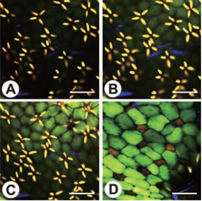
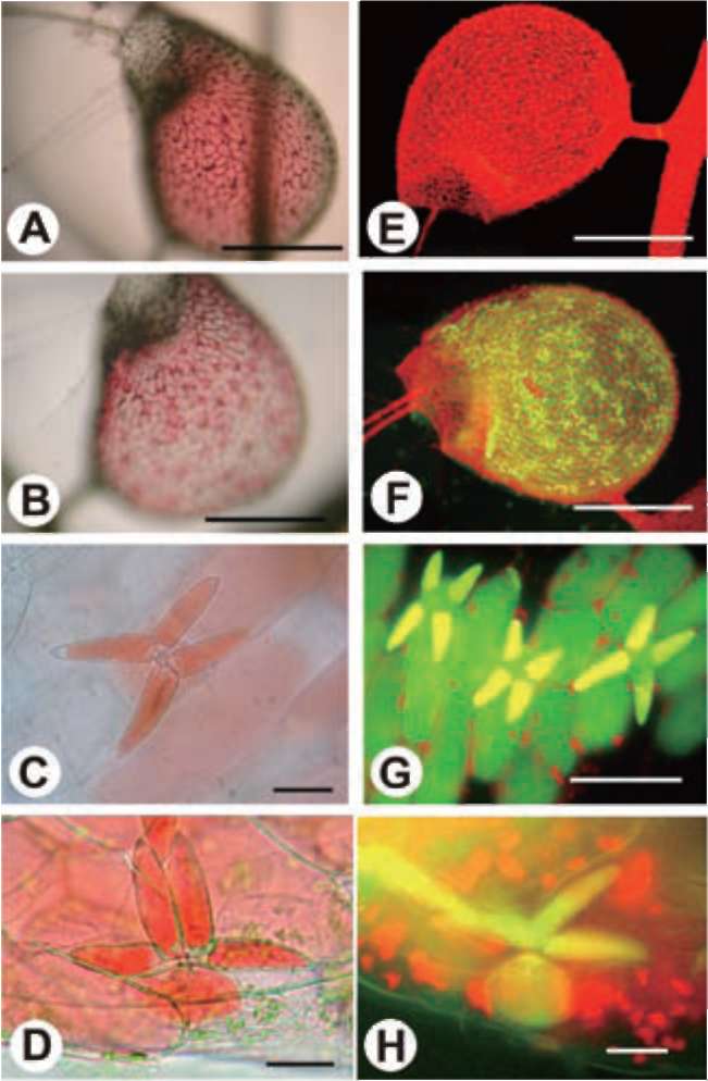
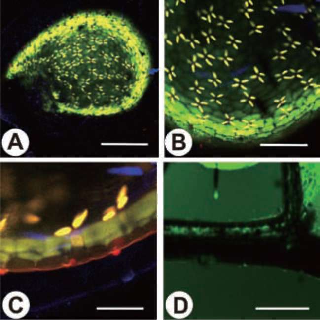
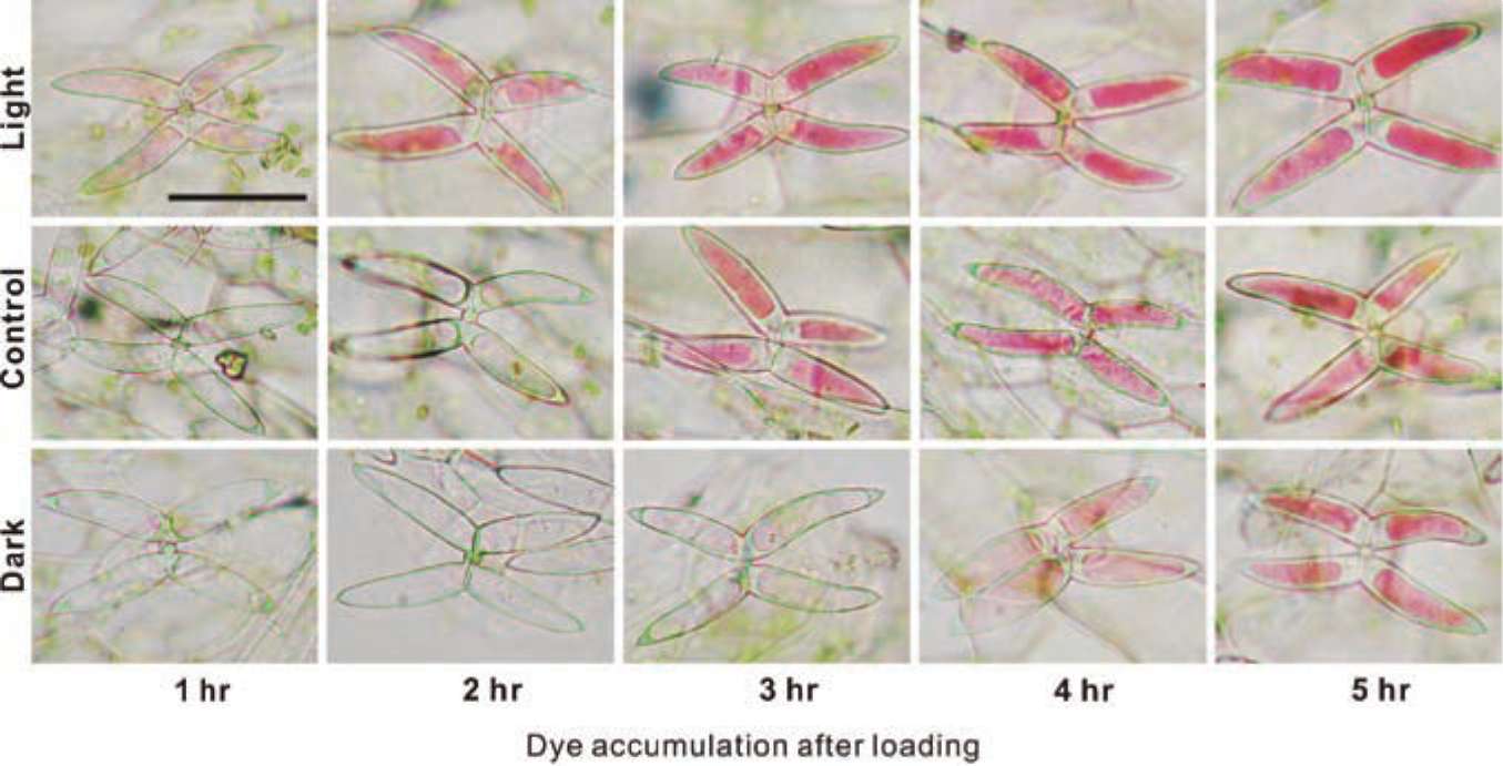
ABSTRACT. To capture prey, Utricularia gibba, an aquatic angiosperm carnivorous plant, is equipped with specialized bladders containing bifid/quadrifid glands for nutrient absorption. Several studies have focused on the nutrient absorption and subsequent transportation in the bladderworts; more specifically, the apoplastic pathway has been demonstrated as employing radioactive uranium salts. Nevertheless, the symplastic path­way has not been unambiguously demonstrated. Herein, we initially used food dyes as tracers to monitor the absorption processes by light microscope. We then confirmed the observed symplastic pathway using another vital tracer carboxyfluorescein diacetate (CFDA). The absorption and transportation of the CFDA inside the traps were observed using epifluorescence microscope and confocal laser scanning microscope. Our data clearly suggested that the tracers were transported through different tissues in the following order: terminal cells, pedestal cells, basal cells, surrounding epidermal cells, nearby leaves, and, finally, stems. The process was found to be light-sensitive, suggesting that it is energy-dependent. The uptake of the fluorescent dye was observed within seconds, while that of food dyes required 2 to 3 h. Thus, CFDA provides better resolution, while the food dyes afford prolonged tracing procedures. Taken together, the findings lead us to conclude that the symplastic pathway is an important transportation process that has never been shown previously in Utricu-laria.
Keywords: Carboxyfluorescein; Symplastic transport; Utricularia gibba.
INTRODUCTION
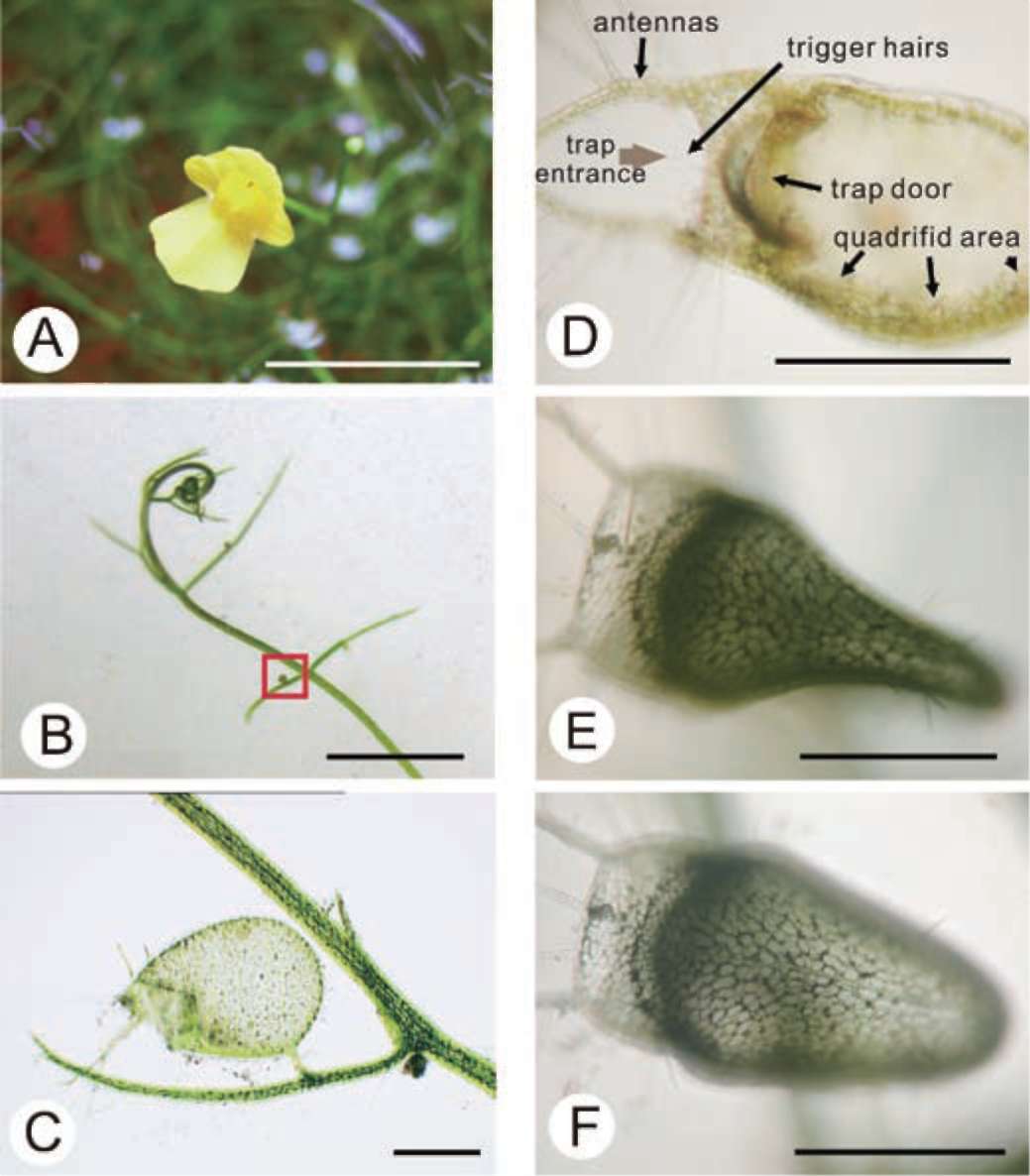
small water animal touches it, the threshold swings open inwardly, causing the water animal to be sucked into the trap along with rushing water. All of this happens in about 1/30 second (Figure 1D). Over a period of half an hour to an hour, the trap mechanism is automatically reset in preparation for the next catch and the pressure inside the trap is kept lower than the outside (Sydenham and Findlay, 1975). Because of this pressure difference, when the trap is viewed from above, the walls are warped inwards and appear concave (Figure 1E and 1F).
The inner surface of the trap is covered by quadrifid hairs. Only the inner surface of the threshold is covered by bifid hairs. Both quadrifids and bifids consist of a basal cell, a pedestal cell, and several terminal cells, which bear a complex architecture (Finerran, 1985). The pedestal cells of these glands have differentiated into transfer cells, and the cell walls are impregnated (Offler et al., 2003 Plachno and Jankun, 2004). The functions of the glands have been gradually clarified from the time of Darwin's early obser­vations with a light microscope (1875) to recent studies based on the electron microscope. Their main functions in­clude water removal from the lumen after prey-catching to maintain negative pressure inside the trap, transportation of nutrients, and the digestion and absorption of objects in-
Plants are considered the producers in the ecosystem, but carnivorous plants can derive nutrients from animals through carnivorous activities (Adamec, 1997). Starting with Darwin's Insectivorous Plants (1875), species of Utricularia have drawn the attention of many botanists. Utricularia gibba is herbaceous, angiospermic (Figure 1A), rootless, branching, with a green or brown stem 0.1­0.2 mm thick and up to 3 m long, with fine green branches bearing tiny bulbous traps, or bladders, and characteristic finely divided foliage (Figure 1B). A single trap is a small ovoid bladder up to 4 mm in length, with an entrance and a stalk that attaches it to the plant (Figure 1C). These sacs are highly sophisticated mechanical traps with a self-re­setting mechanism capable of catching tiny water animals with amazing efficacy. Each trap has some antenna-like hairs on one side of the trap opposite the attaching stem. The ventral part of the trap wall in the entrance forms the threshold. There are trigger hairs on it, and when a

*Corresponding author: E-mail: zhliu@mail.nsysu.edu.tw; Tel: 886-7-5252000 ext. 3617 Fax: 886-7-525-3609.
48
Botanical Studies, Vol. 52, 2011
Figure 1. Morphology of Utricularia gibba and its Traps. (A) A flower of Utricularia gibba. Bar = 1 cm; (B) The Utricularia gibba and its apical meristem visible. The box indicates an immature trap. Bar = 1 cm; (C) The trap is attached to a leaf, ovoid in shape, 1 mm long with a lateral mouth and two antennas attached around the mouth. Bar = 0.1 cm; (D) Longitudinal section of the trap in Utricularia gibba. Bar = 0.1 cm; (E) The trap in set phase. Bar = 0.1 cm; (F) The trap is shown in the re-setting phase when water sucked into the trap along with captured prey. Bar = 0.1 cm.
side the trap. The traps also support a diverse community of microorganisms, including many species of living bac­teria, algae, rotifers, and protozoa (Friday, 1989; Knight and Frost, 1991). Species of Euglena (Euglenophyta) apparently even reproduce in these traps, supporting the hypothesis that Utricularia plants benefit more from the byproducts of this community than from carnivory itself (Richards, 2001; Sirova et al., 2009).
Little is known about the mechanisms of digestion and absorption in Utricularia. It was found that within the empty traps of growing plants, phosphatases always exhib­ited the highest activity, followed by p-glucosidases, while the activities of a-glucosidases, p-hexosaminidases, and aminopeptidases were usually lower by one or two orders
of magnitude (Sirova et al., 2003). Using enzyme-labeled fluorescence, phosphatase activity in the glands was clearly demonstrated (Plachno et al., 2006). Despite the in-situ activity of a few common hydrolases in the trap, the fluid collected from aquatic Utricularia species has been mea­sured, indicating the availability of the enzymes in the traps for direct prey digestion. However, detailed studies on the transport of nutrients in Utricularia plants are quite rare.
Two pathways are involved in the entrance of water and nutrients to a plant: one is the symplastic pathway known to pass through the plasmodesmata, and the other is the apo-plastic pathway that traverses the cell wall and intercellular air spaces in plant tissues without crossing the cytoplasm. Several studies have focused on the nutrient absorption
JUANG et al. ― The symplastic pathway in the trap of the Utricularia gibba                                                                         49
and subsequent transportation in the bladderworts; more specifically, the apoplastic pathway has been demonstrated to use radioactive uranium salts as tracers. The tracers were confined outside of the impermeable lateral cell wall of the pedestal cells. Only in the arms of the quadrifids and bifids and the terminal cells of external glands did the tracers penetrate the cuticle (Fineran et al., 1980).
The symplastic pathway of carnivorous plants has been demonstrated mainly with Nepenthes traps, but only two studies have brought out the symplastic pathway with the fluorescent tracer 5(6)-carboxyfluorescein diacetate (CFDA) (Owen et al., 1999; Schulze et al., 1999). CFDA itself is a membrane-permeable dye that is converted into carboxyfluorescein (CF) by cytosolic esterases. CF is a reliable fluorescent tracer of phloem transport and sym-plastic transport. Schulze et al. (1999) searched for feeding pathways between glands and vascular bundles in Nepen­thes pitchers using fluorescent dyes and clearly demon­strated symplastic transport from the gland into subtending cells. Nevertheless, Nepenthes traps are multilayer struc­tures with smaller and thinner cells that are more difficult to examine. The quadrifid glands of the Utricularia are different from the Nepenthes glands, and the application of CFDA to investigation of the symplastic pathway in Utricularia traps has not yet been undertaken.
In this study, food dyes and the fluorescent compound CFDA were used as tracers to investigate the absorption and transport pathways in the traps of U. gibba. A light mi­croscope, epifluorescence microscope, and confocal laser scanning microscope were used to examine the absorption. Meanwhile, the effects of light on the absorption of food dyes in U. gibba were also studied. Based on our results, we were able to depict the complete symplastic pathway. This is, to the best of our knowledge, the first attempt to apply food dyes and CFDA to the study of Utricularia traps.
The study of the movement of the fluorescent probes of CFDA by epifluorescent microscope and noninvasive imaging techniques with the
confocal laser scanning microscope
To detail the symplastic connections within the quadri-fids, 10 mg of carboxyfluorescent diacetate was dissolved in 1 ml of 0.3 M KOH and then brought to a final con­centration of 1 mM in 20 ml distilled water (pH 6.3). One tenth ml of Eserine (Sigma) was added into the solution to block secreted esterases inside the traps and to prevent cleavage of CFDA before the uptake of the quadrifids. The U. gibba was placed into the solution. The traps were manually triggered to suck in the solution, and the sub­sequent procedures were carried out as mentioned above. The traps that did not suck in the CFDA were used as the control. After various durations of incubation, free-hand sections of the traps were mounted in water and analyzed under blue light (excitation wavelength 450-490 nm, chro­matic beam splitter 510, and barrier filter LP 520 with fluorescent microscope, Olympus BH2-RFCA).
In a non-invasive imaging technique with a confocal la­ser scanning microscope (Nikon EZ-C1, Laser type V-LD 408, Ar 488, G-HeNe 543, Y-HeNe 594, R-HeNe 633),
the CFDA was introduced into the Utricularia traps in the same manner as described above. The whole tissue of the U. gibba was examined without dissection.
Effect of the light and dark pretreatment on the food dye absorption in U. gibba
To study the effect of light on the absorption, ten branches of Utricularia gibba were divided into two groups and cultured in light (fluorescent lamp PL-F, 27W) or in a dark environment for 2 days. The mature traps on the bladderwort were randomized, chosen, and manually triggered to suck in the food dyes. Thereafter, the absorp­tion of food dyes inside the quadrifid glands were ob­served under a light microscope every hour for five hours after free-hand dissection. The absorption of food dye in­tensity inside the quadrifids arms was further analyzed by Multi Gauge 2.0 (Fuji Film).
MATERIALS AND METHODS
plant material
Utricularia gibba, native to southern Taiwan, was cul­tivated outdoors in plastic containers under a natural envi­ronment (temperature around 20°C-30°C). Watering was done with spring water.
RESULTS
Absorption and transport of food dyes
The absorption and transport of food dyes in Utricularia gibba
Initially, food dyes were used as tracers to monitor the absorption processes by a light microscope (Alpha-phot-2 YS2). Amaranth (Food Red 9, C20H11O20N2S3Na3,molecular weight 604.5, Scott-Bathgate LTD. Canada) and Brilliant Blue FCF (Food Blue 2, C37H36N2O9S3Na2, molecular weight 794, Scott-Bathgate LTD. Canada) were used in the experiments. Each of them was diluted to 6.7% with distilled water. The U. gibba traps were triggered manually under the dissecting microscope in the presence of tracers, rinsed in distilled water, and then examined at various time intervals (0 h, 8 h, and 16 h). The traps were then dissected, and the quadrifids were observed.
Amaranth Red can be easily sucked into the traps man­ually. The traps then appear red (Figure 2A and 2B). Over­all, the food dye density inside the traps decreased over time (images at 8-h intervals), and the rates of decline were much the same for both the Amaranth Red and Brilliant Blue food dyes. At this time, the color in the quadrifids gradually increased under light microscope, suggesting that food dyes were absorbed into the quadrifids (Figure 2B). Free-hand sections of the trap in these stages showed quadrifids with prominent red or blue food dye resting on the inner epidermis; one of them already was filled with
50
Botanical Studies, Vol. 52, 2011
red dye, and another one was partially filled up (Figure 2C). In the lateral view of the quadrifid, the arms, stalk, basal epidermal cells, and nearby inner epidermis were filled with a considerable amount of red dye (Figure 2D). Two days later, the dye reached the trap stalk and entered the accompanying leaf. Food dye transport took consid­erably longer than did CF. The transportation pathway of both food dyes and CF occurred predominantly in the same route.
Symplastic transport of carboxyfluorescent
The trap was red in color due to being full of chloro-
Figure 3. Images were taken by confocal laser scanning micro­scope at different planes of the trap wall using CFDA as a tracer. (A) The tips of the terminal cells. Bar = 100 pm; (B) A layer in which terminal cells are clearly observed. Bar = 100 pm; (C) The inner epidermis and the basal epidermal cells are filled with the tracer CF. Bar = 100 pm; (D) The inner epidermis at a plane that quadrifids can't be observed. Bar = 100 pm.
phyll (Figure 2E). The regions exhibiting yellow fluores­cence were the valve and external glands. After applying CFDA to the trap, the quadrifids turned out to be full of fluorescence within minutes under the epifluorescence mi­croscope (Figure 2F). Hand-sections of the traps showed the movement of the fluorescent symplastic tracer (CF) through the quadrifids and into the inner epidermis below (Figure 2G). The quadrifid stalks, pedestal cells, and basal epidermal cells were clearly filled with CF (Figure 2H).
Symplastic transport evidence of CF in the intact traps of Utricularia Gibba can be well-presented non-invasively using a confocal laser scanning microscope. The inner wall of the trap, viewed from the upper part of the quadri-fid arm and gradually down to the inner epidermis, is filled with CF (Figure 3A-3D). The immediate appearance of the trap is shown after manual triggering in the CFDA solu­tion (Figure 4A and 4B). Enlargement of the lateral view of the quadrifid showed that the basal epidermal cell was full of CF extending to the nearby inner epidermis. Trans­port of the tracer was restricted to the endodermal layer of the trap, and it was absent from the outer epidermis (Figure 4C). The upper portion of the stem was clearly labeled with dye (Figure 4D).
Figure 2. The absorption of food dye and the fluorescent com­pound, CFDA, in the trap. (A) The gross image taken immedi­ately after food dye uptake. The dye is loaded evenly within the trap. Bar =1 mm; (B) Image taken 16 h after dye absorption. Dye was accumulated in quadrifids. Bar = 1 mm; (C) Food dye was clearly observed in the terminal cells and the inner epidermis. Bar = 20 μm; (D) As the incubation was prolonged, the pedestal cell was filled with food dye. Bar = 20 μm; (E) Autofluorescence image of the Utricularia trap in control. The red fluorescence is of chlorophyll autofluorescence. Bar = 100 μm; (F) Gross image taken immediately after CFDA suction. Bar = 100μm; (G) Cross section of the Utricularia trap after CFDA absorption. Fluores­cence is observed in the terminal cells and the inner epidermis. Bar = 80 μm; (H) As the incubation time was prolonged, the pedestal cell is filled with fluorescence. Bar = 20 μm.
Effect of light and dark treatments on food dye absorption in the traps of Utricularia gibba
In the 2-day light and dark pretreatments, the effect of light on food dye absorption in the traps of U. gibba was examined. As shown in Figure 5, the food dye was
JUANG et al. ― The symplastic pathway in the trap of the Utricularia gibba
51
absorbed much faster in the 2-day light pretreatment than in the dark pretreatment. The light treatment significantly enhanced the absorption rate in the traps of U. gibba (Fig­ure 6). The absorption of CFDA in the traps of U. gibba
accomplished the absorption within seconds. In contrast to the fluorescent compound CFDA, U. gibba took hours to absorb the food dyes.
DISCUSSION
Utricularia gibba, an aquatic carnivorous plant, be­longs to the family of Lentibulariaceae, the largest fam­ily of carnivorous plants, with three genera (Pinguicula, Utricularia, Genlisea) and about 350 species (Ellison and Nicholas, 2009; Plachno and Swiatek, 2009). According to molecular studies, the genus Genlisea is sister to Utricu-laria, and this pair is sister to the genus Pinguicula (Jobson and Albert, 2002; Jobson et al., 2003). Both Pinguicula and Utricularia develop active traps, but both the physiol-ogy and functioning of the traps differ much between these genera. Pinguicula are active "flypapers" with slightly modified leaves for carnivory, but Utricularia form suc-tion bladders (reviewed by Legendre, 2000). Genlisea species are small, rootless wetland plants, which produce different special kinds of underground corkscrew-shaped traps of foliar origin-eel (lobster-pot) traps. (Reut, 1993). Besides having a similar architecture (basal cell, pedestal cell, and secretory cells), the digestive-absorptive hairs of the Lentibulariaceae present morphological and ultrastruc-tural differences (Plachno et al., 2007). For example, the pedestal cell has a highly developed wall labyrinth only in Utricularia. This is associated with rapid water transport during removal of water from Utricularia bladders (Fin-eran and Lee, 1975; Fineran, 1985). Also, it is clear that the most complicated terminal cell in the digestive hairs of this family has evolved in Utricularia (Fineran and Lee, 1975; Heslop-Harrison and Heslop-Harrison, 1981;
Figure 4. Confocal micrographs of Utricularia gibba after ap­plying CFDA to the trap. (A) The gross appearance of trap after applying CFDA to the inside of the trap. The yellowish appear­ance was shown in the quadrifids. Bar = 300 μm; (B) The same condition with larger magnification. Bar= 500 μm; (C) The quadrifid stalk, pedestal cell and basal epidermal cell were filled with CF. Bar = 100 μm; (D) CF was observed in the stem of U. gibba. Bar = 1 mm.
Figure 5. Effects of light on the accumulation of food dye into quadrifids. Plants were grown under continuous light (48 h, top row), normal condition (16 h light, 8 h dark, middle row), or continuous darkness (48 h, bottom row) before dye loading. Bar = 100 μm.
52
Botanical Studies, Vol. 52, 2011
Fineran, 1985; Plachno et al., 2007). In contrast to the ses­sile hairs in Pinguicula and Genlisea, in Utricularia the quadrifid and bifid terminal cells not only play a role in secretion and absorption but also partially take over the function of the pedestal cell. Several studies have focused on nutrient absorption and subsequent transportation in the bladderwort; more specifically, the apoplastic pathway has been demonstrated using radioactive uranium salts (Fineran et al., 1980). So far only the symplastic pathway of terminal cell in Utricularia is delineated in the present study. Further work with this method to demonstrate the
symplastic pathway in Pinguicula and Genlisea hairs may shed light on the evolution of the carnivorous syndrome in Lentibulariaceae.
Prior to the present study, a new enzyme labeled fluo­rescence (ELF) phosphatase substrate assay was applied to the detection of phosphatase activity in the glandular struc­tures of 47 carnivorous plant species, especially Lentibu-lariaceae, in order to understand their digestive activities (Plachno et al., 2006). In all examined Utricularia species, Plachno et al. found quadrifid glands inside the traps pro­ducing an intense signal of phosphatase activity. The stain­ing of quadrifid glands starts in only the vacuolated tip of the terminal cell and proceeds to the basal part containing the bulk cytoplasm and the nucleus. No further transpor­tation of this fluorescence could be traced. However, the ELF assay developed by Nedoma et al. (2003) specifically labels extracellular enzymes on the cell surface since the ELF hardly penetrates biomembranes (Strojsova et al., 2003), which is different from the vital tracer carboxyfluo-rescein diacetate (CFDA) used in our study.
CFDA is a membrane-permeable, non-fluorescent vi­tal stain that converts into the fluorescent, membrane-impermeant carboxyfluorescien when cleaved by cytosolic esterases. It is often used for the fluorometric quantitation of viable cells (Breeuwer et al., 1995), evaluation of gap-junctional intercellular communication and adhesion, and cytotoxicity assays (Flury and Fluhler, 1994). Since CFDA is strictly confined inside the cytoplasm after cleavage, travels only through the plasmodesmata, and is restricted to the phloem regions, it is also used to trace phloem translocation (Grignon et al., 1989), especially for track­ing phloem loading and unloading (Oparka et al., 1994). The permeability of the biological membrane is strongly dependent on the nature of the transported solute. CFDA itself is a membrane-permeable dye due to its hydrophobic property. When it is absorbed into cells, it is converted into carboxyfluorescein by cytosolic esterases (Grignon et al., 1989). CF is hydrophilic, and its fluorescence persists in the quadrifids and stem phloem for several days. The absorption and transportation of the CFDA inside the traps were observed easily using an epifluorescence microscope and confocal laser scanning microscope.
Brilliant blue FCF is water-soluble and dissociated to a mono or bivalent anion in the water (Flury and Fluhler, 1994). Therefore, anionic blue dyes cannot pass through the biological membrane easily. The food dyes appeared in the quadrifids 16 h after absorption. In contrast, CFDA, a weak acid, as an uncharged compound that can easily permeate the biological membrane, was taken in to the quadrifids within seconds (Figure 2F).
The experiment of the light effect on food dye absorp­tion in the traps of U. gibba clearly showed that the light treatment significantly enhanced the absorption rate in the traps of U. gibba. This current result supports previ­ous work showing that bladder function requires greater metabolic cost, i.e., relating to ion and water pumping (Sydenham and Findlay, 1975; Laakkonen et al., 2006)
Figure 6. Quantitative analysis of the effect of light on the rate of dye accumulation in quadrifids. The absorption rates were mean ± SD from three independent experiments.
Figure 7. Model of the symplastic pathway in the quadrifid glands of Utricularia gibba. The trap wall is made up of two cell layers. On the inner epidermis rests countless quadrifids, in contact with the trap fluid. The outer epidermis bears scattered and mostly sessile, dome-shaped hairs referred to as external glands. Each glandular hair consists of six cells. The first is the basal epidermal cell which lies directly beneath the hair and is similar to adjacent epidermal cells. Then is the middle or ped­estal cell which rests on the basal epidermal cell. The four other cells of the gland are the four terminal cells (or quadrifid arms). Solutes (food dyes or fluorescent compound CFDA) and water may follow a symplastic route. The compoundsfirst enter the quadrifid armsthen the stalk and then the pedestal cellFrom the pedestal cell the absorbed solute is transported via the compound plasmodesmata to the underlying basal epidermal cell and from there to the surrounding inner epidermis⑤
JUANG et al. ― The symplastic pathway in the trap of the Utricularia gibba
53
and digestion of prey (Sirova et al., 2003). Moreover, mu­tations of the mitochondrially encoded cytochrome c oxi-dase in Utricularia, which could result in faster reaction kinetics of this enzyme, may be an adaptation for greater respiratory capacity associated with the high energetic demands of bladder traps (Laakkonen et al., 2006; Albert et al., 2010). However, the extent of such an influence on traps, whether during the resetting of Utricularia bladders or just in the post-firing state, is not known. Also, we did not work on traps with or without living prey.
As the symplastic pathway in Lentibulariaceae has not been unambiguously demonstrated before, herein we initially used food dyes as tracers to monitor the absorp­tion processes in Utricularia gibba. We then confirmed the above symplastic pathway using another vital tracer, CFDA. Our data clearly suggested that the tracers were transported in the following order: terminal cells, stalks, pedestal cells, basal cells, surrounding epidermal cells, nearby leaves, and, finally, stems, and also that it is ener­gy-dependent. These findings lead us to conclude that the symplastic pathway is an important transportation process that has never been investigated in this carnivorous plant.
Fineran, B.A. 1985. Glandular trichomes in Utricularia: a re­view of their structure and function. Israel J. Bot. 34: 295­330.
Flury, M. and H. Fluhler. 1994. Brilliant Blue FCF as a dye trac­er for solute transport studies~A toxicological overview, J. Environ. Qual. 23: 1108-1112.
Friday, L.E. 1989. Rapid turnover of traps in Utricularia vul-garis L. Oecologia 80: 272-277.
Grignon, N., B. Touraine, and M. Durand. 1989. 6(5)-Carboxy-fluorescein as a tracer of phloem sap translocation. Am. J. Bot. 76: 871-877.
Heslop-Harrison, Y. and J. Heslop-Harrison. 1981. The diges­tive glands of Pinguicula: structure and cytochemistry. Ann. Bot. 47: 293-319.
Jobson, R.W., J. Playford, K.M. Cameron, and V.A. Albert. 2003. Molecular phylogenetics of Lentibulariaceae inferred from plastid rps16 intron and trnL-F DNA sequences: im­plications for character evolution and biogeography. Syst. Bot. 28: 157-171.
Jobson, R.W. and V.A. Albert. 2002. Molecular rates parallel diversification contrasts between carnivorous plant sister lineages. Cladistics 18: 127-136.
Knight, S.E. and T. M. Frost. 1991. Bladder control in Utricu­laria macrorhiza: lake-specific variation in plant investment in carnivory. Ecology 72: 728-734.
Laakkonen, L., R.W. Jobson, and V.A. Albert. 2006. A New
Model for the Evolution of Carnivory in the Bladderwort Plant (Utricularia): Adaptive Changes in Cytochrome c Ox-idase (COX) Provide Respiratory Power. Plant Boil. (Stuttg) 8(6): 758-764.
Legendre, L. 2000. The genus Pinguicula L. (Lentibulariaceae): an overview. Acta Bot. Gall. 147: 77-95.
Nedoma, J., A. Strojsova, J. Vrba, J. Komarkova, and K. Simek. 2003. Extracellular phosphatase activity of natural plankton studied with ELF97 phosphate: fluorescence quantification and labeling kinetics. Environ. Microbiol. 5: 462-472.
Offler, C.E., D.W. McCurdy, J.W. Patrick, and M.J. Talbot.
2003. Transfer cells: cells specialized for a special purpose.
Annu. Rev. Plant Biol. 54: 431-54. Oparka, K.J., C.M. Duckett, D.A.M. Prior, and D.B. Fisher.
1994. Real-time imaging of phloem unloading in the root tip of Arabidopsis. Plant J. 6(5): 759-766.
Owen, T.P., K.A. Lennon, M.J. Santo, and A.Y. Anderson. 1999. Pathways for nutrient transport in the pitchers of the carnivorous plant Nepenthes alata. Ann. Bot. 84: 459-466.
Plachno, B.J. and A. Jankun. 2004. Transfer cell wall architec­ture in secretory hairs of Utricularia intermedia Traps. ActaBiol. Cracoviensia Series Bot. 46: 193-200.
Plachno, B.J., L. Adamec, I.K. Lichtscheidl, M. Peroutka, W. Adlassnig, and J. Vrba. 2006. Fluorescence labelling of phosphatase activity in digestive glands of carnivorous plants. Plant Biol. (Stuttg) 8(6): 813-820.
Plachno, B.J., M. Kozieradzka-Kiszkurno, and P. Swiatek. 2007. Functional utrastructure of Genlisea (Lentibulariaceae) di-
Acknowledgments. We are grateful to Professor Yiu-Kay Lai and Margaret Dah-Tsyr Chang of National Tsing Hua University. Their helpful suggestions and comments in regard to the nature of the research, experimental design, and manuscript had a positive impact of this project.
LITERATURE CITED
Adamec, L. 1997. Mineral nutrition of carnivorous plants: A re­view. Bot. Rev. 63: 273-299.
Albert, V.A., R.W. Jobson, T.P. Michael, and D.J. Taylor. 2010. The carnivorous bladderwort (Utricularia, Lentibulariace-ae): a system inflates. J. Exp. Bot. 61(1): 5-9.
Breeuwer, P., J.L. Drocourt, N. Bunschoten, M.H. Zwieter-ing, F.M. Rombouts, and T. Abee. 1995. Characteriza­tion of uptake and hydrolysis of fluorescein diacetate and carboxyfluorescein diacetate by intracellular esterases in Saccharomyces cerevisiae, which result in accumulation of fluorescent product. Appl. Environ. Microbiol. 61(4): 1614­1619.
Darwin, C. 1875. Insectivorous plants. J. Murray, London. (The Writings of Charles Darwin on the web, http://pages.britishlibrary.net/charles.darwin3/
insectivorous/insect_fm.htm
)
Ellison, A.M. and J.G. Nicholas. 2009. Energetics and the evo­lution of carnivorous plants―Darwin's 'most wonderful plants in the world'. J. Exp. Bot. 60: 19-42.
Fineran, B.A. and M.S.L. Lee. 1975. Organization of quadrifid and bifid hairs in the trap of Utricularia monanthos. Proto-plasma 84: 43-70.
Fineran, B.A. and J.M. Gilbertson. 1980. Application of lantha­num and uranyl salts as tracers to demonstrate apoplastic pathways for transport in glands of the carnivorous plant Utricularia monanthos. Eur. J. Cell Biol. 23(1): 66-72.
54
Botanical Studies, Vol. 52, 2011
gestive hairs. Ann. Bot. 100(2): 195-203.
Plachno, B.J. and P. Swiatek. 2009. Functional anatomy of the ovule in Genlisea with remarks on ovule evolution in Lentibulariaceae. Protoplasma 236: 39-48.
Reut, M.S. 1993. Trap structure of the carnivorous plant Genli-sea (Lentibulariaceae). Bot. Helvet. 103: 101-111.
Richards, J.H. 2001. Bladder function in Utricularia purpurea (Lentibulariaceae): is carnivory important? Am. J. Bot. 88:170-176.
Schulze, W., W.B. Frommer, and J.M. Ward. 1999. Transporters for ammonium, amino acids and peptides are expressed in pitchers of the carnivorous plant Nepenthes. Plant J. 17:637-646.
Sirova, D., J. Borovec, B. Cerna, E. Rejmankova, L. Adamec, and J. Vrba. 2009. Microbial community development in the traps of aquatic Utricularia species. Aquatic Bot. 90:129-136.
Sirova, D., L. Adamec, and J. Vrba. 2003. Enzymatic activities in traps of four aquatic species of the carnivorous genus Utricularia. New Phytol. 159: 669-675.
Sydenham, P.H. and G.P. Findlay. 1975. Transport of solutes and water by resetting bladders of Utricularia. Aust. J. Plant Physiol. 2: 335-351.
Strojsova, A., J. Vrba, J. Nedoma, J. Komarkova, and P. Znach-or. 2003. Seasonal study of extracellular phosphate expres­sion in the hytoplankton of a eutrophic reservoir. European
J. Phycol. 38: 295-306.
絲葉狸藻捕蟲囊的共質體输送
莊淳喬1 莊迪喬2 劉景煌3
1國立臺灣大學生命科學院生命科學系
2美國史丹佛大學
3國立中山大學生物系
水生開花食蟲植物絲葉狸藻{Utricularia gibba)是非常獨特的,它具備捕蟲囊捕捉水中小生物,及
囊內具有吸收營養的四爪及二爪腺毛。目前有很多文獻探討狸藻的營養吸收途徑,例如有文獻報導利用
放射性元素鑭追蹤以顯示其非共質體輸送途徑,但尚未有文獻報導其共質體運輸途徑。本研究先以食用
色素為追蹤物質,在複式顯微鏡觀察絲葉狸藻捕蟲囊的吸收途徑,接著採用追蹤物質螢光染劑(carboxy-
fluorescein diacetate, CFDA)
,以螢光顯微鏡及共軛焦雷射顯微鏡來觀察囊內CFDA輸送途徑,以確認所
觀察為共質體運輸途徑。CFDA本身具有細胞膜通透性,被囊內的酶分解成不可穿透膜的carboxyfluo-
rescein (CF)
,此為觀察韌皮部運輸的可靠螢光追蹤物質。由實驗結果中,我們認為囊內物質的輸送路徑
如下:四爪腺毛尾端、基座、底層表皮、囊內表皮、囊柄、葉及莖。因為光合作用可以增加腺毛對色素
的吸收速率,所以我們認為腺毛吸收物質是需要能量的反應。腺毛可以很快的吸收螢光染劑而食用色素
則較慢,因此前者較適合觀察物質在腺毛的輸送路徑,後者較適合長久觀察。目前為止並沒有狸藻捕蟲
囊內共質體輸送路徑的直接證據而我們是首先以螢光染劑及食用色素,清楚地將囊內共質體輸送路徑
完整的表現出來。
關鍵詞:葉狸藻;螢光染劑;共質體運輸。