Botanical Studies (2011) 52: 55-72.
The XopE2 effector protein of Xanthomonas campestris pv. vesicatoria is involved in virulence and in the suppression of the hypersensitive response
Rong-Hwa LIN1'3'4, Chih-Wen PENG2, Yuan-Chuen LIN3, Hwei-Ling PENG4, and Hsiou-Chen HUANG3*
1Biotechnology Center, National Chung Hsing University, Taichung, 40227, Taiwan
2Department of Life Science, Tzu-Chi University, Hualien 97004, Taiwan
3Graduate Institute of Biotechnology, National Chung Hsing University, Taichung, 40227, Taiwan
4Department of Biological Science and Technology, National Chiao Tung University, Hsin Chiu 30050, Taiwan
(Received May 14, 2010; Accepted June 29, 2010)
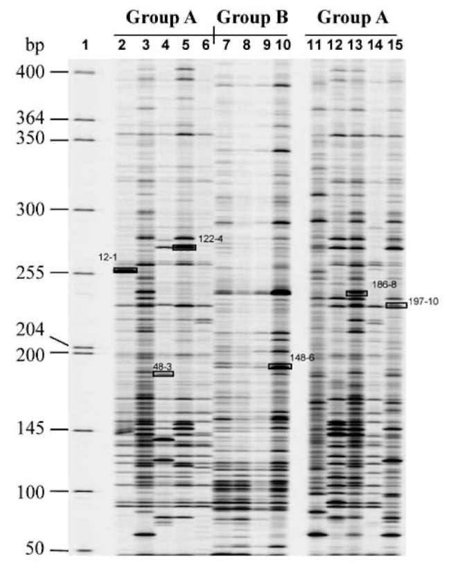
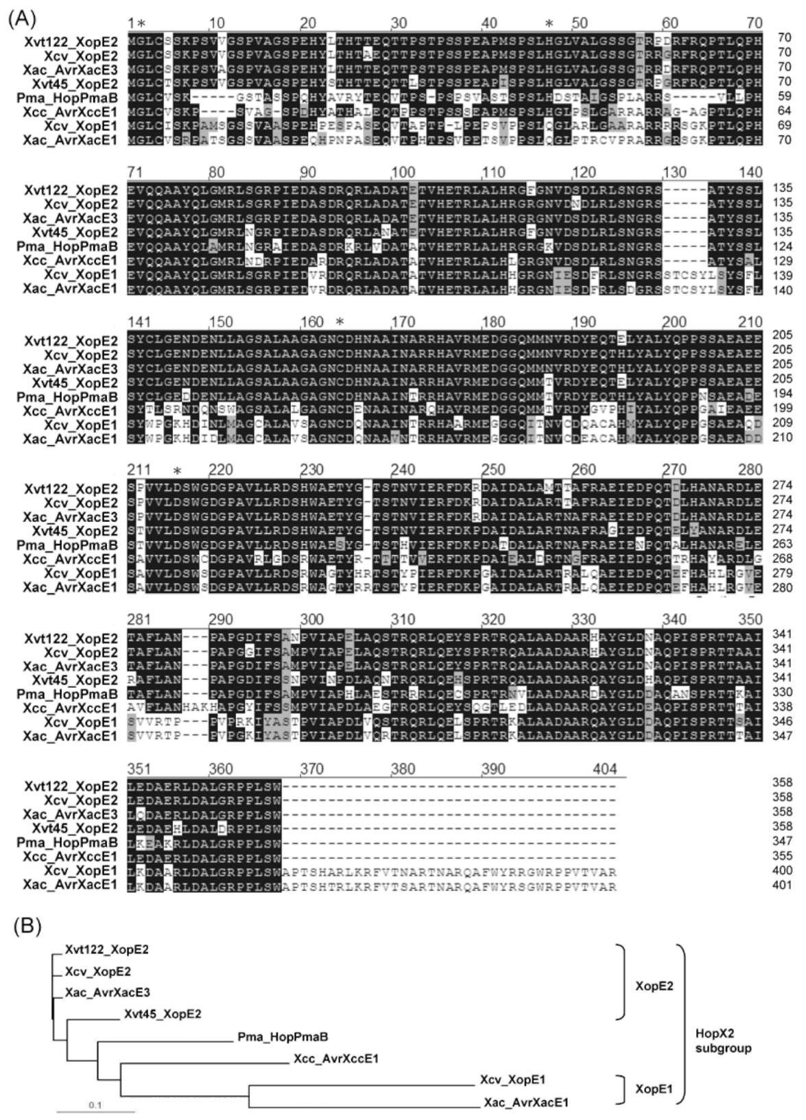
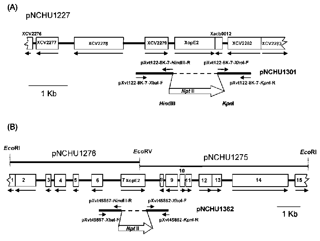
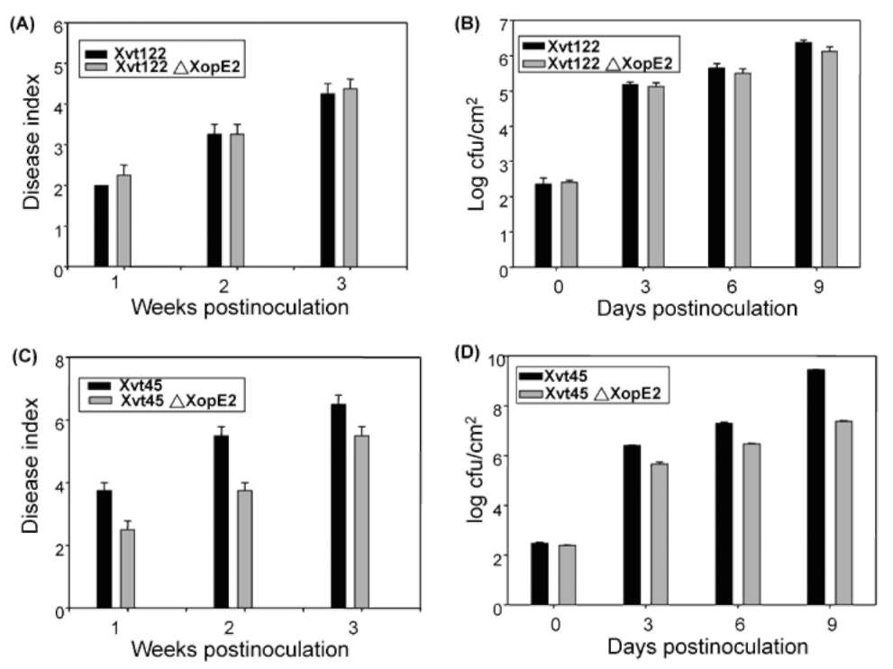
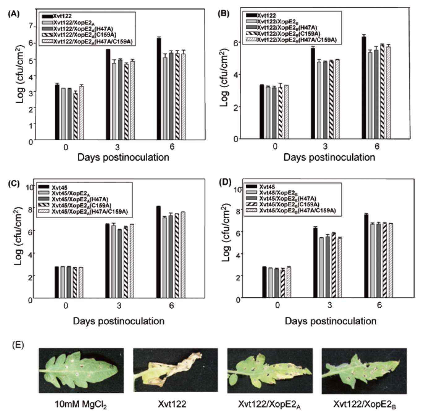
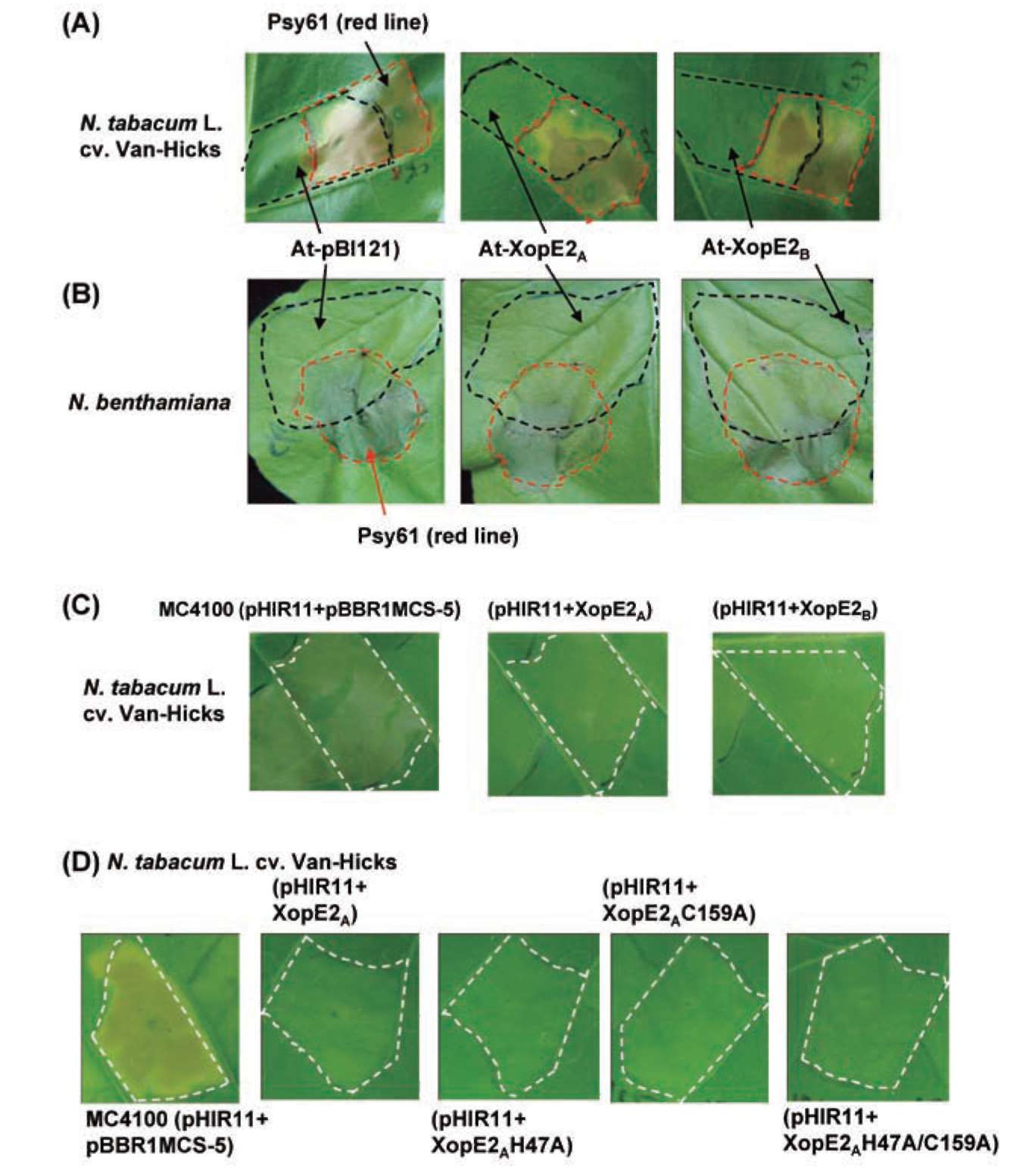
ABSTRACT. Pathogenicity of Xanthomonas campestris pv. vesicatoria (Xcv) causing bacterial spot disease on tomato (Lycopersicon spp.) and pepper (Capsicum spp.) requires the type III secretion system (T3SS) and T3SS effectors. In this study, we employed the AFLP technique to investigate the diversity of X. campestris pv. vesicatoria isolated in Taiwan, and consequently a XopE2 homologue was identified in all fourteen Xcv strains that have been classified into two groups. Phylogenic analysis of XopE2 amino acid sequences indi­cated that XopE2 of Xcv Xvt122 (group A) has a closer genetic distance to XopE2 of Xcv 85-10 than to that of Xcv Xvt45 (group B). Interestingly, although it was suggested that Xvt45 contains duplicated xopE2 genes, one being located on the genome and the other located on a large plasmid, a single copy deletion of xopE2 within the genome caused a substantial reduction in virulence, but no effect of xopE2 mutation on virulence of Xcv 85-10 and Xvt122 was observed. Furthermore, our results revealed that XopE2 of Xcv Xvt122 or Xcv Xvt45 was able to suppress HR in a T3SS-dependent manner and the heterologously-expressed XopE2 was sufficient to modulate the virulence on susceptible tomato plants. Their biological functions are not dependent on the consensus catalytic triad (159th cysteine) and thiol-protease His residue (47th histidine) of XopE2.
Keywords: Amplified restriction fragment length polymorphism; Bacterial spot; Hypersensitive response; Type III secretion system; XopE2 effector.
Abbreviations: AFLP, Amplified restriction fragment length polymorphism; T3SS, type III secretion system; HR, hypersensitive response; avr, avirulence; PCD, programmed cell-death.
INTRODUCTION
nicity)/ hrc (hypersensitive response and conserved) gene clusters. The T3S machinery secretes proteins into the ex­tracellular milieu (e.g. harpin or pilus proteins) and trans­locates effector proteins (e.g. Xop or Avr proteins) into the plant cell (Grant et al., 2006; Gurlebeck et al., 2006; Kay and Bonas, 2009). Some effectors designated as avirulence (avr) proteins are specifically recognized in resistant plants containing corresponding resistance (R) genes, usually resulting in a hypersensitive response (HR) that restricts bacterial growth via programmed cell-death (PCD) reac­tions (Klement, 1982; Staskawicz et al., 2001; Grant et al., 2006; Mudgett, 2005). The T3S mutants that are impaired in growth in planta also fail to cause disease symptoms in susceptible plants and lose the capacity to induce the HR in non-hosts or resistant hosts, indicating that T3S effec­tors may play essential roles in the interaction of bacteria with plants (Kay and Bonus, 2009).
Plants are armed with an elaborate network of defense mechanisms to protect themselves from the invasion of microorganisms. On the other hand, bacterial pathogens have evolved sophisticated strategies to conquer their plant hosts, i.e., they suppress the basic defense mecha­nisms in order to successfully establish the initiation of invasion (Nurnberger et al., 2004; Schechter et al., 2006). Similar to most Gram-negative phytopathogenic bacteria, Xanthomanas sp. possesses a conserved type III secretion (T3S) system which contains a needle-like structure and is encoded by hrp (hypersensitive response and pathoge-

*Corresponding author: E-mail: hchuang@dragon.nchu.edu. tw; Telephone: 886-4-22852155; Fax: 886-4-22853527.
56
Botanical Studies, Vol. 52, 2011
Individual Xanthomonas strains secrete a repertoire of 15 or more T3 effectors, but only a few effectors were shown to be major virulence factors because their muta­tion led to a dramatic loss of virulence (Thieme et al., 2005; Gurlebeck et al., 2006; Kay and Bonas, 2009). For instance, AvrBs2 of X. campestris pv. vesicatoria causing bacterial spots on pepper and tomato significantly con­tributes to the symptom development, epiphytic survival, and growth in planta (Gurlebeck et al., 2006). The XopX (Xanthomonas outer proteins) of X. campestris pv. vesica-toria promotes lesion development and growth of nonhost pathogens in Nicotiana benthamiana, suggesting that XopX suppresses basal plant defense (Metz et al., 2005). The members of Xanthomonas avrBs3/pthA gene family, such as apl1, avrXa7, and avrXa10, can suppress nonhost HR and down-regulate the expression of basal defense-associated genes, such as RbohB, PR1 , and PAL (Fujikawa et al., 2006). In the genome sequence of X. campestris pv. vesicatoria 85-10, two new type III effector proteins, XopE1 and XopE2 belonging to the HopX family of Pseu-domonas syringae were recently identified (Thieme et al., 2007). The N-termini of XopE1 and XopE2 encompass a putative N-myristoylation motif that mediates host cell membrane targeting. Interestingly, this conserved N-my-ristoylation motif of XopE1 was found to be essential for the induction of cell-death reactions in N. benthamiana, whereas such a membrane targeting signal motif is dis­pensable for the induction of the avirulence activity in So-lanum pseudocapsicum by XopE2 (Thieme et al., 2007).
The causal agent of bacterial spot symptoms on tomato (Lycopersicon spp.) and pepper (Capsicum spp.) was orig­inally identified to be X. campestris pv. vesicatoria (Jones and Stall, 1998). However, in the past two decades, strains of X. campestris pv. vesicatoria were determined to be composed of two genetically and phenotypically distinct groups, group A and group B, based on their amylolytic activities (Beaulieu et al., 1991), protein, and DNA poly­morphisms (Bouzar et al., 1994; Jones and Stall, 1998; Stall et al., 1994; Vauterin et al., 1995). Furthermore, Vauterin et al. (1995 and 2000) reclassified and defined the group A and group B into species X. axonopodis pv. vesicatoria and X. vesicatoria, respectively. According to more refined technology, recently at least four taxo-nomically distinct xanthomonads that cause bacterial spot symptoms on pepper and tomato plants have been identi­fied, e.g X. euvesicatoria (=X. axonopodis pv. vesicatoria), X. vesicatoria, X. perforans, and X. gardneri (Jones et al., 2000; Stall et al., 2009). Nowadays, X. campestris pv. vesicatoria (X. euvesicatoria) strain 85-10 is often used as a model isolate for the study of the involvement of T3SS and its effectors in pathogenesis (Thieme et al., 2007; Kay and Bonas, 2009). To date, 17 type III effector proteins have been verified experimentally in X. campestris pv. vesicatoria 85-10 and its closely related strains (Gurlebeck et al., 2006; Thieme et al., 2007).
Amplified restriction fragment length polymorphism (AFLP) has been developed as an efficient technique for fingerprinting plant or microbial genomes (Vos et al.,
1995; Folkertsma et al., 1996; Lin et al., 1996; O'Neill et al., 1997). For the study on genetic diversity of strains of X. axonopodis pv. manihotis (Xam) causing bacterial blight disease on cassava, the AFLP primer combinations EcoRI+T/Msel+A were shown to be the most efficient in discriminating between pathogenic and nonpathogenic Xam strains and the sequence analysis of polymorphic bands obtained showed significant homology with genes involved in pathogenic fitness and regulators of virulence (Gonzalez et al., 2002). In X. campestris pv. vesicatoria 85-10, 30 HrpG-induced (hgi) and five HrpG-repressed (hgr) cDNA fragments were identified using cDNA-AFLP technique (Noel et al., 2001). In Taiwan, bacterial spot dis­eases of tomato and pepper are very destructive, especially in rainy seasons, and are mainly caused by X. campestris pv. vesicatoria group A (=X. euvesicatoria = X. axonopo-dis pv. vesicatoria,) and X. campestris pv. vesicatoria group B (=X. vesicatoria). In this study, we applied AFLP analysis to investigate the diversity between X. campestris pv. vesicatoria (Xcv) group A and B strains isolated in Taiwan. The virulence-associated locus xopE2 was cloned and Southern blot analysis revealed that xopE2 was highly conserved in Xanthomonas spp. Comparisons of the DNA sequences and genomic annotation surrounding xopE2 suggested that the gene organization of the xopE2 locus in the genome could be considered as a unique feature to discriminate the members of X campestris pv. vesicato-ria group A from that of group B. The XopE2 mutant of group B exhibited reduced growth and symptom forma­tion, while such phenotypes were not seen in the group A-derived XopE2 mutant. Furthermore, our results sug­gested that XopE2 from both groups of Xcv can efficiently suppress the hypersensitive response (HR) induced by the avirulence effector HopPsyA of P. syringae pv. syringae in a T3SS-dependent manner.
MATERIALS AND METHODS
Bacterial strains, growth conditions, and DNA manipulation
Bacterial strains and plasmids used in this study are listed in Table 1. Escherichia coli strains were cultured in Luria-Bertani (LB) medium at 37°C and Xanthomonas spp. strains were grown at 28°C in LB broth or 523 agar plate (sucrose 10 g/l, casein hydrolysate 8 g/l, yeast extract 4 g/l, K2HPO4 2 g/l, MgSO4'7H2O 0.3 g/l). The concentra­tions of antibiotics used were as follows: ampicillin (Amp), 100 |ig/ml; Chloramphenicol (Cm), 12.5 |ig/ml; gentamy-cin (Gm), 10 |ig/ml; kanamycin (km), 50 |ig/ml; and tet­racycline (Tc), 20 [ig/ml. Plasmids were introduced into bacteria by electroporation (GenePulser, Bio-Rad). DNA manipulations and PCR were carried out according to stan­dard procedures (Sambrook et al., 1989). All the primers used in this study are listed in Table 2. Colony hybridiza­tion and Southern blotting analysis were performed with DIG Luminescent Detection Kit (BM) according to the manufacturer's instructions. DNA sequencing was done with ABI 3700 DNA sequencer (in Biotechnology center,
LIN et al. — Biological functions of Xcv XopE2 in tomato and tobacco plants                                                                                       57
National Chung Hsing University). Database searches were performed using gapped BLASTN and BLASTP (http://www.ncbi.nlm.nih.gov/).
pNCHU1229 and pNCHU1300. Subsequently, a 2.5 Kb XbaI-XhoI fragment containing nptII gene was isolated from pNCHU1229 and subcloned into pNCHU1300 gen-erating pNCHU1301 containing the up- and down-stream regions of xopE2 in which the xopE2 coding sequence was replaced with nptII gene (Figure 4A). To make xopE2 mutant of Xct45, the plasmid pNCHU1362 (Figure 4B) primer pairs pXvt45857-XbaI-F/pXvt45857-HindIII-R and pXvt45852-XhoI-F/pXvt45852-KpnI-R (Table 2). Fi-nally, the resultant plasmid pNCHU1301 or pNCHU1362 was introduced into Xanthomonas strains by electropora-tion followed by subculturing 5 days in liquid broth to promote homologous recombination as reported (Huang et al., 1988). Putative mutants were screened for the Km-resistant and Gm-sensitive phenotypes. The genome types presented in Xvt122AxopE2 and Xvt45AxopE2 were veri-fied by Southern hybridization.
AFLP analysis
The AFLP analysis was carried out according to the manufacturer's instruction (AFLP Expression Analy­sis Kit, LI-COR Biosciences). Briefly, a total of 250 ng genomic DNA was digested with TaqI/MseI enzymes, and then ligated with the respective adapters. The pre-amplification (pre-amp) was performed using 1:10 dilution of the above ligation mixture as the template and the pre-amp primers (primer MseI-N/primer TaqI-N), while MseI primer/IRDyeTM 700-labeled TaqI primers and 1:10 dilu­tion of the pre-amp DNA as template were used for the selective amplification. The PCR program was one cycle of 94°C for 30 sec, 65°C for 30 sec, and 72°C for 1 mm, followed by 12 cycles of 0.7°C/per cycle down-gradient annealing temperature; and then additional 23 cycles of 94°C for 30 sec, 56°C for 30 sec, and 72°C for 1 mm. The amplified products were denatured at 95°C, chilled on ice and resolved by a 6.5% polyacrylamide denaturing gel (KBPlusTM). Electrophoresis was carried out for 4 h at 45 V/cm and 45°C using 0.8 x TBE buffer. The polymorphic bands were viewed with LI-COR OdysseyTM scanner. The bands of interest were then excised from gels using a razor blade, resuspended in 20 [l TE buffer, and the suspension was subjected to a series of freeze-thaws and finally the DNA was leaky out from the gel and collected by centrifu-gation at 15,000 g for 20 min at 4°C. The DNA was re-amplified by PCR and cloned into pGEM-T easy vector (Promega). Sequence of the DNA inserts was determined and analyzed.
Site-directed mutagenesis
The plasmids, pNCHU1913 and pNCHU1917 con­taining xopE2 genes of Xvt122 and Xvt45 respectively, were used as the templates for the crossover PCR-based mutagenesis. Briefly, the primer pairs pXvt45-E3-F-XhoI (including start codon) / prH47A-1 (the 47th histidine was substituted by alaine) and prH47A-3 (the complementary sequence of prH47A-1)/ pXvt45-E3-R-XbaI (including stop codon) were used for amplification of the DNAs containing two partial xopE2 fragments. The DNAs were then used as templates for reamplification with the primer pair pXvt45-E3-F-XhoI / pXvt45-E3-R-XbaI, and the resulting xopE2 mutant fragment were cloned into the pDrive vec­tor (GIAGEN PCR Cloning Kit), resulting in recombinant plasmids pNCHU1914 (XopE2A H47A) and pNCHU1918 (XopE2B H47A) respectively. Similar procedure with different primer pairs was applied and the resulting plasmids containing different xopE2 mutations were ob­tained and designated as pNCHU1915 (XopE2A C159A),
pNCHU1919 (XopE2B C159A), pNCHU1916 (XopE2A H47A/C159A), and pNCHU1920 (XopE2e H47A/ C159A). The DNA fragments containing various site-directed mutations of xopE2 in the above-mentioned plas-mids were isolated by XhoI/XbaI digestion and subcloned into the broad host range vector pBBR1MCS-5 (Kovach et al., 1995). The resulting plasmids include pNCHU1921 (XopE2A), pNCHU1922 (XopE2A H47A), pNCHU1923 (XopE2A C159A), and pNCHU1924 (XopE2A H47A/ C159A), pNCHU1925 (XopE2B), pNCHU1926(XopE2B H47A), pNCHU1927(XopE2B C159A), and pNCHU1928 (XopE2B H47A/C159A).
Construction of the genomic Library
The genomic library was constructed using Copy-ControlTM BAC Cloning Kit (Epicentre) according to the manufacturer's instruction. Briefly, genomic DNA was partially digested with EcoRI and the resulting DNA frag­ments 10 to 25 Kb in length were collected and cloned into pCC1BAC. The transformants were selected on LB plates supplemented with chloramphenicol and incubated at 37°C overnight until colonies reached a diameter of 1 mm. Colony hybridization was applied to screen the clones of interest.
Generation of xopE2 knocked out strains
To create xopE2 deletion in Xcv Xvt122, an nptII gene lacking a Rho-independent transcription terminator (Beck et al., 1982; Alfano et al., 1996) was used to replace the coding sequence of xopE2. Briefly, 1 kb XbaI-HindIII fragment and 0.9 kb XboI-KpnI fragment that encom­passed the flanking region of xopE2 were amplified using the primer pairs pXvt122-8K-7-XbaI-F and pXvt122-8K-7-HindIII-R, and pXvt122-8K-7-XhoI-F and pXvt122-8K-7-KpnI-R, respectively. The amplified DNAs were then cloned into pCPP2988 and pBBR1MCS-5 to generate
Plant bioassays
For bacterial multiplication assays in susceptible tomato (Bonny Best L305) leaves, the bacteria were suspended in distilled water at 104 ~105 cfu /ml for syringe infiltration. Tomato plants were incubated in a humid growth chamber (RH = 90%) with a light intensity of 150 μE/cm2 and a
58
Botanical Studies, Vol. 52, 2011
Table 1. Bacterial strains and plasmids used in this study.

Designation

Relevant characteristics

Source or reference

Strains


E. coli
DH10B
endA1 hsdR17 recA1 relAA(argF-lacZYA)U169080d lacZAM15
Life sciences technologies (Gaithersburg, MD)
EPI300™
E. coli carrying inducible trfA gene for amplification of high copy number. Used for genomic DNA library.
CopyControlTM BAC
cloning kit (Epicentre)
MC4100
F-araD139A(argF-lacZYA)U169 relA rpsL150flb-5301 ptsF25 deoCl thi, Smr
Casadaban (1976)
A. tumefaciens LBA4404
Wild type
Life technologies
X. c. pv. vesicatora
Xvt12, Xvt28, Xvt48, Xvt122, Xvt185,
Wild types isolated from tomato, classified into group A, no amylolytic activity
Hsu (1998)
Xvp169, Xvp182, Xvp186, Xvp194, Xvp197
Wild types isolated from pepper, classified into group A, no amylolytic activity
Hsu (1998)
Xvt45, Xvt46, Xvt147, Xvt148
Wild types isolated from tomato, classified into group B, with amylolytic activity
Hsu (1998)
Plasmids
pBluescript II SK+
ColE1 mcs-lacZ, Apr
Stratagene
pBBR1MCS-5
A broad host range vector containing lac promoter, compatible to IncP, IncQ, or IncW group plasmids, Gmr
Kovach et al. (1995)
pCC1BACTM
Used for construction of genomic DNA library, Cmr
Epicentre
pGEM-T easy
T/A cloning vector, Apr
Promega Inc.
pDrive
T/A cloning vector carrying T7 & SP6 RNA polymerase, Kmr, Apr
Qiagen
pCPP2988
pBluescriptII SK- carrying 1.5 kb HindIII-SalI terminator-lacking nptII gene fragment from pRZ102
Aflano et al. (1996)
pHIR11
P.s. pv. sringae 61 hrp/hrc/hrmA cluster in pLAFR3, Tcr
Huang et al. (1988)
pNCHU1068
1.3 kb pavrXacE3-F/pavrXacE3-R-generated fragment containing xopE2 from Xcv Xvt122 cloned in pGEM-T easy
This study
pNCHU1070
1.1 kb pavrXacE3-F/pavrXacE3-R -generated fragment containing xopE2 from Xcv Xvt45 cloned in pGEM-T easy
This study
pNCHU1200
1.1 kb pavrXacE3-F-XbaI/pavrXacE3-R-SmaI-generated fragment containing xopE2 from Xcv Xvt45 cloned in pBI121
This study
pNCHU1201
1.3kb pavrXacE3-F-XbaI/pavrXacE3-R-SmaI-generated fragment containing xopE2 from Xcv Xvt122 cloned in pBI121
This study
pNCHU1226
30 kb EcoRI fragment containing xopE2 from Xcv Xvt45 cloned in pCC1BACTM
This study
pNCHU1227
7 kb EcoRI fragment containing xopE2 from Xcv Xvt122 cloned in
pCC1BACTM
This study
pNCHU1275
7.8 kb EcoRI-EcoRV fragment from pNCHU1226 subcloned in pBluescript II SK
This study
pNCHU1276
5.8 kb EcoRI-EcoRV fragment from pNCHU1226 subcloned in pBluescript II SK
This study
pNCHU1229
1 kb pXvt122-8K-7-X^aI-F/pXvt122-8K-7-HindIII-R-generated XbaI-HindIII fragment containing upstream region of xopE2 from
Xvt122 cloned in pCPP2988
This study
pNCHU1300
0.9 kb pXvt122-8K-7-XhoI-F/pXvt122-8K-7-ATpnI-R-generated XhoI-KpnI fragment containing downstream region of xopE2 from Xvt122 cloned in pBBR1MCS-5
This study

LIN et al. — Biological functions of Xcv XopE2 in tomato and tobacco plants
59
Table 1. (Continuing)

Designation

Relevant characteristics

Source or reference

pNCHU1301

2.5 kbXfcaI-XhoI fragment from pNCHU1229 cloned in pNCHU1300, creating Xvt122 xopE2 non-polar mutant.

This study
pNCHU1360
1 kb pXvt45852-XhoI-F/Xvt45857-^pnI-R-generated XhoI-KpnI
fragment containing downstream region of xopE2 from Xvt45 cloned
in pBBR1MCS-5
This study
pNCHU1361
1 kb pXvt45857-XfcaI-F/pXvt45857-HindIII-R-generated XfcaI-HindIII fragment containing upstream region of xopE2 from Xvt45 cloned in
pCPP2988
This study
pNCHU1362
2.5 kbXbal-Xhol fragment from pNCHU1361 cloned in pNCHU1360, creating Xvt45 xopE2 non-polar mutant.
This study
pNCHU1921 (pNCHU1925)
1.1 kb pXvt45E3-F-XhoI/pXvt45E3-R-XhaI-generated fragment
containing xopE2 from pNCHU1068 (pNCHU1070) and cloned in
pBBR1MCS-5
This study
pNCHU1922 (pNCHU1926)
1.1 kb pXvt45E3-F-XhoI/prH47A-1 and prH47A-3/pXvt45E3-R-XhaI-
generated XopE2(H47A) fragment from pNCHU1068 (pNCHU107)
and cloned in pBBR1MCS-5
This study
pNCHU1923 (pNCHU1927)
1.1 kb pXvt45E3-F-XhoI/prC159A-1 and prC159A-3/pXvt45E3-R-XhaI- This study generated XopE2(C159A) fragment from pNCHU1068 (pNCHU107)
and cloned in pBBR1MCS-5
pNCHU1924 (pNCHU1928)
1.1 kb pXvt45E3-F-XhoI/prC159A-1 and prC159A-3/pXvt45E3-R-XhaI-generated XopE2(H47AC159A) fragment from pNCHU1914 (pNCHU1918) and cloned in pBBR1MCS-5
This study

Table 2. Primers used in this study.

Primer

Sequence

Restriction enzyme

pavrXacE3-F

5'-GTGAGGCGAAGCGAAGCGGA-3'

pavrXacE3-R
5'-TCACCAACTCAAGGGGGGGC-3'
pavrXacE3-F-XbaI
5'-AGCCTCTAGAACCATGGGGCGGAGCGAA-3'
XbaI
pavrXacE3-R-SmaI
5'-ATTCACCCCGGGTTTCACCAACTCAAGGG-3'
SmaI
pXvt122-8K-7-XbaI-F
5'-ATCGCCTCTAGACATGCGATGGAGAACC-3'
XbaI
pXvt122-8K-7-HindIII-R
5'-GCGATGAAGCTTTCGAGTTCGCCAACGG-3'
HindIII
pXvt122-8K-7-XhoI-F
5'-TGACGCTCGAGCAAGCCGGATGAGCG-3
XhoI
pXvt122-8K-7-KpnI-R
5'-GGCCGGTACCGCCTGGACGAACTCG-3'
KpnI
pXvt45857-XbaI-F
5'-GCGGTCTAGACCGTTTGCCCGAGCTG-3'
XbaI
pXvt45857-HindIII-R
5'-CCGAAAGCTTGGCTGGGATGGCGAAG-3'
HindIII
pXvt45852-XhoI-F
5'-GACGCTCGAGTAAACCGGATGAGCG-3'
XhoI
pXvt45857- KpnI-R
5'-TGGCGGTACCGATCAACGCAACCTTG-3'
KpnI
pXvt45-E3-F-XhoI
5'-CGCCACTCGAGCCTCTACAGTCACTG-3'
XboI
pXvt45-E3-R-XbaI
5'-GGTTTTCTAGAGCGTCACCAACTCAAG-3'
XhaI
prH47A-1
5'-CACCAAGCCAGCCAGGCTGGGTG-3'
prH47A-3
5'-CACCCAGCCTGGCTGGCTTGGTG-3
prC159A-1
5'-TGTGGTCAGCGTTGCCTGCC-3'
prC159A-3
5'-GGCAGGCAACGCTGACCACA-3'

60
Botanical Studies, Vol. 52, 2011
photoperiod of 16 h at 25-28°C. Three 0.6-cm-diameter leaf disks of each treatment were sampled 0, 3, 6, and 9 dpi (days post inoculation) and were collected and blend­ed in 200 fil 10 mM MgCl?. Population of bacteria grown in leaf disk was measured by serial dilution methods. For symptom formation analysis, tomato plants were grown to the five-leaf stage followed by dipping in bacteria sus­pensions (5 x 10⁷ cfu/ml) containing 0.025% silwet L-77. Symptom formation was scored with 0-11 scales according to the percentage of diseased area appearing in the plants: The scales are: 0, 0%; 1, 1-3%; 2, 3-6%; 3, 6-12%; 4, 12­25%; 5, 25-50%; 6, 50-75%; 7, 75-88%; 8, 88-94%; 9, 94­97%; 10, 97-100%; 11, 100% infected area of plantlets (Horsfall and Barratt, 1945).
For the HR suppression assay with a transient expres­sion of XopE2 on tobacco leave mediated by Agrobacte-rium infection, A. tumefaciens LBA4404 containing the pBI121 binary vector harboring wild type xopE2 gene was infiltrated into tobacco leaves. In brief, overnight cultures of A. tumefaciens LBA4404 and its derivative strains were washed and resuspended in 5 mM MES (pH 5.6) to OD600 of 0.4. The bacterial suspensions were incubated with 200 fiM acetosyringone 2 h prior to infiltration into leaves of N. tabacum L. cv. Van-Hicks or N. benthamiana plants (Kang et al., 2004). The leaves were then challenged with an incompatible bacterium P. syringae pv. syringae 61 (Psy61) after 24 h of the Agro-infiltration. Moreover, to confirm the suppression of HR by XopE2 via the T3SS, the plasmid pBBR1MCS-5 harboring full length xopE2 or xopE2 mutant gene was transformed into E. coli strain MC4100 (pHIR11 containing a functional T3SS and an effector hopPsyA gene) (Huang et al., 1988; Alfano et al., 1997) and 5 x 10⁸ cfu/ml of the transformants were infil-trated into tobacco (N. tabacum L. cv. Van-Hicks) leaves to assess the XopE2-mediated suppression of HR. The appearance of HR on tobacco leaves was examined 24-48 hpi (hour post inoculation).
Xvt122-4 and Xvp197-10 were absent in group B group, while 186-8 was present in all group B strains and some group A strains, and Xvt48-3 was present in some strains isolated from tomato but not from pepper. Except for the fragments (12-1, 148-6, and 186-8) with no similarity found, the results of the sequence analysis revealed that Xvt48-3 encodes a type I site-specific deoxyribonuclease with 98% sequence homology to that of Xcv 85-10 (acces­sion no. YP_362244); Xvp197-10 encodes a putative trans-posase of TL5044/Tn3926 (accession no. YP_001972246); Xvt122-4 encodes a protein with 85% sequence homology to AvrXacE3 of X. axonopodis pv. citri 306 (accession
no. AAM39257) and 92% homology to XopE2 of Xcv 85-10 (accession no. CAJ23957). The XopE2 of Xcv 85­10, a novel type III effector, has recently been shown to play a critical role in triggering cell death in solanaceous plants (Thieme et al., 2007). Whether Xvt122-4 (XopE2 homolog) functions differentially between the two groups was subsequently investigated.
The xopE2 is conserved in all the X. campestris pv. vesicatoria strains
To investigate whether the xopE2 contained in Xvt122
RESULTS
AFLP analysis of the X. campestris pv. vesicatoria strains
A total of fourteen strains of X. campestris pv. vesi-cataoria (Xcv) isolated from tomato (Xvt) or pepper cultivars (Xvp) have previously been grouped into A or B group based on amylolytic activity assay (Table 1) (Hsu, 1998). Herein, AFLP was employed to search for a ge­netic marker that will allow further classification of these strains. As shown in Figure 1, the DNA banding patterns resulted from the AFLP analysis appeared to be highly polymorphic among the strains in group A rather than in group B. Nevertheless, six polymorphic fragments desig­nated Xvt12-1, Xvt48-3, Xvt122-4, Xvt148-6, Xvp186-8,
and Xvp197-10, were isolated and cloned into pGEM-T easy vector prior to sequence determination. The fragment Xvt12-1 appeared to be unique in the strains from which the band was isolated, Xvt148-6 was absent in group A,
Figure 1. AFLP fingerprints of the Xanthomonas campestris pv. vesicatoria strains. The genomic DNA isolated from each of the bacteria was digested with EcoRI/MseI and then subjected to PCR using primers IR700/MseI-GA and IR700/Mse-GT. Lanes 1: DNA marker; 2 to15: Xvt12, Xvt28, Xvt48, Xvt122, Xvt185, Xvt45, Xvt46, Xvt147, Xvt148, Xvp169, Xvp182, Xvp186, Xvp194, and Xvp197. The six polymorphic bands isolated for sequence determination are marked with rectangles.
LIN et al. — Biological functions of Xcv XopE2 in tomato and tobacco plants
61
is group A-specific as shown in the AFLP analysis (Figure 1), Southern blot analysis of the genomic DNA isolated from all 14 strains (Figure 2A) hybridized with the xopE2 probe was carried out. Interestingly, the xopE2 gene ap­peared to be present in all fourteen strains as well as in X. axonopodis pv. citri XW19 and X. campestris pv. oryzae 84 (Figure 2B). On the other hand, the XopE2 encoding gene was not found in X. campestris pv. campestris 70 and X. campestris pv. diffenbachiae 49. Some of the group A strains as well as X. campestris pv. oryzae 84 appeared to carry a single copy of xopE2, while other group A strains (Xvp169, Xvp182, Xvp197) and all the group B strains carried two copies of xopE2 (Figure 2B).
The xopE2 (XACb0011, previously named avrXacE3) of X. axonopodis pv. citri 306 has been shown to be plas-mid encoded (da Silva et al., 2002). To determine whether any copy of the xopE2 genes are derived from a plasmid, plasmids from the fourteen strains and X. axonopodis pv. citri XW19 were isolated and subjected to Southern hy­bridization analysis. As shown in Figure 2C, a copy of the xopE2 gene in group B strains and some group A strains (Xvp169, Xvp182, Xvp197) is located on the plasmid. Consistent with the finding for X. axonopodis pv. citri 306, the xopE2 gene of X. axonopodis pv. citri XW19 appeared to be plasmid-encoded (Figure 2C).
A-group Xvt122 and Xcv 85-10 than between the B-group
Xvt45 and Xcv 85-10 (Figure 3B).
The genomic sequences surrounding xopE2 could be used to distinguish the members of group A from group B
Although avrPphE, a member of the HopX1 subfamily, and its homologues are present in all races of P. syringae
XopE2 of the group A strains has closer genetic distance to XopE2 of X. campestris pv. vesicatoria 85-10 than to that of the group B strains
Since xopE2 sequence was shown to be present in two groups of Xcv, we further investigated whether the sequence variation existed in the two groups to validate the result of AFLP analysis. The intact xopE2 gene frag­ments from 14 Xcv strains were cloned by PCR using primer pairs pavrXacE3-F/pavrXacE3-R. The amino acid sequence analysis of XopE2 proteins revealed about 99%-100% identity among the strains in the same group, while 93% sequence identity was found between the group A (e.g. XopE2 of Xvt122) and B (e.g. XopE2 of Xvt45). In addition, all 14 XopE2 proteins contain a conserved N-myristoylation motif (G2) and a catalytic triad, which are also present in the HopX protein family (Figure 3A; Nim-chuk et al., 2007). The comparison of the XopE2 between Xcv 85-10 and X. axonopodis pv. citri 306, Xcv Xvt122, Xcv Xvt45 resulted in amino-acid sequence identity of 98%, 97%, and 92%, respectively. Less sequence identities were observed between the HopPmaB (HopX2 subgroup) of P. syringae pv. maculicola (Pma) and the XopE2 of Xcv 85-10, Xcv Xvt122 and Xcv Xvt45 (79%, 78%, and 77% amino-acid identity, respectively) and the AvrXccE1 of X. campestris pv. campestris (71%). On the other hand, the XopE2 of Xcv Xvt122 and Xcv Xvt45 shared 62% and 61% amino-acid identity with the XopE1 of Xcv 85-10. Phylogenetic analysis of the XopE2 using vector NTI (In-formax) and SDSC Biology Workbench (http://workbench.sdsc.edu) revealed a closer genetic distance between the
Figure 2. Southern blot analysis of the Xanthomonas campestris pv. vesicatoria strains. The genomic DNA was isolated, sub­jected to EcoRI digestion, separated on 1% agarose gel, and the gel stained with ethidium bromide (A) or subjected to Southern blot hybridization with the Dig-labeled XopE2 (B). Lane 1, XacXW19 (X. axonopodis pv. citri XW19); Lane 2, 1-Kb ladder; Lanes 3-19, XcvXvt45, Xvt46, Xvt147, Xvt148, Xvt12, Xvt28, Xvt48, Xvt122, Xvt185, Xvp169, Xvp182, Xvp186, Xvp194, Xvp197, Xcc70 (X. c. pv. campestris 70), XcdA49 (X. c. pv. diffenbachiae 49), and Xco84 (X. c. pv. oryzae 84) respectively. (C) The plasmid DNA was isolated, separated on 0.7 % agarose gel, and hybridized with the Dig-labeled XopE2. Lanes 1-10, XcvXvt12, Xvt28, Xvt48, Xvt122, Xvt185, Xvp169, Xvp182, Xvp186, Xvp194, and Xvp197; 11, l/Hindlll DNA marker; Lanes 12-16, Xvt45, Xvt46, Xvt147, Xvt148, and XacXW19.
62
Botanical Studies, Vol. 52, 2011
Figure 3. Sequence comparison between XopE2 and its homologues. (A) Amino acid sequence alignment of XopE2 and its homo-logues according to CLUSTAL W. The conserved residues are highlighted and the GenBank accession numbers for the following proteins are CAJ23957 (Xcv XopE2, X. campestris pv. vesicatoria 85-10), AAM39257 (Xac AvrXacE3, X. axonopodis pv. citri 306),
AAL84240 (Pma HopPmaB, P. syringae pv. maculicola), AAM40923 (Xcc AvrXccE1, X. c. pv. campestrisATCC33913), CAJ21925
(Xcv XopE1, X. c. pv. vesicatoria 85-10), AAM35178 (Xac AvrXacE1, X. axonopodis pv. citri 306). The asterisk indicates the N-my-ristoylation motif (G2), the consensus catalytic triad, C159, D211 (Nimchuk et al., 2007) and the Thiol-Protease His residue (H47) based on PSORT II prediction. (B) Phylogenetic analysis using vector NTI (Informax). Each of the horizontal branched distance is proportional to the estimated numbers of amino acid substitutions.
LIN et al. — Biological functions of Xcv XopE2 in tomato and tobacco plants
63
pv. phaseolicola, the DNA sequence variations of those homologues were responsible for the different level of virulence in certain cultivars of bean plants (Stevens et al., 1998). To address whether the sequence variation of xopE2 (HopX2 subfamily) could also result in differential virulence and to determine the genomic organization of the xopE2 in two groups, genomic libraries for Xvt122 (as a representative strain of group A) and Xcv Xvt45 (as a rep­resentative strain of group B) were constructed. By colony hybridization of the libraries using a Dig-labeled xopE2 fragment as a probe, two clones, pNCHU1227 (from Xvt122) and pNCHU1226 (from Xvt45) were obtained. The 30-kb DNA insert from pNCHU1226 was digested with EcoRI/EcoRV and then the 7.5-Kb and 5.8-Kb frag-ments were subcloned into pBluescript II SK+ (Stratagene) to generate plasmids pNCHU1275 and pNCHU1276, respectively. The sequences of inserts in pNCHU1227, pNCHU1275 and pNCHU1276 were then determined.
As shown in Figure 4A, the insert in pNCHU1227 (accession no. HM125707) encompasses 5 open reading frames (ORFs) flanking the xopE2 gene of Xvt122. The gene organization appeared to be identical to that of Xcv 85-10 (Thieme et al., 2005) except that the XCV2281 is replaced with a hypothetical protein encoding gene XACb0012 (accession no. AAM39258). On the other hand, the flanking sequences of Xvt45 xopE2 are differ-ent from that of the group A strain Xvt122. As shown in Figure 4B, in addition to the xopE2, the genomic DNA (accession no. HM125708) contained in pNCHU1275 and pNCHU1276 encodes nine ORFs and two incom-plete ORFs which share significant similarities with the known bacterial genes. These include the genes encod-ing DNA-methyltransferase (Vibrio alginolyticus 12G01, EAS74231), phage-related integrase (Stenotrophomonas sp. SKA14, EED40494), conserved hypothetical protein (Stenotrophomonas sp. SKA14, EED37125), hypothetical
Figure 4. Organization of xopE2 gene and its neighboring genes. (A) The insert from Xcv Xvt122 cloned into pNCHU1227 contains the xopE2 and its flanking genes including an incomplete XCV2276 (hypothetical protein), XCV2277 (putative secreted protein),
XCV2278 (pectate lyase precursor), XCV2279 (cointegrate resolution protein T), XCV2280 (XopE2), XACb0012 (hypothetical pro­tein), XCV2282 (hypothetical protein), and incomplete XCV2283 (hypothetical protein). (B) The xopE2 and the flanking genes of Xcv Xvt45 encoded on pNCHU1275 and pNCHU1276 are shown. The ORFs are: 1, DNA-methyltransferase (Vibrio alginolyticus 12G01); 2, phage-related integrase (Stenotrophomonas sp. SKA14); 3, conserved hypothetical protein (Stenotrophomonas sp. SKA14); 4, hy­pothetical protein Xvt45-1 (Xcv Xvt45); 5, hypothetical protein XALc0184 (X. albillineans); 6, hypothetical protein Xvt45-2 (Xcv Xvt45); 7, XopE2 (Xcv 85-10); 8, hypothetical protein SSKA14_4431(Stenotrophomonas sp. SKA14); 9, hypothetical protein XF2127 (Xylella fastidiosa 9a5c); 10, hypothetical protein XF2126 (X. fastidiosa 9a5c); 11, hypothetical protein Xvt45-3 (Xcv Xvt45); 12, hy­pothetical protein Xvt45-4 (Xcv Xvt45); 13, hypothetical protein XALc0195 (X. albillineans); 14, putative primase (Stenotrophomonas phage S1); 15, putative terminase small subunit (Stenotrophomonas phage S1). The arrows show the direction of transcription of the ORF. The construction maps for nonpolar mutations of the xopE2 genes of Xvt122 and Xvt45 are also shown below the gene organiza­tion maps.
64
Botanical Studies, Vol. 52, 2011
protein XALc0184 (X. albillineans, CBA14730), hypo­thetical protein SSKA14_4431 (Stenotrophomonas sp. SKA14, ACF52220), hypothetical protein XF2127 (Xylella fastidiosa 9a5c, AAF84926), hypothetical protein XF2126 (X. fastidiosa 9a5c, AAF84925), hypothetical protein XALc0195 (X. albillineans,CBA14740), putative primase (Stenotrophomonas phage S1, ACJ24725), and putative terminase small subunit (Stenotrophomonas phage S1, ACJ24727). In addition, a consensus nucleotide sequence 5'-TTCG-N16-TTCG-3', the plant-mducible promoter box or PIP box (Fenselau and Bonas, 1995), was identified up-stream of the non-coding sequence of both xopE2 genes. The presence of the locus coding for cointegrate resolution protein T, XCV 2279 (Figure 4A) or phage-related inte-grase (Figure 4B) suggests that the xopE2 allele may be a result of horizontal gene transfer from other bacteria.
(carrying xopE2 of Xvt45; designated XopE2B) were gen­erated. After introducing pNCHU1921 or pNCHU1925 into Xcv Xvt122 and Xcv Xvt45, four recombinant bac-terial strains were generated and named Xcv Xvt122/XopE2A, Xcv Xvt122/XopE2B, Xcv Xvt45/XopE2A, and Xcv Xvt45/XopE2B. Moreover, XopE2 contains a con-sensus catalytic triad (159th cysteine and 211th aspartic acid) of the HopX family type III effectors (Nimchuk et al., 2007) and a thiol-protease His residue (47th histidine) predicted by PSORT II (http://psort.ims.u-tokyo.ac.jp). To determine the role of the triad residues and the thiol-protease His residue in virulence, site-directed mutagen-esis was employed to replace the C159 and H47 on XopE2-expressing pNCHU1921 (XopE2a) or pNCHU1925 (XopE2B) with alanine. The resulting XopE2 site-specific mutant plasmids were designated XopE2A-H47A (pNCHU1922), XopE2A-C159A (pNCHU1923), XopE2A-H47A/C159A (pNCHU1924), XopE2B-H47A (pNCHU1926), XopE2B-C159A (pNCHU1927), and XopE2B-H47A/C159A (pNCHU1928). In virulence assays on tomato inoculated with Xcv and its derivatives, a substantial reduction in bacterial numbers, by 1 order of magnitude compared to the population of Xcv Xvt122, was observed for Xcv Xvt122/XopE2A, Xvt122/XopE2A-H47A, Xvt122/XopE2A-C159A and Xvt122/XopE2A-H47A/C159A on tomato Bony Best L305 at 3 and 6 dpi as shown in Figure 6A. An iden-tical pattern of growth was also observed for Xcv Xvt122/XopE2B, Xvt122/ XopE2B-H47A, Xvt122/ XopE2B-C159A and Xvt122/XopE2B-H47A/C159A (Figure 6B). The growth Xvt45/XopE2A-H47A, Xvt45/XopE2A-C159A and Xvt45/reduction was seen only at 6 dpi for Xvt45/XopE2A, XopE2A-H47A/C159A (Figure 6C). However, the reduction in growth on the tomato Bony Best L305 leaves at 3 dpi was also observed for Xvt45/XopE2B, Xvt45/XopE2B-H47A, Xvt45/XopE2B-C159A and Xvt45/XopE2B-H47A/C159A (Figure 6D). As shown in Figure 6E, the yellow-ing but not wilting symptom was observed for the leaves inoculated with Xcv Xvt122/XopE2A or Xcv Xvt122/XopE2B compared to that inoculated with the wild type. Taken together, results indicate that the overexpression of xopE2 allele appeared to ultimately reduce the growth of bacteria in planta and disease severity, and the consensus catalytic triad and a thiol-protease His residue in XopE2 is not required for this growth effect.
The xopE2 mutant in group B strain but not in group A strain reduced virulence in its tomato host
The XopE2 of X. campestris pv. vesicatoria 85-10 has recently been shown to be able to trigger avirulence activ­ity in Solanum pseudocapsicum, suggesting a critical role in pathogenesis (Thieme et al., 2007). To study the bio­logical functions of XopE2 protein in two groups of Xcv on their hosts, the xopE2 nonpolar mutants of two groups were generated using marker-exchange mutagenesis by transforming Xvt122 (group A) and Xvt45 (group B) with pNCHU1301 (Figure 4A) and pNCHU1362 (Figure 4B), respectively. Southern blot analysis confirmed the xopE2 deletion in Xvt122AxopE2, while one copy of the xopE2 remained intact in Xvt45AxopE2 (data not shown).
The xopE2 deleting effect on Xvt122AxopE2 or Xvt45AxopE2 was then assessed on the basis of the bacte­rial growth and symptom development in a susceptible tomato cultivar (Bony Best L305). As shown in Figure 5A, the Xvt122AxopE2 mutant retained its ability to develop disease symptoms similar to that of its wild type Xvt122 on tomato Bony Best L305. Both growth of Xcv Xvt122 and Xvt122AxopE2 increased from 102 cfu/cm2 to 105 cfu/ cm2 at 3 dpi and up to 106~107 at 9 dpi (Figure 5B). In con­trast, the xopE2 mutation in Xvt45 caused 10% to 20% re­duction in symptom formation and the affected phenotypes were persistent for three weeks (Figure 5C). As shown in Figure 5D, the bacterial population of Xvt45AxopE2 dropped about ten-fold compared to the wild type Xvt45 at 3 dpi. A hundred-fold reduction in growth at 9 dpi was observed for Xvt45AxopE2 compared to that of Xvt45 (Figure 5D).
The XopE2-mediated HR suppression requires a type III secretion system
To verify whether the XopE2 confers the ability to suppress the HR on nonhost tobacco plants induced by P. syringae pv. syringae 61 (Psy61), the Agrobacterium-mediated transient expression of XopE2 system was employed. The recombinant A. tumefaciens LBA4404 was transformed with the control plasmid pBI121 and the XopE2 expressing plasmid, pNCHU1201 (XopE2A) or pNCHU1200 (XopE2B), and the resulting bacteria were designated as At-pBI121, At-XopE2A and At-XopE2B, re­spectively. Aliquot of the diluted Psy61 was infiltrated into tobacco leaves as a challenge inoculum 24 h after inocula-
The overexpressed XopE2 is sufficient to modulate the virulence of X. campestris pv. vesicatoria on tomato
To investigate whether the overexpression of XopE2 al-lele in two groups of Xcv could alter the virulence in their hosts, two recombinant plasmids pNCHU1921 (carrying xopE2 of Xvt122; designated XopE2A) and pNCHU1925
LIN et al. — Biological functions of Xcv XopE2 in tomato and tobacco plants                                                                            65
Figure 5. Symptom development and the bacterial growth on tomato leaves. (A) and (C) Leaves of tomato cultivar Bony Best L305 were dipped into 108 cfu/ml of bacteria culture and the disease symptom formation was scored following the rule described in the Materials and Methods. The disease index was the mean of the analysis of four independent plants. (B) and (D) The tomato leaves were infiltrated with 104 cfu/ml of bacteria suspension, and the growth was quantified at 0, 3, 6, 9 days after inoculation. The bacterial growth was determined as the mean of the bacterial number obtained from three independent plants. The experiments were repeated three times with similar results.
tion of A. tumefaciens derivatives. The HR symptom was investigated in the area of leaves where the infiltrations overlapped. As shown in Figure 7A (in N, tabacun) and B (in N. benthamiana), the Agro-mediated expression of XopE2 led to 12 to 24 h delay in HR induced by low titer of Psy61 inoculum (5x10⁶ cfu/ml).
The cosmid pHIR11, carrying the hrp/hrc cluster of Psy61 encoding a functional T3SS and the effector HopPsyA, enables nonpathogenic bacteria, such as P. fluo-rescens 55 and E. coli MC4100, to elicit an HR in tobacco and several other plants (Huang et al., 1988; Alfano et al., 1997). To study if XopE2 could suppress the HR induced by the T3SS effector HopPsyA, E. coli MC4100 [pHIR11] was transformed with pBBR1MCS-5, pNCHU1921 (Xo-pE2A), or pNCHU1925 (XopE2B). As shown in Figure 7C, an inoculation of tobacco (N, tabacum) leaves with 5 x10⁸ cfu/ml of the E. coli MC4100[pHIR11/pBBR1MCS-5] elicited a typical HR at 2 dpi. The HR was not observed for the leaves inoculated with either E. coli MC4100[pHIR11/XopE2A] or MC4100[pHIR11/XopE2B] (Figure 7C). Be-sides, the mutation effect of the catalytic triad residues and a thiol-protease His residue on HR suppression was also evaluated. As shown in Figure 7D, all the XopE2 mutants MC4100[pHIR11/ XopE2A-H47A], MC4100[pHIR11/ Xo-pE2A-C159A], and MC4100[pHIR11/ XopE2A-H47A/C159A] could inhibit the pHIR11-dependent HR. Taken together, the results indicate that XopE2 proteins derived from the
two groups of Xcv have the ability to suppress the HR via the T3SS and the ability is not dependent on the consensus catalytic triad and a thiol-protease His residue.
DISCUSSION
In this study, AFLP analysis using two selective primer combinations TaqI-GA IR700 and MseI-NN (NN means AC, AG, CA, CT, GA, GT, TC, TG) was applied to evalu­ate the diversity among 14 strains of X. campestris pv. vesicataoria (Xcv), which had been classified into A (=X. axonopodis pv. vesicatoria) and B (=X. vesicatoria) groups based on their amylolytic activities (Table 1). The polymorphic patterns shown in the 8 AFLP maps (one of them shown in Figure 1) appeared to be highly polymor­phic among the strains in group A, but relatively similar in group B (unpublished, Lin). Nevertheless, one of poly­morphic fragments was cloned and identified to be xopE2 gene, encoding a T3S effector of X. campestris pv. vesica-toria 85-10 (Thieme et al., 2007). The features of XopE2 protein from both groups shown in this study revealed (i) the amino acid sequence variation of xopE2 and its flank-ing genomic organization are dramatically distinct between both groups; (ii) xopE2 mutation affects on the virulence of Xcv group B on its tomato host; (iii) overexpression of XopE2 in both groups reduces the bacterial multiplica­tion and symptom severity; (iv) XopE2 from both groups
66
Botanical Studies, Vol. 52, 2011
suppresses the HR elicited by P. syirngae pv. syringae in tobacco leaves.
The taxonomy and evolutionary relationships among the strains of the genus Xanthomonas causing bacterial spot diseases on tomato and pepper are considerably con­troversial (Jones et al., 2000). The strains classified in X. campestris pv. vesicatoria (Xcv) were reclassified into two
genetically distinct groups by Stall et al. (1994) and Vau-terin et al. (1990), in subsequent revision, these two groups were renamed as X. axonopodis pv. vesicatoria (Xav) or X. vesicatoria respectively (Jones et al., 2000). Since the strains of Xav (composed of A and C group) were found to be very heterogeneous, the strains belonging to C-group were later considered as a subspecies of Xav (Jones et
Figure 6. The overexpression of XopE2 suppressed the formation of symptom and bacterial growth on tomato leaves. The expression plasmids carrying XopE2A (pNCHU1921) or the XopE2A with site-directed mutation XopE2A-H47A (pNCHU1922), XopE2A-C159A (pNCHU1923), or XopE2A-H47A/C159A (pNCHU1924) were used to transform Xcv Xvt122 (group A) or Xcv Xvt45 (group B). The XopE2B expressing plasmids including pNCHU1925, pNCHU1926 (XopE2B-H47A), pNCHU1927 (XopE2B-C159A ), and pNCHU1928 (XopE2B-H47A/C159A) were also transformed into Xcv Xvt122 (group A) and Xcv Xvt45 (group B) individually. Bacterial growth was determined at 0, 3, 6 days after tomato leaves were inoculated with 105 cfu/ml Xvt122 or the derivative strains (A) and (B), or with 5 × 10⁴ cfu/ml of Xvt45 and the derivative strains (C) and (D). The leaves were photographed 3 weeks after inoculation (E). The experiments were carried out twice and similar results were obtained.
LIN et al. — Biological functions of Xcv XopE2 in tomato and tobacco plants
67
Figure 7. Suppression of the HR by the XopE2 of Xcv Xvt122 or Xcv Xvt45. The Agrobacterium tumefaciens LBA4404 carrying pBI121 or xopE2 expressing plasmids was injected into the leaf of Nicotiana tabacum L. cv. Van-Hicks (A) or N. enthamiana (B) and the infiltrated areas were encircled with black dashed line. The leaves was challenged with 5 × 10⁶ cfu/ml of P. syringae pv. syringae 61 (Psy61) 24 h after the infiltration. The second infiltrated sites were encircled with red dashed lines and photographs were taken 6 days after the challenge inoculation. (C) The tobacco leaves were infiltrated with 5 × 10⁸ cfu/ml of E. coli MC4100 (pHIR11/ pBBR1MCS-5), MC4100 (pHIR11/ XopE2A), and MC4100 (pHIR11/ XopE2B) respectively and photographs were taken 48 h after the inoculation. (D) The tobacco leaves were infiltrated with 5 × 10⁸ cfu/ml of E. coli MC4100 (pHIR11/pBBR1MCS-5), MC4100 (pHIR11/ XopE2A); MC4100 (pHIR11/ XopE2A-H47A), MC4100 (pHIR11/ XopE2A-C159A), and MC4100 (pHIR11/XopE2A-H47A/ C159A) individually, and photographs were taken 4 days after the inoculation. The experiments were repeated three times with similar results.
68
Botanical Studies, Vol. 52, 2011
al., 2000). The strains of Xcv group A (=Xav) used in this study are also more genetically diverse based on AFLP analysis, so it is worthy to evaluate whether some of Xav strains collected in Taiwan can be reclassified into C-group.
The gene organization surrounding xopE2 in Xvt122 (belonging to A-group) or in Xvt45 (belonging to B-group) are different, this can be very useful in discriminating both groups by using PCR technology. For example, we can de­sign feasible primers according to the sequence of xopE2 gene and its flanking sequences. Moreover, the sequences surrounding xopE2 in both strains (Xvt122 and Xvt45) containing loci coding for cointegrate resolution protein T, XCV 2279 (Figure 4A) or phage-related integrase (Figure 4B), respectively, indicate that the xopE2 gene was trans­ferred from other bacteria via a horizontal transfer event. This event was believed to play some roles in the evolution of pathogenicity (Hacker and Kaper, 2000). For example, phytobacterial avr genes are acquired through horizontal gene transfer and are integrated into the bacterial genome because they confer some selective advantages in full viru­lence, symptom development or maximum growth rates in planta (Alfano and Collmer, 2004; Rohmer et al., 2004). Effect of xopE2 mutation in B-group Xvt45 on virulence (Figure 5) echoes this significance of gene transfer event.
The HopX family including AvrPphE (recently renamed HopX), XopE1, XopE2, and homologs from subspecies of the plant pathogen genera Pseudomonas, Ralstonia, and Xanthomonas, is composed of two subfamilies HopX1 and HopX2 (Rohmer et al., 2004; Lindeberg et al., 2005; Nimchuk et al., 2007; Thieme et al., 2007). XopE1 and XopE2 from Xcv 85-10 (=Xcv groupA= X. axonopodis pv. vesicatoria) share 69% amino acid sequence identity and belong to the HopX2 subfamily (Thieme et al., 2007). As shown in this study, xopE2 is present not only in the chromosomes of all strains of Xcv B-group (=X. vesicato-ria), but also in the plasmid (Figure 2). In contrast, except for strains Xvp169, Xvp182, and Xvp197, most strains of Xcv groupA tested here only have a chromosomal copy of xopE2. Interestingly, xopEl seems to be absent in B-group strains tested in this study as inferred from the result of Southern blot assay probed with xopEl gene cloned from A-group Xvt122 (unpublished data, Lin). Altogether, presence or absence of xopEl and presence of a copy of plasmid-borne xopE2 appear to be determined by different evolutionary paths in these two groups.
The xopE2 mutation in A-group Xvt122 does not affect bacterial growth and symptom formation in susceptible tomato (cultivar Bony Best L305) plants. The same holds true for the Xcv 85-10 xopE2 mutant on susceptible pep­per plants (ECW) and a mutation in the X. campestris pv. campestris HopX2 homolog avrXccEl (Castaneda et al., 2005; Thieme et al., 2007). No contribution of the xo-pE2A and its homolog to bacterial virulence may be due to functional redundancy with repertories of T3S effectors (Alfano and Collmer, 2004, Kay and Bonas, 2009). Sur-prisingly, an xopE2 mutation of B-group Xvt45 strain re-
duces symptom development and bacterial multiplication in susceptible tomato (cultivar Bony Best L305), although Xvt45 contains another copy of plasmid-borne xopE2. It implies that B-group strains of Xcv possess different T3S effectors (e.g. no xopEl gene) and the xopE2 gene product encoded in the plasmid was not able to comple­ment the function of chromosome-derived xopE2 gene product. Moreover, overexpression of XopE2 proteins in both groups reduces the bacterial growth by one order of magnitude and symptom formation on their susceptible host (Figure 6), suggesting that XopE2 behaves like its homolog AvrPphE which sequence variations in alleles of several P. syringae pv. phaseolicola races were responsible for the different level of virulence in certain cultivars of bean plants (Stevens et al., 1998) and may act as a 'rec­ognition rheostat' as proposed by Jones and Dangl (1996) and Mansfield et al. (1997), and functions in Xcv-tomato/ pepper interactions through an unknown mechanism.
Effectors functioning in virulence by suppressing the ef­fector-triggered immunity (ETI) in susceptible or nonhost plants had been reported in many cases (Abramovitch et al., 2003; Jamir et al., 2004; Kang et al., 2004; Fujikawa et al., 2006; Cunnac et al., 2009). The ETI is typically distin­guished from PTI [PAMP (pathogen-associated molecular patterns)-triggered immunity] by elicitation of HR-asso-ciated localized programmed cell death (PCD) (Cunnac et al., 2009). Suppression of PCD is one of the mechanisms for plant-pathogenic bacteria to escape inhibitions imposed by the HR such as HopPsyA, and to ensure its survival in the host plant (Tsiamis et al., 2000; Abramovitch et al., 2003; Jamir et al., 2004; Kang et al., 2004; Fujikawa et al., 2006).To date, the biological functions of HopX family members were reported to be capable of eliciting cell death in Nicotiana spp. or suppressing ETI (Cunnac et al., 2009). For example, HopX1 of P. syringae pv. tomato DC3000 is capable of suppressing HR induced by HopPsyA in tobac­co (Guo et al., 2009). In this study, XopE2 proteins from both groups were also shown to be capable of suppressing the HR of Nicotiana spp. induced by HopPsyA of P. syrin-gae pv. syringae 61 and the reaction occurred within the plant cells after delivery of the XopE2 proteins by TTSS (Figure 7). In Xcv85-10, XopE1 is capable of eliciting cell death in N. benthamiana, whereas XopE2 can trigger cell death in Solanum pseudocapsicum (Thieme et al., 2007). Taken together, biological functions of XopE2 protein are more complex than expected since it plays at least two functions in different nonhost backgrounds.
The members of HopX family are modular proteins composed of a conserved potential cysteine-based cata­lytic triad of the TGase (transglutaminase) superfamily (Makarova et al., 1999; Nimchuk et al., 2007). Mutation in the residues of this putative catalytic triad of HopXPph race4 including C179, H215, and D233, abolished avirulence activity on R2-expressing bean cultivars and also prevented initia­tion of cell death in Arabidopsis following transient condi­tional expression assays. Like HopXPph race4, both HopXPto DC3000 and HopXPsy B728a also triggered full R2-mediated
LIN et al. — Biological functions of Xcv XopE2 in tomato and tobacco plants                                                                                    69
HR that was dependent on the proposed catalytic cysteine residue in each allele (Nimchuk et al., 2007). However, the mutation in the putative conserved catalytic amino acids (C159) and (or) predicted Thiol protease His region (H47) of XopE2 still display wild types phenotypes in terms of reduction in virulence on the susceptible tomato plants and suppression of HR (shown in Figures 6 and 7), implying that the target recognized by XopE2 inside the plants may be completely different from that for HopX or other functional domain(s) in XopE2 is (are) responsible for biological activities. XopE2 from the two Xcv groups also contains a conserved N-myristoylation motif which was previously shown to drive effectors to the host plasma membrane (Nimchuk et al., 2000; Shan et al., 2000;
Robert-Seilaniantz et al., 2006; Thieme et al., 2007). This motif in Xcv85-10 XopJ is required for triggering cell-death reaction in N. benthamiana, in contrast, the mutant derivative XopE1(G2A) of Xcv85-10 triggers more faster and stronger cell-death reaction (Thieme et al., 2007). Whether the biological functions of XopE2 shown in this study require this motif awaits further evaluation.
Bouzar, H., J.B. Jones, G.V. Minsavage, R.E. Stall, and J.W. Scott. 1994. Proteins unique to phenotypically distinct groups of Xanthomonas campestris pv. vesicatotia revealed by silver staining. Phytopathology 84: 39-44.
Castaneda, A., J.D. Reddy, B. El-Yacoubi, and D.W. Gabriel.
2005. Mutagenesis of all eight avr genes in Xanthomonas campestris pv. campestris had no detected effect on patho-genicity, but one avr gene affected race specificity. Mol. Plant-Microbe Interact. 18: 1306-1317.
Cunnac, S., M. Lindeberg, and A. Collmer. 2009. Pseudomonas syringae type III secretion system effectors: repertoires in search of functions. Curr. Opin. in Microbiol. 12: 1-8.
Da Silva, A.C.R., J.A. Ferro, F.C. Reinach, C.S. Farah, L.R.
Furian, R.B. Quaggio, C.B. Monteiro-Vitorello, M.A. Van Sluys, N.F. Almeida, L.M.C. Alves, A.M. do Amaral, M.C. Bertolini, L.E.A. Camargo, G. Camarotte, F. Cannavan, J. Cardozo, F. Chambergo, L.P. Ciapina, R.M.B. Cicarelli, L.L. Coutinho, J.R. Cursino-Santos, H. El-Dorry, J.B. Faria, A.J.S. Ferreira, R.C.C. Ferreira, M.I.T. Ferro, E.F. Formighieri, M.C. Franco, C.C. Greggio, A. Gruber, A.M.
Katsuyama, L.T. Kishi, R.P. Leite, E.G.M. Lemos, M.V.F. Lemos, E.C. Locali, M.A. Machado, A.M.B.N. Madeira, N.M. Martinez-Rossi, E.C. Martins, J. Meidanis, C.F.M. Menck, C.V. Miyaki, D.H. Moon, L.M. Moreira, M.T.M. Novo, V.K. Okura, M.C. Oliveira, V.R. Oliveira, H.A. Perei-ra, A. Rossi, J.A.D. Sena, C. Silva, R.F. de Souza, L.A.F. Spinola, M.A. Takita, R.E. Tamura, E.C. Teixeira, R.I.D. Tezza, M. Trindade dos Santos, D. Truffi, S.M. Tsai, F.F. White, J.C. Setubal, and J.P. Kitajima. 2002. Comparison of the genomes of two Xanthomonas pathogens with differing host specificities. Nature 417: 459-463.
Folkertsma, R.T., J.N. Rouppe van der Voort, K.E. de Groot, P.M. van Zandvoort, A. Schots, F.J. Gommers, J. Helder, and J. Bakker. 1996. Gene pool similarities of potato cyst nematode populations assessed by AFLP analysis. Mol. Plant-Microbe Interact. 9: 47-54.
Fenselau, S. and U. Bonas. 1995. Sequence and expression analysis of the hrpB pathogenicity operon of Xanthomonas campestris pv. vesicatoria which encode eight proteins with similarity to components of the Hrp, Ysc, Spa, and Fli se­cretion systems. Mol. Plant-Microbe Interact. 8: 845-854.
Fujikawa, T., H. Ishihara, J.E. Leach, and S. Tsuyumu. 2006. Suppression of defense response in plants by the avrBs3/ pthA gene family of Xanthomonas spp. Mol. Plant-Microbe
Interact. 19: 342-349.
Gonzalez, C., S. Restrepo, J. Tohme, and V. Verdier. 2002. Characterization of pathogenic and nonpathogenic strains of Xanthomonas axonopodis pv. manihotis by PCR-based DNA fingerprinting techniques. FEMS Microbiol. Lett.
215: 23-31.
Grant, S.R., E.J. Fisher, J.H. Chang, B.M. Mole, and J.L. Dangl.
2006. Subterfuge and manipulation: Type III effector pro­teins of phytopathogenic bacteria. Annu. Rev. Microbiol. 60: 425-449.
Guo, M., F. Tian, Y. Wamboldt, and J.R. Alfano. 2009. The
majority of the type III effector inventory of Pseudomonas
Acknowlegments. We are grateful to Dr. Jaw-Fen Wang (AVRDC, Taiwan) for providing tomato (Bony Best L305) seeds and Xanthomonas campestris pv. vesicatoria strains. This work was supported by the NSC grants NSC96-2752-B-005-003-APE and NSC94-2317-B-005-012.
LITERATURE CITED
Abramovitch, R.B., Y.-J. Kim, S. Chen, M.B. Dickman, and G.B. Martin. 2003. Pseudomonas type III effector AvrPtoB induces plant disease susceptibility by inhibition of host programmed cell death. EMBO J. 22: 62-69. Alfano, J.R.,
D.W. Bauer, T.M. Milos, and A. Collmer. 1996.
Analysis of the role of the Pseudomonas syringae pv. syrin-gae HrpZ harpin in elicitation of the hypersensitive response in tobacco using functionally non-polar hrpZ deletion muta­tions, truncated HrpZ fragments, and hrmA mutations. Mol.
Microbiol. 19: 715-728. Alfano, J.R., H.-S. Kim, T.P. Delaney, and A. Collmer. 1997.
Evidence that the Pseudomonas syringae pv. syringae hrp-linked hrmA gene encodes an Avr-like protein that acts in an hrp-dependent manner within tobacco cells. Mol. Plant-
Microbe Interact. 10: 580-588.
Alfano, J.R. and A. Collmer. 2004. Type III secretion system ef­fector proteins: Double agents in bacterial disease and plant defense. Annu. Rev. Phytopathol. 42: 385-414.
Beaulieu, C., G.V. Minsavage, B.I. Canteros, and R.E. Stall. 1991. Biochemical and genetic analysis of a pectate lyase gene from Xanthomonas campestris pv. vesicatoria. Mol.
Plant-Microbe Interact. 4: 446-451.
Beck, E., G. Ludwig, E.A. Auerswaid, B. Reiss, and H. Schaller. 1982. Nucleotide sequence and exact localization of the neomycin phosphotransferase gene from transposon Tn5.
Gene 19: 327-336.
70
Botanical Studies, Vol. 52, 2011
syringae pv. tomato DC3000 can suppress plant imminity.
Mol. Plant-Microbe Interact. 22: 1069-1080.
Gurlebeck, D., F. Thieme, and U. Bonas. 2006. Type III effector proteins from the plant pathogen Xanthomonas and their role in the interaction with the host plant. J. Plant. Physiol. 163: 233-255.
Hacker, J. and J.B. Kaper. 2000. Pathogenicity island and the evolution of microbes. Annu. Rev. Microbiol. 54: 641-679.
Hsu, S.Y. 1998. Variability and grouping of strains of Xanthomo-nas campestris pv. vesicatoria from Taiwan. Department of Plant Pathology, National Chung Hsing University, Master thesis.
Horsfall, J.G. and R.W. Barratt. 1945. An improved grading sys­tem for measuring plant disease. Phytopathology 35: 655. (Abstract)
Huang, H.-C., R. Schuurink, T.P. Denny, M.M. Atkinson, C.J. Baler, I. Yucel, S.W. Hutchson, and A. Collmer. 1988.
Molecular cloning of a Pseudomonas syringae pv. syringae gene cluster that enables Pseudomonas fluorescens to elicit the hypersensitive response in tobacco. J Bacteriol. 170:
4748-4756.
Jamir, Y., M. Guo, H.S. Oh, T. Petnicki-Ocwieja, S. Chen, X. Tang, M.B. Dickman, A. Collmer, and J.R. Alfano. 2004.
Identification of Pseudomonas syringae Type III effec­tors that can suppress programmed cell death in plants and
yeast. Plant J. 37: 554-565.
Jones, A.M. and J.L.Dangl. 1996. Logjam at the Styx: pro­grammed cell death in plants. Trends Plant Sci 1: 114-119.
Jones, J.B. and R.E. Stall. 1998. Diversity among xanthomonads pathogenic on pepper and tomato. Annu. Rev. Phytopathol. 36: 41-58.
Jones, J.B., H. Bouzar, R.E. Stall, P.D. Almira, P.D. Roberts, B.W. Bowen, J. Sudberry, P.M. Strickler, and J. Chun. 2000. Systematic analysis of xanthomonads (Xanthomonas spp.) associated with pepper and tomato lesions. Int. J. Syst. Evol. Microbiol. 50: 1211-1219.
Kang, L., X. Tang, and K.S. Mysore. 2004. Pseudomonas type III effector AvrPto suppresses the programmed cell death induced by two nonhost pathogens in Nicotiana ben-thamiana and Tomato. Mol. Plant-Microbe Interact. 17:
1328-1336.
Kay, S. and U. Bonas. 2009. How Xanthomonas type III effec­tors manipulate the host plant. Curr. Opin. in Microbiol. 12:37-43.
Klement, Z. 1982. Hypersensitivity. In M.S. Mount and G.H. Lacy (eds.), Phytopathogenic Prokaryotes, Vol. 2. Academic
Press, New York, pp. 149-177. Kovach, M.E., P.H. Elzer, D.S. Hill, G.T. Rovertson, M.A.
Farris, R.M. Roop II, and K.M. Peterson. 1995. Four new derivatives of the broad-host-range cloning vector pB-BR1MCS, carrying different antibiotic-resistance cassettes.
Gene 166: 175-176.
Lin, J.J., J. Kuo, and J. Ma. 1996. A PCR-based DNA finger­printing technique: AFLP for molecular typing of bacteria.
Nucleic Acids Res. 24: 3649-3650.
Lindeberg, M, J. Stavarinides, J.H. Chang, J.R. Alfano, A. Col-lmer, J.L. Dangl, J.T. Greenberg, J.W. Mansfield, and D.S. Guttman. 2005. Proposed guidelines for a unified nomen­clature and phylogenetic analysis of type III Hop effector proteins in the plant pathogen Pseudomonas syringae. Mol. Plant-Microbe Interact. 18: 275-282.
Makarova, K.S., L. Aravind, and E.V. Koonin. 1999. A su-perfamily of archaeal, bacterial, and eukaryotic proteins homologous to animal transglutaminases. Protein. Sci. 8:
1714-1719.
Mansfield, J., M. Bennett, C. Bestwick, and A. Woods-Tor. 1997. Phenotypic expression of gene-for-gene interaction involv­ing fungal and bacterial pathogens: variation from recogni­tion to response. In I.R. Crute, E.B. Holub, and J.J. Burdon (eds.), The Gene-for Gene Relationship in Plant-Parasite Interactions. Wallingford: CAB International, pp. 265-291.
Metz, M., D. Dahlbeck, C.Q. Morales, B.A. Sady, E.T. Clark, and B.J. Staskawicz. 2005. The conserved Xanthomonas campestris pv. vesicatoria effector protein XopX is a viru­lence factor and suppresses host defense in Nicotiana ben-thamiana. Plant J. 41: 801-814.
Mudgett, M.B. 2005. New insights to the function of phyto-pathogenic bacterial type III effectors in plants. Annu. Rev.
Plant Biol. 56: 509-531.
Noel, L., F. Thieme, D. Nennstiel, and U. Bonas. 2001. cDNA-AFLP analysis unravels a genome-wide hrpG-regulon in the plant pathogen Xanthomonas campestris pv. vesicatoria.
Mol. Microbiol. 41: 1271-1281.
Nimchuk, Z.L., E.J. Fisher, D. Desveaux, J.H. Chang, and J.L. Dangl. 2007. The HopX (AvrPphE) family of Pseudomonas syringae Type III effectors requires a catalytic triad and a novel N-terminal domain for functon. Mol. Plant-Microbe Interact. 20: 346-357.
Nimchuk, Z.L., E. Marois, S. Kjemtrup, R.T. Leister, F. Kata-giri, and J.L. Dangl. 2000. Eukaryotic fatty acylation drives plasma membrane targeting and enhances function of sev­eral Type III effector proteins from Pseudomonas syringae.
Cell 101: 353-363.
Nurnberger, T., F. Brunner, B. Kemmerling, and L. Piater. 2004. Innate immunity in plants and animals: striking similarities and obvious differences. Immunol. Rev. 198: 249-266.
O'Neill, N.R., P. van Berkum, J.J. Lin, J. Kuo, G.N. Ude, W. Kenworthy, and J.A. Saunders. 1997. Application of ampli­fied restriction fragment length polymorphism for genetic characterization of Colletotrichum pathogens of alfalfa. Phytopathology 87: 745-750.
Robert-Seilaniantz, A., L. Shan, J.M. Zhou, and X. Tang. 2006.
The Pseudomonas syringae pv. tomato DC3000 Type III ef­fector HopF2 has a putative myristoylation site required for its avirulence and virulence functions. Mol. Plant-Microbe Interact. 19: 130-138.
Rohmer, L., D.S. Guttman, and J.L. Dangl. 2004. Diverse evo­lutionary mechanisms shape the type III effector virulence factor repertoire in the plant pathogen Pseudomonas syrin-
LIN et al. — Biological functions of Xcv XopE2 in tomato and tobacco plants
71
gae. Genetics 167: 1341-1360.
Sambrook, J., T.I. Maniatis, and E.F. Fritsch. 1989. Molecular Clonging: A Laboratory Manual, 2nd edn. Cold Spring Har­bor, NY: Cold Spring Harbor Laboratory Press.
Schechter, L.M., M. Vencato, K.L. Jordan, S.E. Schneider, D.J. Schneider, and A. Collmer. 2006. Multiple approaches to a complete inventory of Pseudomonas syringae pv. tomato DC3000 type III secretion system effector proteins. Mol. Plant-Microbe Interact. 19: 1180-1192.
Shan, L., V.K. Thara, G.B. Martin, J.-M. Zhou, and X. Tang.
2000. The Pseudomonas AvrPto protein is differentially recognized by tomato and tobacco and is localized to the plant plasma membrane. Plant Cell 12: 2323-2337.
Stall, R.E., C. Beaulieu, D. Egel, N.C. Hodge, R.P. Leite, G.V.
Minsavage, H. Bouzar, J.B. Jones, A.M. Alvarez, and A.A. Benedict. 1994. Two genetically diverse groups of strains are included in Xanthomonas campestris pv. vesicatoria.
Int. J. Syst. Bacteriol. 44: 47-53.
Stall, R.E., J.B. Jones, and G.V. Minsavage. 2009. Durability of resistance in tomato and pepper to xanthomonads causing bacterial spot. Annu. Rev. Phytopathol. 47: 265-284.
Staskawicz, B.J., M.B. Mudgett, J. Dangl, and J.E. Galan. 2001.
Common and contrasting themes of plants and animal dis­eases. Science 292: 2285-2289.
Stevens, C., M.A. Bennett, E. Athanassopoulos, G. Tsiamis, J.D. Taylor, and J.W. Mansfield. 1998. Sequence variations in alleles of the avirulence gene avrPphE.R2 from Pseudo-monas syringae pv. phaseloicola lead to loss of recogni­tion of the AvrPphE protein within bean cells and a gain in cultivar-specific virulence. Mol. Microbiol. 29: 165-177.
Thieme, F., R. Koebnik, T. Bekel, C. Berger, J. Boch, D. Buttner, C. Caldana, L. Gaigalat, A. Goesmann, S. Kay, O. Kirchner, C. Lanz, B. Linke, A.C. McHardy, F. Meyer, G. Mittenhu-
ber, D.H. Nies, U. Niesbach-Klosgen, T. Patschkowski, C.
Ruckert, O. Rupp, S. Schneiker, S.C. Schuster, F.-J. Vorholt-
er, E. Weber, A. Puhler, U. Bonas, D. Bartels, and O. Kaiser. 2005. Insights into genome plasticity and pathogenicity of the plant pathogenic bacterium Xanthomonas campestris pv. vesicatoria revealed by the complete genome sequence. J. Bacteriol. 187: 7253-7266.
Thieme, F., R. Szczesny, A. Urban, O. Kirchner, G. Hause, and U. Bonas. 2007. New type III effectors from Xanthomonas campestris pv. vesicatoria trigger plant reactions dependent on a conserved N-myristoylation motif. Mol. Plant-Microbe Interact. 20: 1250-1261.
Tsiamis, G., J.W. Mansfield, R. Hockenhull, R.W. Jackson, A. Sesma, E. Athanassopoulos, M.A. Bennett, C. Stevens, A. Vivian, J.D. Taylor, and J. Murillo. 2000. Cultivar-specific avirulence and virulence functions assigned to avrPphF in Pseudomonas syringae pv. phaseolicola, the cause of bean
halo-blight disease. EMBO J. 19: 3204-3214.
Vauterin, L., J. Swings, K. Kersters, M. Gillis, T.W. Mew, M.N. Schroth, N.J. Palleroni, D.C. Hildebrand, D.E. Stead, E.L. Civerolo, A.C. Hayward, H. Maraite, R.E. Stall, A.K. Vi-daver, and J.F. Bradbury. 1990. Towards an improved tax-onomy of Xanthomonas. Int. J. Syst. Bacteriol. 40: 312-316.
Vauterin, L., B. Hoste, K. Kersters, and J. Swing. 1995. Re-classification of Xanthomonas. Int. J. Syst. Bacteriol. 45:
472-489.
Vauterin, L., J. Rademaker, and J. Swings. 2000. Synopsis on the taxomomy of the genus Xanthomonas. Phytopathology
90: 677-682.
Vos, P., R. Hoger, M. Bleeker, T. van de Lee, M. Hornes, A. Frijter, J. Pot, J. Peleman, M. Kuiper, and M. Zabeau. 1995. AFLP: a new technique for DNA fingerprinting. Nucleic Acids Res. 23: 4407-4414.
72
Botanical Studies, Vol. 52, 2011
茄科細菌性斑點病菌XopE2有效蛋白具有毒性與抑制
過敏性反應的功能
林榕華1,3,4 彭致文2 林元春3 彭慧玲4 黃秀珍3
1國立中興大學生物科技發展中心
2慈濟大學生命科學系
3國立中興大學生物科技學所
4國立交通大學生物科技所
JXcmthomonas campestris pv. vesicatoria (Xcv)引起的茄科細菌性斑點病,需透過第三型分泌系統
的調控機制和其分泌的有效蛋白effectors)引發其病原性。在此研究中,利用增幅限制片段核酸多型性
技術AFLP)調查台灣茄科植物細菌性斑點病菌的變異性,並選殖出一 xopE2的同源基因。本實驗中所
採用的14株菌株依據澱粉分解酶有無,可將菌株區分為A B兩菌群。根據胺基酸序列所作樹狀親源
分析'顯示A群菌株Xvt122其與I campestris pv. vesicatoria 85-10的親源關係較之B群菌株Xvt45
近。雖然Xcv Xvt45存在有2xopE2基因,一套座落在基因組上,另一套則位於質體上,剔除基因
組上的xopE2會降低病原菌的感染力。然而,Xcv X85-10Xvt122xopE2突變株並不會影響其病原
性。此外,根據實驗結果顯示,無論是選殖自Xcv Xvt122Xcv Xvt45XopE2皆能透過第三型分泌
系統抑制由HopPsyA所引發的過敏性反應而異質表現XopE2則會降低其在感病番茄品系的毒性
這些生物性功能和XopE2所具有的保留性三元催化胺基酸consensus catalytic triad)(159^ cysteine)His
硫醇蛋白酶胺基酸thiol-protease His residue)(47^ histidine)無關。
關鍵詞:增幅限制片段核酸多型性分析;茄科細菌性斑點病;第三型分泌系統;XopE2蛋白;過敏性反應。