Botanical Studies (2011) 52: 73-78.
Programmed cell death induced by heat shock in mung bean seedlings
Valentina P. EGOROVA1,2, Yih-Shan LO1, and Hwa DAI1*
(Received June 4, 2010; Accepted July 13, 2010)
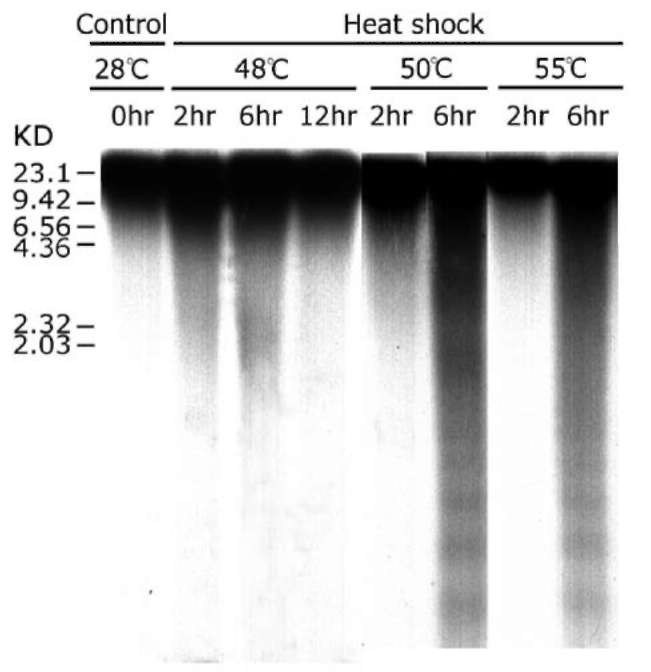
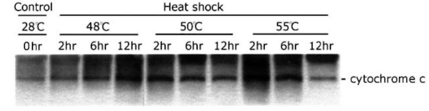
ABSTRACT. The effect of heat shock (HS) on death, weight and membrane leakage of mung bean seed­lings as well as on early biochemical markers of PCD (cytochrome c release from mitochondria and internu-cleosomal DNA fragmentation) was studied. It was found that heat shock ranging from 48-55C stimulated the releasing of cytochrome c from mitochondria to cytosol and the nuclear DNA fragmentation occurred during the first six hours of heat treatment. Sublethal heat shock (48°C) gives rise to non-ladder DNA degradation. Lethal heat shock (50 and 55C) generates two types, ladder and non-ladder, DNA degradation. Internu-cleosomal (ladder) DNA fragmentation and cytochrome c releasing suggest the programmed cell death occurs in mung bean seedling induced by heat shock.
Keywords: Cytochrome c; DNA fragmentation; Heat shock; Programmed cell death.
INTRODUCTION
nucleosomal DNA degradation has been observed during certain development events (Young and Gallie, 2000; He and Kermode, 2003), after induction by different stresses (Koukalova et al., 1997), and during pathogen-induced death (Mittler and Lam, 1997). DNA is first cleaved into large fragments of about 300 and/or 50 kb (Oberhammer et al., 1993), and these are further digested between nu-cleosomes resulting in DNA fragments that are multimers of about 180 bp monomers (Lagarkova et al., 1995). It is currently believed that these changes in genomic DNA are incompatible with cell survival and mark the point of no return in the execution stage of the PCD pathway.
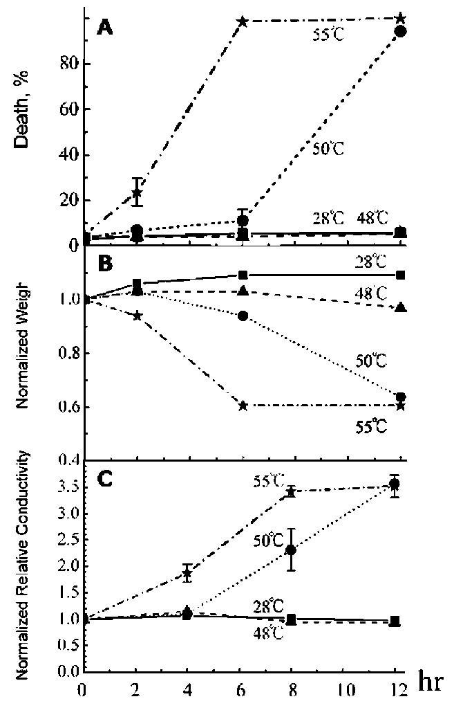
In this work, we studied the effect of heat shock (HS) on death, weight and membrane leakage of mung bean seedlings as well as on early biochemical markers of PCD (cytochrome c release in cytosol and DNA fragmentation). It was found that all temperatures applied for heat shock (48-55°C) increase the releasing of cytochrome c from mitochondria to cytosol and causes a concomitant nuclear DNA fragmentation during the first six hours of heat shock at 50°C and 55°C. The sublethal heat shock (48C) gave rise to non-ladder DNA fragmentation and lethal heat shock (50C and 55C) exhibited both ladder and non-ladder DNA degradation.
Programmed cell death (PCD) occurred during plant developmental processes such as flower development, embryogenesis, seed germination, and vessel and trachea formation was reported before (Pennell and Lamb, 1997; Mittler, 1998; Egorova et al., 2010). In addition, PCD can be also triggered in plant cells infected by pathogens, wound by physical treatments or damaged by low dosage of toxic compounds (Buckner et al., 2000).
One of the earliest markers of animal PCD is the releas­ing of cytochrome c from mitochondria to cytosol (Reed, 1997; Reape and McCabe, 2010). By analogy with mam­malian apoptosis, plant mitochondria have been suggested to play a pivotal role in the integration of environmental and developmental signals that trigger cell death (Lam et al., 2001). Few studies, however, have explored the pos­sible involvement of cytochrome c and mitochondria in plant PCD (Balk et al., 1999; Balk and Leaver, 2001; Yu et al., 2002; Reape and McCabe, 2010). Nuclear DNA
degradation, which occurs following the initial stage of chromatin condensation, is an early feature of both animal and plant PCD (Krishnamurthy et al., 2000). The orderly degradation of genome during PCD is contrast to random decay of DNA that follows other types of cell death such as injury-induced necrosis (Kerr et al., 1995). In plants,
MATERIALS AND METHODS
Plant material

2Present address: Institute of Bioorganic Chemistry, Belarus National Academy of Sciences, Kuprevich St. 5/2, 220141 Minsk, Belarus.
*Corresponding author: E-mail: bodaihwa@gate.sinica.edu. tw; Tel: 886-2-27871176; Fax: 886-2-27838609.
Mung bean seeds (Vigna radiata L. (Wilczek) cv. Tain­an) were grown as previously described (Dai et al., 1998). To induce HS, three-day-old seedlings were incubated for 12 h in a dark growth chamber (humidity 67%) at 48°C, 50C, and 55C (Altschuler and Mascarenhas, 1982). Con-
74
Botanical Studies, Vol. 52, 2011
trol seedlings were incubated at 28C. Fifteen seedlings from the HS-induced and control treatments, respectively, were harvested at successive time points, frozen in liquid nitrogen, and stored at -80C ready for further extraction and subfractionation in order to carry out DNA and immu-noblotting analysis.
Isolation of proteins
HS exposed and control seedlings were frozen in liquid nitrogen and ground to a fine powder. After adding of the extraction buffer: 220 mM mannitol, 68 mM sucrose, 50 mM PIPES-KOH, pH 7.4, 50 mM KCl, 5 mM EGTA, 2 mM MgCl2, 1 mM dithiothreitol, and 1 tablet of EDTA-free protease inhibitor (Rocher) for each 50 ml of buffer, the samples were incubated for 30 min at 4。C (Bossy-Wetzel et al., 1998). Cell debris was pelleted by a quick centrifugation step during which the rotor was stopped as soon as it reached 6000 x g. The supernatant was recen-trifuged at 14000 x g for 15 min to remove mitochondria, and the remaining cytosolic supernatant was stored at -80°C for further analysis. A modified Lowry method was used for protein quantitation in the cytosolic fractions (Larson et al., 1986). Equal amounts of total protein were used in all the experiments.
Viability assay
Seedling death was assessed by both morphological (wilting symptoms) and physiological criteria. Quantita­tive experiments were repeated three times and more then 500 seedlings were examined in each experiment. The percentage of dead seedlings was calculated from the total seedling scored after subtracting the basal amount of dead seedlings (which was assessed in the same manner prior to the beginning of each experiment). Only seedling popula­tions with less then 5% initial death were chosen for assay (Elbaz et al., 2002).
Western blot analysis
Fresh weight determination
30 fg of total cytosolic protein was separated on 12.5% SDS-PAGE gel. The protein was transferred to a PVDF membrane (Millipore, Bredford, MA) and immu-no-blotting analysis against cytochrome c antibody (BD Biosciences) was the same as previously described (Dai et al., 1998).
Fifty mung bean seedlings were collected and weighted in 0, 2, 6, and 12 h along heat shock at 48C, 50C, and 55C (28C as control). The average weight of 50 seed­lings was determined and standard error (SE) was less than 8% among three repeats.
Membrane leakage
RESULTS AND DISCUSSION
Measurement of ion leakage from leafs was performed essentially as described (Xu and Hanson, 2000) with some modification. For each measurement, eight leaves (two primary leaves from four independent seedlings) were floated in 15 ml of double distilled water for 4 h at room temperature. This solution was tested for the "sample con­ductivity". Then the leaves were frozen in 15 ml of double distilled water at -20C overnight and this solution was use to obtain the "subtotal conductivity". Membrane leakage is represented by the "relative conductivity", which was cal­culated as sample conductivity divided by total conductiv­ity (the last one is sum of sample conductivity and subtotal conductivity). Conductivity of the solutions was measured with conductivity meter (model 30, Denver Instrument, USA).
It was known from previous report that plants could activate either HS responses (Altschuler and Mascarenhas, 1982) or PCD (McCabe et al., 1997; Vacca et al., 2006) depending on the severity of heat shock. Temperature known to induce the HS response (45C) had no influ­ence on plant survival (Lebel et al., 1993). However, plant cells subjected to 55C for 10 min can activate a cell death program that results in the series distinctive morphological and biochemical changes (Balk et al., 1999).
The degree of HS effect on plants is generally exempli­fied by the changes in a set of physiological parameters, including weight and cellular membrane leakage (Mittler and Lam, 1995; Panavas et al., 1998) as well as by the percentage of plant death. The time dependences of these changes were examined in our work. Proper number of mung bean seedlings was pooled at each time point during each individual HS experiment, and at least three indepen­dent HS experiments were carried out for analyzing the value of each parameter.
Exposure of three-day-old mung bean seedlings to 12-h HS at 48C did not affect viability of the seedlings throughout the experiment (Figure 1A, the percentage dead seedlings remains less then 5%), while the treatment of seedlings for 12 h at 50C as well as for 6 and 12 h at 55C induced death of almost all seedlings: the percentage of dead seedlings reached 92% at 50C and 100% at 55C (Figure 1A). The survived seedlings showed no visible wilting after 12-h HS at 48。C, 6-h HS at 50。C and 2-h HS at 55C. Furthermore, they continued to grow normally
DNA extraction and degradation analysis
At successive time points during 12-h HS, three seed­lings (approximately 700 mg) were ground to a fine pow­der in liquid nitrogen, and DNA was isolated according to a modified cetyltrimethylammonium bromide method (Fulton et al., 1995). DNA samples were digested with DNase-free RNase for 1 h at 37C, and the DNA content was estimated. 15 fig DNA was analyzed by electro-phoresis on a 2% (w/v) agarose gel in a standard TAE buffer. The DNA was transferred to a nylon membrane (Hybond-N, Amersham, Ireland) and hybridized with a radioactive probe prepared from mung bean total genomic DNA. The Southern blot analysis was the same as de­scribed previously (Dai et al., 1998).
EGOROVA et al. ― Programmed cell death induced by heat shock
75
after being returned to 28C .
As shown in Figure 1B, fresh weight of seedlings ex­posed to 12-h HS at 48C only decreased slightly. On the other hand, a fast decline in fresh weight was observed along 50C and at 55C treatment (Figure 1B). This result is consistent with the traditional concept that the loss of water content in seedling tissues contributes to wilting symptoms and finally brings about the death of seedlings (Kimpei and Key, 1985).
Plant cell death is also associated with an increase in cellular membrane leakage (Mittler and Lam, 1995) which can be measured by ion leakage (Xu and Hansen, 2000). In seedlings exposed to 48C for 12-h HS, membrane leakage
Figure 2. Southern blot analysis of genomic DNA from 3-day-old mung bean seedlings exposed to 2, 6 and/or 12 h-heat shock, respectively. Temperature and time periods of exposure to heat shock are indicated. Fifteen micrograms of DNA was fractionat­ed on a 2% (w/v) agarose gel, transferred to a nylon membrane, and hybridized to radioactive probe prepared from mung bean total genomic DNA.
remains constant as control through 12 h treatment period. Heat shock at 50C on seedlings, the conductivity kept as low as control for first 4 h treatment, a quick increases was observed after 4 h heat incubation. When temperature el­evates up to 55C, the conductivity increase continuously up to 12 h heat treatment (Figure 1C). This data show a close correlation and a good agreement with death per­centage (Figure 1A) as well as with decrease in weight of seedlings (Figure 1B) effected by HS on plant seedlings.
One of the hallmarks of plant PCD is the organized degradation of the genome DNA caused by internu-cleosomal fragmentation resulting in the appearance of ladder DNA fragments formed by 180-200 bp multimers resolved by agarose gel electrophoresis (Barry and Eas-man, 1992; Collins et al., 1992; He and Kermode, 2003). To examine whether similar PCD events may occur in heat shocked mung bean seedlings, DNA was purified gently from seedlings after the heat treatment of consecu-tive time points (Figure 2) and analyzed on agarose gel. In order to increase the sensitivity of detection, DNA was transferred to nylon membrane and Southern hybridization was carried by using purified mung bean nuclear DNA as a probe. The result in Figure 2 shows that the majority of DNA molecules from control seedlings as well as from heat shocked seedlings stay in the well exhibited its high molecular weight. Besides the main high molecular weight band, all temperatures of heat shock treatment induce DNA degradation and this degradation is most prominent
Figure 1. Influence of heat shock on three-day old mung bean seedlings. Temperature and duration used for heat shock treat­ment are shown in the figures. Vertical bars represent SE (n=3). Panel A. The average percentage of dead seedlings. For each treatment, 500 seedlings was examined for each time point. Three repeated experiments were carried out. Death of seedlings was assessed morphologically; Panel B. Effects of heat shock on seedling fresh weight. For each time point of every heat treat­ment, 50 seedlings were used; Panel C. Change in relative con­ductivity during heat shock caused by leakage of ions from leaf disks. Relative conductivity is the ratio of sample conductivity to total conductivity (see text).
76
Botanical Studies, Vol. 52, 2011
Figure 3. Immunodetection of cytochrome c in the cytosolic fraction of 3-day-old mung bean seedlings during heat shock. Each sample containing mitochondria-free cytosolic proteins was separated by SDS-PAGE followed by a Western blot analysis against anti-cytochrome c antibody.
at 6 h heat shock treatment. The degradation does not form a ladder DNA in the case of 48C heat shock treatment. The reversal of DNA degradation was shown after further 6 h 48C heat treatment. These data together with the data on seedling survival, seedling weight and leaves conduc­tivity show in Figure 1 indicate that HS at 48oC induces a weak reversible DNA degradation.
Both internucleosomal DNA fragmentation (ladder of nucleosomal multimers) as well degradation of non-nu-cleosomal fragmentation (non-ladder) are clearly observed at 6-h of heat treatment at 50C and 55C. This result sug­gests that heat shock for 6 h at 50C and at 6 hr at 55C trigger PCD on molecular level in some cells of seedlings or in some seedlings.
It has been demonstrated the translocation of cyto-chrome c from the mitochondria to the cytosol upon induc­tion of PCD in plant (Yu et al., 2002; Reape et al., 2008;
Taken together, our result presents that: 1. Heat shock at 48C will not change seedling survival, leaf conductivity but some minor change is shown in seedling weight. Heat shock at 50°C caused significant change of above three criteria after 6 h treatment time. The drastic change oc­curred right after heat treatment at 55C. 2. For 48°C treat­ment, no DNA fragmentation was found but smear DNA Reape and McCabe, 2010). To investigate cytochrome c relocation in cytosol during heat stress, control and HS exposed seedlings were fractionated to remove mitochon-dria from cytosol by centrifugation. The Western blot analysis on cytosol purified from different temperatures treated seedlings reveal that the cytochrome c is increased in cytosol for all heat shock treatments at different time in-tervals (compare lanes of heat shock to lane of 28。C con-trol in Figure 3). Obviously, the cytochrome c increasing in cytosol of 48。C treated sample do not cause the DNA fragmentation. However, the increasing of cytochrom c was correlated with DNA fragmentation of 50。C/55。C, 6 h treated seedlings. The cytochome c was increasing with prolonged 48°C heat treatment but neither DNA fragmen-tation was observed nor increasing of DNA degradation exhibited consecutively correlated to increasing amount of cytochrome c. The immunoblotting analysis was repeated 3 times and similar data were obtained. In order to obtain the accurate result, the amount of cytosol protein was measured three repeats and the average was taken in above experiment.
degradation appears during 2 and 6 h heat treatment. The reversal of DNA degradation was exhibited at 12 h of heat treatment. 3. The internucleosomal DNA fragmentation was occurred during heat shock at 50C /55C for 6 h. 4. Induction of cytochrome c was shown in all heat treating seedlings. For 48C treatment, no DNA fragmentation was detected caused by increasing of cytosolic cytochrome c. There is also no correlation between amount of DNA degradation in smear form and amount of cytochrome c increasing. For 50C and 55°C heat shock treatments, the correlation between increasing of cytosolic cytochrome c and DNA fragmentation at 6 h heat shock time point was found. We conclude here that additional cytochrome c re­leasing from mitochondria to cytosol during heat shock in mung bean seedling may not be a sole factor essential for DNA fragmentation in PCD.
Acknowledgements. This research was supported by re­search grants from the National Science Council and from Academia Sinica, ROC. VE was supported by a postdoc­toral grant from National Science Council, ROC.
LITERATURE CITED
Altschuler, M. and J.P. Mascarenhas.1982. Heat shock proteins and effects of heat shock in plants. Plant Mol. Biol. 1: 103-115.
Balk, J., C.J. Leaver, and P.F. McCabe. 1999. Translocation of cytochrome c from the mitochondria to the cytosol occurs during heat-induced programmed cell death in cucumber plant. FEBS Lett. 463: 151-154.
Balk, J. and C.J. Leaver. 2001. The PET1-GMS mitochondrial
mutation in sunflower is associated with premature pro­grammed cell death and cytochrome c release. Plant Cell 13: 1803-1818.
Barry, M.A. and A. Eastman. 1992. Endonuclease activity dur­ing apoptosis: role of the cytosolic Ca2+ and pH. Biochem Biophys. Res. Com. 186: 778-789.
Bossy-Wetzel, E., D.D. Newmeyer, and D.R. Green. 1998.
Mitochondrial cytochrome c release in apoptosis occurs upstream of DEVD-specific caspase activation and inde­pendently of mitochondrial transmembrane depolarization. EMBO J. 17: 37-49.
Buckner, B., G.S. Johal, and D. Janick-Buckner. 2000. Cell
EGOROVA et al. ― Programmed cell death induced by heat shock                                                                                                        77
death in maize. Physiol. Plant 108: 231-243.
Collins, I.R.J., B.V. Harmon, G.S. Gobe, and J.F.R. Kerr. 1992.
Intenucleasomal DNA cleavage should not be the sole criterion for identifying apoptosis. Int. J. Radiat. Biol. 61:
451-453.
Dai, H., Y.S. Lo, W.N. Jane, L.W. Lee, and K.S. Chiang. 1998.
Population heterogeneity of higher-plant mitochondria in structure and function. Eur. J. Cell Biol. 75: 198-209.
Egorova, V.P., Q. Zhao, Y.S. Lo, W.N. Jane, N. Cheng, S.Y. Hou, and H. Dai. 2010. Programmed cell death of the mung bean cotyledon during seed germination. Bot. Stud. 51: 439-449.
Elbaz, M., A. Avni, and M. Weil. 2002. Constitutive caspase-like machinery executs programmed cell death im plant cell. Cell Death Differ. 9: 726-733.
Fulton, T.M., J. Chunwongse, and S.D. Tanksley. 1995. Micro-prep protocol for extraction of DNA from tomato and other
herbaceous plants. Plant Mol. Biol. Rep. 13: 207-209.
He, X. and A.R. Kermode. 2003. Nuclease activities and DNA fragmentation during programmed cell death of megaga-metophyte cells of white spruce (Picea glauca) seeds. Plant
Mol. Biol. 51: 509-521.
Kerr, J.F.R. and G.S. Gobe. 1995. Anatomical methods in cell death. In L.M. Schwartz and B.A. Osborn (eds.), Cell Death Methods in Cell Biology. Academic Press, San Diego, pp.1-27.
Kimpei, J.A. and J.L. Key. 1985. Presence of heat shock mRNAs in field grown soybeans. Plant Physiol. 79: 672-678.
Koukalova, B., A. Kovarik, J. Fajkus, and J. Siroky. 1997. Chromatin fragmentation associated with apoptotic changes in tobacco cells exposed to cold stress. FEBS Lett. 414:
289-292.
Krishnamurthy, K.V., R. Krishnaraj, R. Chozhavendan, and F.S. Christopher. 2000. The programmed cell death in plants and animals-A comparison. Curr. Sci. 79: 1169-1180.
Lagarkova, M.A., O.V. Jarovaia, and S.V. Razin. 1995. Large-scale fragmentation of mammalian DNA in the course of apoptosis proceeds via excision of chromosomal DNA loops and their oligomers. J. Biol. Chem. 270: 20239-20241.
Lam, E., N. Kato, and M. Lawton. 2001. Programmed cell death, mitochondria and the plant hypersensitive response. Nature
411: 848-853.
Larson, E., B. Howlett, and A. Jagendorf. 1986. Artificial reduc-tant enhancement of the Lowry method for protein determi­nation. Anal. Biochem. 155: 243-248.
Lebel, E.G., J. Masson, A. Bogucki, and J. Paszkowski. 1993. Stress-induced intrachromosomal recombination in plant
somatic cells. Proc. Natl. Acad. Sci. USA 90: 422-426.
Mittler, R. and E. Lam. 1995. Identification, characterization, and purification of a tobacco andonuclease activity induced upon hypersensitive response cell death. Plant Cell 7:
1951-1962.
Mittler, R. and E. Lam. 1997. Characterization of nuclease activ­ities and DNA fragmentation induced upon hypersensitive response cell death and mechanical stress. Plant Mol. Biol. 34: 209-221.
Mittler, R. 1998. Cell death in plants. In R.A. Lockshin, Z. Zak-eri and J.L. Tilly (eds.), When Cells Die: A Comprehensive Evaluation of Apoptosis and Programmed Cell Death. Wiley-Liss, New York, pp. 147-174.
Oberhammer, F., W. Wilson, C. Dive, I.D. Morris, J.A. Hick-man, E. Wakeling, P.R. Walker, and M. Sikorska. 1993.
Apoptotic death in epithelial cells: cleavage of DNA to 300 and/or 50 kb fragments prior to or in the absence of internu-
cleosomal fragmentation. EMBO J. 12: 3679-3684.
Panavas, T., P.P. Reid, and B. Rubinstein. 1998. Programmed cell death of daylily petals: Activation of wall-based en­zymes and effect of heat shock. Plant Physiol. Biochem. 36:379-388.
Pennell, R.G. and C. Lamb. 1997. Programmed cell death in
plants. Plant Cell 9: 1157-1168.
Reape, T.J. and P.F. McCabe. 2010. Apoptotic-like regulation of programmed cell death in plants. Apoptosis 15: 249-256.
Reape, T.J., E.M. Molony, and P.F. McCabe. 2008. Programmed
cell death in plants: distinguishing between different modes.
J. Exp. Bot. 59: 435-444.
Reed, J.C. 1997. Cytochrome c: can't live with, can't live with­out it. Cell 91: 559-562.
Vacca, R.A., D. Valenti, A. Bobba, R.S. Merafina, S. Passarella, and E. Marra. 2006. Cytochrome c is released in a reac­tive oxygen species-dependent manner and is degraded via caspase-like proteases in tobacco bright-yellow 2 cells en route to heat shock-induced cell death. Plant Physiol. 141:
208-219.
Xu, Y. and R. Hanson. 2000. Programmed cell death during pollination-induced petal senescence in Petunia. Plant
Physiol. 122: 1323-1333.
Young, T.E. and D.R. Gallie. 2000. Programmed cell death dur­ing endosperm development. Plant Mol. Biol. 44: 283-301.
Yu, X.N., T.D. Perdue, Y.M. Heimer, and A.M. Jones. 2002.
Mitochondrial involvement in traheary element pro­grammed cell death. Cell Death Differ. 9: 189-198.
78
Botanical Studies, Vol. 52, 2011
熱處理誘發綠豆幼苗的細胞凋亡
伊娃蒂娜1,2 羅意珊1 戴華1
1中央研究院植物暨微生物學研究所
2Institute of Bioorganic Chemistry, Belarus National Academy of Sciences, Kuprevich St. 5/2, 220141 Minsk, Belarus
熱處理影響綠豆幼苗的死亡、重量及其膜滲漏。同時也可引起早期細胞凋亡的生化指標,包括細胞
色素C在細胞質中的增加及核DNA之片段化。在48°C55°C之熱處理下,皆有誘導綠豆幼苗細胞質
中細胞色素C增加的現象。核DNA片段化則在50°C55°C熱處理六小時的綠豆幼苗中發生。未達致
死量之48°C熱處理可造成DNA非片段化的降解。致死性之熱處理(50°C55°C處理六小時)可造成
DNA片段化降解及非片段化降解。本研究證實熱處理可誘發綠豆幼苗的細胞凋亡。
關鍵詞:細胞色素C DNA片段化熱處理細胞凋亡。