Botanical Studies (2011) 52: 161-171.
BIOCHEMISTRY
The extrinsic proteins of an oxygen-evolving complex in marine diatom Cylindrotheca fusiformis
Gow-Jen SHIEH1, Lee-Feng CHIEN2 *, and Rong-Long PAN3
1National Taiwan University Hospital Yun Lin Branch, Dou Liou City 64005, Taiwan
2Department of Life Sciences, National Chung Hsing University, Tai Chung 40227, Taiwan
3Department of Life Sciences, National Tsing Hua University, Hsin Chu 30043, Taiwan
(Received July 3, 2009; Accepted October 8, 2010)
ABSTRACT. Diatom Cylindrotheca fusiformis's oxygen-evolving complex (OEC) exhibited much lower 2,6-
dichlorophenylindophenol (DCIP) photoreduction activity (the electron transfer rate during O2 evolution) when compared to that of spinach. The protein composition of the OEC in C. fusiformis diatom thylakoids consist of only two extrinsic proteins with apparent molecular masses of 33 kDa and 15 kDa after Tris or NaCl washing. The 33 kDa protein was mainly responsible for O2 evolving activity and for about 60% of the DCIP photore-duction activity, whereas the 15 kDa protein's photoreduction activity was approximately 40%. Beside the two extrinsic proteins, we found that Ca2+, Cl- and Mn ions also participate in the O2 evolution of C. fusiformis. The concentrations of Ca2+ and Cl- required for O2 evolution in diatom thylakoids were higher than those for spinach, suggesting that the binding affinity of Ca2+ and Cl- in diatom OEC is lower than that of spinach. The 33 kDa and 15 kDa proteins may coincidently function in Ca2+ trapping due to a 23% diminishment in content when diatom thylakoids were depleted of both proteins. These two proteins were also involved in the binding of Mn ions. Analysis of amino acid occurrence frequencies confirmed that both 33 kDa and 15 kDa proteins were extrinsic.
Keywords: Diatom thylakoids; Electron transfer; Extrinsic proteins; Oxygen-evolving complex; Photoreduc-tion; Photosystem II.
Abbreviations: BSA, bovine serum albumin; Chl, chlorophyll; DCIP, 2,6-dichlorophenylindophenol; EGTA, ethyleneglycol-bis-(aminoethyl ether) N,N,A^',N'-tetraacetic acid; MES, 2-(N-morpholino)ethanesulfonic acid; OEC, oxygen-evolving complex; PS, photosystem; SDS-PAGE, sodium dodecyl sulfate polyacrylamide gel electrophoresis; Tris, tris(hydroxymethyl)aminomethane.
INTRODUCTION
The involvement of an oxygen-evolving complex (OEC) with several extrinsic proteins has been clearly shown in studies of O2 evolution in photosynthetic organ­isms (Seidler, 1996; Barber, 2002; Bricker and Ghanota-kis, 2003; Ferreira et al., 2004; Roose et al., 2007; De Las Rivas et al., 2007; Barber, 2008). These extrinsic proteins of the OEC are located in the lumen of thylakoid mem­branes and differ greatly among species. In higher plants, there are at least three extrinsic proteins of 33 kDa (PsbO), 23 kDa (or 24 kDa) (PsbP) and 17 kDa (or 18 kDa) (PsbQ); in other organisms, there may be between two and five ex­trinsic proteins with different molecular weights (De Las Rivas and Barber, 2004; Enami et al., 2008). Along with these extrinsic polypeptides, tetranuclear manganese (Mn4)

*Corresponding author: E-mail: lfchien@dragon.nchu. edu.tw; Phone: +886-4-22840416 ext. 609; Fax: +886-4­22874640.
cluster and cation calcium (Ca2+) and anion chloride (Cl-) cofactors also participate in O2 evolution (Ono and Inoue, 1983; Andersson et al., 1984; Pstorius and Schmid, 1984;Coleman and Govindjee, 1987; Ono and Inoue, 1988; De-bus, 1992; Loll et al., 2005; Yocum, 2008).
It has been demonstrated that washing PSII spinach par­ticles in either Tris or NaCl inactivates O2 evolution and the partial or complete release of three extrinsic proteins (Yamamoto et al., 1981; Ono and Inoue, 1983). We found that Tris washing completely removed 33 kDa, 23 kDa and 17 kDa proteins, while NaCl washing merely removed 23 kDa and 17 kDa proteins (Ono and Inoue, 1983). The 33 kDa component is involved in stabilizing Mn in the cata­lytic center of the OEC (Enami et al., 1994; Bricker and Frankel, 1998; De Las Rivas and Heredia, 1999). Mn is es­sential for water oxidation, and there is evidence that four Mn ions exist per O2-evolving center in algae and higher plants (Hsu et al., 1987; Yamamoto, 1988; Ferreira et al., 2004; Barber and Murry, 2008; Pushkar et al., 2008). We found that two Mn ions of the Mn4-cluster were released in the absence of the 33 kDa protein (Miyao and Murata,
162
Botanical Studies, Vol. 52, 2011
1984b). Upon Tris washing, a complete disassociation of the Mn4-cluster occurred, while NaCl washing caused the release of a small amount of Mn ions from the Mn4-cluster (Ghanotakis et al., 1984a; Enami et al., 1994). 23 kDa and 17 kDa proteins are required for Ca2+ and/or Cl- binding, which is in turn necessary for optimal O2 evolution (An-dersson et al., 1984; Miyao and Murata, 1984a; Miyao and Murata, 1985). Exogenous Ca2+ was able to stimulate O2 evolution of inactive PSII particles depleted of the 23 kDa and 17 kDa proteins (Ghanotakis et al., 1984b; Miqyass et al., 2007). Also, numerous functions have been attributed to the OEC anion Cl- (Yocum, 2008), including its role in activating O2 evolution (Coleman and Govindjee, 1987). Several lines of evidence also suggest that Cl- functions as a ligand to the Mn4-cluster and is required for the S-states transition (Popelkova and Yocum, 2007). Although OEC:Cl- stoichiometry is believed to be 1:1, we found that the Cl- binding site existed in both low and high affin-ity states (Lindberg and Andreasson, 1996; Tiwari et al., 2007; Yocum, 2008).
In contrast to the numerous studies on higher plants, there is little information about OECs in lower eukaryotic organisms such as diatoms. Compared with common high­er plants, marine diatoms can grow in the ocean at high salt concentration. The mechanism of O2 evolution in marine diatoms may well be distinct from that of common higher plants (Martinson et al., 1998; Nagao et al., 2007; Okumura et al., 2008). There are, however, higher plants that grow at high salt concentrations: the vast group of halophytes (Flowers and Colmer, 2008). In some of these plants, the OEC is modified with respect to that of common higher plants OEC made of 33 kDa, 23 kDa and 17 kDa subunits (Pagliano et al., 2009). We found that although the PSII of the halophyte Salicornia veneta lacks the 17 kDa protein and contains a reduced amount of the 23 kDa protein, there was no loss of functional activity. Similarly, we detected some modifications in the OEC composition of diatoms living in the oceans. For this reason, we tested the com-position and functionality of the OEC from diatom Cylin-drotheca fusiformis. The protein composition of this OEC was clarified by Tris- and NaCl-washing, showing that the C. fusiformis OEC contained only 33 kDa and 15 kDa extrinsic proteins and not 23 kDa protein. This showed that both 33 kDa and 15 kDa proteins of OEC were involved in O2 evolution, the 33 kDa proteins playing the main role. Both the 33 kDa and 15 kDa proteins were associated with Mn binding and Ca2+ trapping.
MATERIALS AND METHODS
Diatom culture
The C. fusiformis diatom was generously provided by Professor Chia-Wai Li (National Tsing Hua University). For continuous culture, diatoms were grown in artificial sea wa­ter at 25°C under an 8-h day/16-h night cycle with illumina­tion of about 24 faE m-1 sec-1 (1,200 Lux). Spinach (Spinacia oleracea L.) was obtained fresh from the local market.
Thylakoids isolation
Diatom cells in late log phase were collected by centrif-ugation at 4,000 xg for 6 min. Pelleted cells were washed twice with buffer I made of 20 mM Tris-HCl (pH 7.5), 0.3 M sucrose, 10 mM NaCl and 5 mM MgCl2. The washed cells were resuspended in buffer II made of 25 mM MES (pH 6.6), 0.3 M sucrose, 50 mM NaCl and 10 mM CaCl?. The cells were then sonicated with an ultrasonicator (Heat-system-ultrasonics Inc. model W-225) at 4°C set at 7.5 pulses per 50% duty cycle for 20 sec. The unbroken cells were removed by centrifugation at 4,000 xg for 10 min. The sonication procedure was repeated 5-7 times to obtain the maximum yield of thylakoids. After sonication, the thylakoids were spun down at 15,000 xg for 30 min. The pelleted particles were resuspended in buffer II and applied to 35-55% (w/v) sucrose gradient and then centrifuged at 13,000 x g for 1 h. The thylakoids, which are brown in color, were located at the band of about 45% (w/v) sucrose concentration. This band containing thylakoids was col-lected by a needle, washed twice with buffer II, then col-lected by centrifugation at 15,000 xg for 30 min. Spinach thylakoids were prepared as previously described (Pan et al., 1987) and finally resuspended in storage medium [25 mM HEPES-NaOH (pH 7.5), 10 mM NaCl, 10% (v/v) glycerol, 2.5% (v/v) dimethyl sulfoxide, 2 mM dithiothre-itol, 20 mg BSA mL-1, and 2 mg chlorophyll (Chl) mL-1]. After preparation, the Chl contents of the diatom and spin-ach thylakoids were measured using the respective meth-ods of Jeffery (1986) and Arnon (1949).
Photoreduction of DCIP in thylakoids
The photoreduction of DCIP was measured spectropho-tometrically at 590 nm as the electron transport rate dur­ing O2 evolution in thylakoids (Pan et al., 1987; Eastman et al., 1997). Thylakoids (8 fig Chl mL-1) and DCIP (33.3 fM) were applied into an assay medium containing 20 mM MES (pH 6.6) and 0.3 M sucrose with addition of dif­ferent ion concentrations under an illumination of 150 fE m-1 sec-1. If required, thylakoids were washed twice with buffer II containing 100 fM EGTA to remove Ca2+ or buf­fer III made of 25 mM MES (pH 6.6) and 0.3 M sucrose to remove Cl-.
Tris and NaCl thylakoids washing
For salt washing, thylakoids (200-300 fg Chl mL-1) were incubated with 1.2 M Tris-HCl (pH 8.3) or 0.8 M NaCl for 30 min at 4°C under room light of 10-20 fE m-1 sec-1 (Yamamoto et al., 1981). Thylakoids were then col-lected at 15,000xg for 30 min and then resuspended in buffer II. The Tris- or NaCl-washed supernatants were dialyzed against 20 mM sodium phosphate buffer (pH 7.0) and then concentrated by ultrafiltration (Amicon model 8010). For SDS-PAGE analysis, the thylakoid proteins of the supernatants were precipitated with 80% (v/v) acetone.
Gel electrophoresis
Protein components were analyzed with 13.5% SDS-
SHIEH et al. — OEC in marine diatom
163
PAGE. The polypeptidic bands were visualized either by coomassie blue or silver staining. The relative amount of the protein component on SDS-PAGE was determined by a densitometric analysis (Lee et al., 2006).
Purification of 33 kDa and 15 kDa proteins from diatom thylakoids
The 33 kDa protein was extracted from diatom thy-lakoids with 1.2 M Tris-HCl (pH 8.3) buffer after 0.8 M NaCl treatment, and the 15 kDa protein was extracted with 0.8 M NaCl. The crude extracts were concentrated by ultrafiltration. The concentrations of 33 kDa and 15 kDa proteins were determined by a modified Lowry method NaCl treatment, and the 15 kDa protein was extracted with 0.8 M NaCl. The crude extracts were concentrated by ultrafiltration. The concentrations of 33 kDa and 15 kDa proteins were determined by a modified Lowry method (Larson et al., 1980).
Measurement of Ca and Mn content in diatom thylakoids
The contents of Ca and Mn in diatom thylakoids were determined with an Inductively Coupled Plasma Emission Spectrometer (ICPAES). To remove salts, the Tris- or Na-Cl-washed diatom thylakoids were dialyzed with distilled deionized water overnight. Thylakoids were collected at 15,000 x g for 30 min and then dissolved with pure HNO3 and H2SO4 (4:1) just prior to measurement.
Reconstitution of Tris- and NaCl-washed diatom thylakoids
The Tris- or NaCl-washed diatom thylakoids were incu­bated with 33 kDa and/or 15 kDa proteins with or without
addition of 40 mM MnCl2 in buffer II at 4°C stirring for 1 h under room light of 10-20 fE m-1 sec-1. The protein to Chl content ratio was 2:1 (fg proteins versus fg Chl) in the reaction solution. The reconstituted thylakoids were then collected by centrifugation at 15,000 xg for 30 min.
Amino acid composition analysis
To prepare the 33 kDa and 15 kDa proteins for amino acid analysis, polypeptide bands on SDS-PAGE were visualized with 4 M sodium acetate in the dark (Hig-gins and Dahmus, 1979). The 33 kDa and 15 kDa bands were cut, chopped into pieces, homogenized, and then immersed into distilled water and stirred overnight. The eluted proteins were precipitated with 80% (v/v) acetone and dissolved with 6 M HCl in new tubes at 110°C for 24 h in the presence of 1% (v/v) phenol. The hydrolytes were subjected to analysis by an LKB4150 Alpha Acid analyzer (Taipei Regional Analytical Instrumentation Center).
RESULTS
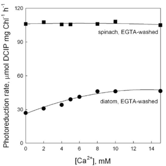
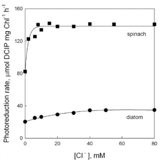
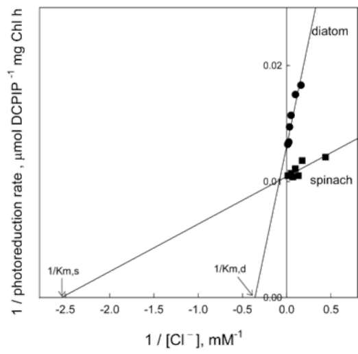
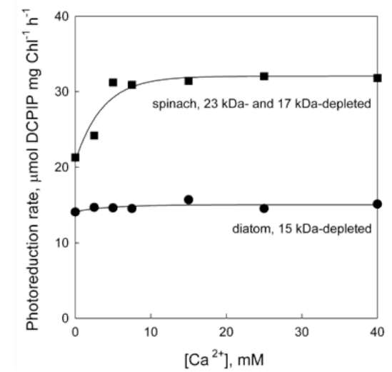
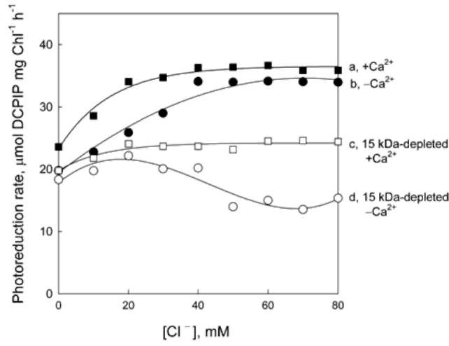
Effect of Tris and NaCl washing on electron transfer in the OEC of diatom thylakoids
The polypeptidic components in the OEC of diatom th-ylakoids were analyzed by SDS-PAGE and compared with those of spinach. In contrast to spinach thylakoids (Figure 1A, lane 1), diatom thylakoids showed two proteins of 33 kDa and15 kDa (molecular weights estimated in accor­dance with protein markers) but no 23 kDa protein (Figure
Figure 1. SDS-PAGE analysis of thylakoid membranes. (A) Lane 1, spinach control membranes; lane 2, diatom control membranes; lane 3, Tris-washed diatom membranes; lane 4, NaCl-washed diatom membranes. (B) Lane 1, Tris-washed diatom extract; lane 2, NaCl-washed diatom extract; lane 3, Tris-washed diatom extract after NaCl treatment.
164
Botanical Studies, Vol. 52, 2011
1A, lane 2). After washing with 1.2 M Tris (Figure 1A, lane 3) or 0.8 M NaCl (Figure 1A, lane 4), some proteins were depleted from the diatom thylakoids, in particular the 33 kDa and 15 kDa proteins. Subsequently, the 33 kDa and 15 kDa proteins were found in the crude extract of the Tris-washed supernatant (Figure 1B, lane 1). The 15 kDa protein was found in the NaCl-washed extract (Figure 1B, lane 2) and the 33 kDa protein in the NaCl-Tris-washed extract (Figure 1B, lane 3). These results suggested that the diatom O2 evolving center contained 33 kDa and 15 kDa proteins but lacked 23 kDa protein.
In order to study the functionality of electron transfer during O2 evolution in the diatom OEC, the photoreduc-tion of DCIP was measured (Table 1). The results showed that the diatom thylakoids exhibited a much lower DCIP photoreduction rate than spinach thylakoids. The photore-duction activity of the Tris-washed (i.e., 33 kDa- and 15 kDa-depleted) diatom thylakoids was completely inhib­ited; as it was in Tris-washed (i.e., 33 kDa-, 23 kDa- and 17 kDa-depleted) spinach thylakoids. The photoreduction activity of the NaCl-washed (i.e., 15 kDa-depleted) diatom thylakoids was reduced to 60% of the control; however, that of the NaCl-washed (i.e., 17 kDa- and 23 kDa-deplet-ed) spinach thylakoids was reduced to 36% of the control.
Requirement of Ca2+ and Cl- for electron trans­fer in the OEC of diatom thylakoids
It has been reported that Ca2+ and Cl- binding to OEC is related to the 23 kDa protein in higher plants (Ifuku et al., 2005; Miqyass et al., 2007); we decided to assess the impact of Ca2+ and Cl- on the diatom OEC lacking the 23 kDa protein.
To clarify the effect of Ca2+ on electron transfer during O2 evolution in the OEC of diatom and spinach thylakoids, the DCIP photoreduction was examined at different CaCl2 concentrations (Figure 2). Before measuring, diatom and spinach thylakoids were washed with buffer II containing the chelator EGTA to remove endogenous Ca2+ (Stevens and Lukin, 2003; Semin et al., 2008). The data showed that the photoreduction activity of EGTA-washed diatom thy-lakoids was reduced to 42% of the control, whereas that of EGTA-washed spinach thylakoids was not significantly af­fected. This suggested that the Ca2+ binding in the diatoms' OEC may be not as tight as it is in spinach. Re-addition of
Figure 2. Effect of Ca2+ on photoreduction rate of EGTA-washed thylakoid membranes. DCIP photoreduction rates of 100 fM EGTA washed thylakoid membranes were measured at dif­ferent designated CaCl2 concentrations in the presence of 40 mM NaCl. (•), diatom; (■), spinach.
Figure 3. Effect of Cl- on photoreduction rate of thylakoid mem­branes. DCIP photoreduction rates of diatom or spinach thyla-koid membranes were measured at different designated NaCl concentrations. (•), diatom; (■), spinach.
Table 1. Effect of Tris or NaCl washing on DCIP photoreduction rate of diatom and spinach thylakoids.

Diatom
Spinach
Thylakoids

DCIP photoreduction ratea (%)

Protein releasedb (%)

DCIP photoreduction ratea (%)

Protein releasedb

(%)


33 kDa 15 kDa


33 kDa

23 kDa

17 kDa
Control
100
100 100
100
100
100
100
Tris-washed
0
95 90
0
100
100
100
NaCl-washed
60
10 90
36
1
91
100

a100% represented the photoreduction rate of 42.7 fmole DCIP mg Chl-1 h-1 in diatom or 160 fmole DCIP mg Chl-1 h-1 in spinach. bThe protein released was estimated from SDS-PAGE of Tris- or NaCl-washed diatom (Figure 1A) and spinach (data not shown) thylakoids after analyzed by a densitometer.
SHIEH et al. — OEC in marine diatom
165
more than 10 mM exogenous Ca2+ into EGTA-washed dia­tom thylakoids restored the photoreduction activity.
We also tested the effect of Cl- on electron transfer during O2 evolution in the OEC of diatom and spinach thylakoids (Figure 3). Before measuring, the diatom and spinach thylakoids were washed with buffer III to remove Cl-. We found that the DCIP photoreduction activity of diatom thylakoids increased with increasing Cl- concen­trations, reaching an optimal rate at 40 mM Cl-, whereas spinach thylakoid activity reached an optimal rate at 15 mM Cl-. These results showed that the electron transfer rate during O2 evolution of diatom thylakoids was about three times slower than for spinach. The double reciprocal plots revealed that the Mechelis-Menton constant for the Cl- requirement of diatom (Km,d) was about 5 mM, while that of spinach (Km,s) was 0.8 mM (Figure 4). This indi­cated that the binding affinity of Cl- for diatom thylakoids was lower than that for spinach thylakoids.
Effect of Ca2+ and Cl- on electron transfer in the OEC of 15 kDa-depleted diatom thylakoids
To understand whether the 15 kDa protein of the diatom OEC could compensate for the 23 kDa protein of the high­er plant OEC for Ca2+ binding, the electron transfer activ­ity was examined by re-adding Ca2+ into 15 kDa-depleted (NaCl-washed) diatom thylakoids (Boussac et al., 1985; Ifuku et al., 2005). The data showed that the DCIP pho-toreduction rates of the 15 kDa-depleted diatom thylakoids were not changed by re-adding exogenous Ca2+, whereas those of the 23 kDa- and 17 kDa-depleted (NaCl-washed) spinach thylakoids could be stimulated by re-adding Ca2+ above 5 mM (Figure 5).
Furthermore, addition of 10 mM Ca2+ to diatom thy-lakoids stimulated DCIP photoreduction activity and re­duced the Cl- requirement from 40 mM to 20 mM (Figure 6, curve a, b). Addition of 10 mM Ca2+ to 15 kDa-depleted (NaCl-washed) diatom thylakoids (Figure 6, curve c) did not stimulate DCIP photoreduction activity; however, it prevented a further reduction (Figure 6, curve d). This suggested that the effect of Ca2+ on O2 evolution of diatom thylakoids was dependent on the presence of the 15 kDa protein. The requirement of Cl- for O2 evolution in the 15 kDa-depleted diatom thylakoids was diminished by adding exogenous Ca2+, implying that the function of the 15 kDa protein may be partially replaced by Ca2+.
Figure 4. Double reciprocal plots of the dependence of the pho­toreduction rate on the concentrations of Cl-. (•), diatom; (■), spinach. Km,d, Mechelis-Menton constant for Cl- requirement of diatom; Km,s, Mechelis-Menton constant for Cl- requirement of spinach.
Figure 5. Effect of Ca2+ on photoreduction rate of 15 kDa-depleted diatom thylakoid. DCIP photoreduction rates of 0.8 M NaCl washed diatom thylakoid membranes were measured at different designated CaCl2 concentration in the presence of 40 mM NaCl. (■), NaCl-washed, 23 kDa- and 17 kDa-depleted, spinach thylakoids; (•), NaCl-washed, 15 kDa-depleted, diatom thylakoids.
Table 2. Ca and Mn abundance and DCIP photoreduction rate in 33 kDa-depleted and/or 15 kDa-depleted diatom thylakoids.

Diatom thylakoids
DCIP photoreduction ratea
Ca abundance1"
Mn abundance
(%)
(atoms/400 Chl)
(atoms/400 Chl)

Control

100

1

3.8
33 kDa-depleted, 15 kDa-depleted
0
0.23
2.1
(Tris-washed)
15 kDa-depleted (NaCl-washed)
60
0.29
3.1

a100% represented the photoreduction rate of 36.4 famole DCIP mg Chl-1 h-1 in diatom.
bThe Ca abundance of the control was set to 1 atom per 400 Chl molecules in accordance with Pushkar et al. (2008) so that all the original data were divided by a factor of 436 which was obtained in the background.
166
Botanical Studies, Vol. 52, 2011
protein (Table 2). This was consistent with the results in Figure 5, showing that the reduced O2 evolving activity of the 15 kDa-depleted diatom thylakoids could not be re­stored by re-adding exogenous Ca2+.
The number of Mn atoms in the two types of diatom thylakoids was also determined (Table 2). This analysis showed that 33 kDa- and 15 kDa-depleted (Tris-washed) diatom thylakoids contained 2.1 Mn atoms per 400 Chl molecules, while 15 kDa-depleted (NaCl-washed) diatom thylakoids contained 3.1 Mn atoms. This demonstrated that both 33 and 15 kDa proteins were involved in Mn binding in the diatom OEC.
Further characterization of the 33 kDa and 15 kDa proteins
To study further how the 33 kDa and 15 kDa proteins cooperated in the diatom OEC, reconstitution experiments were carried out (Ono and Inoue, 1984). We showed earli­er that Tris washing released 33 kDa and 15 kDa proteins, extracted two out of four Mn atoms, and reduced the O2 evolving activity to zero (Table 2). By reconstituting Tris-washed thylakoids with the 33 and 15 kDa proteins, the re­duced O2 evolving activity could be restored to 33% of the control (Table 3). Moreover, NaCl washing released the 15 kDa protein, extracted one out of four Mn atoms, and re­duced the DCIP photoreduction activity to 48% of the con­trol (Table 2). By reconstituting NaCl-washed thylakoids with the 15 kDa protein, the reduced DCIP photoreduction activity could be restored to 66% of the control (Table 3). There was no increase in activity by further addition of Mn2+.
We analyzed the amino acid compositions of the 33 kDa and 15 kDa proteins of the diatom OEC and the 33 kDa and 17 kDa proteins of the spinach OEC (Table 4). The nonpolar R group composition of the diatom 33 kDa protein (40.1%) was larger than that of spinach (35.5%). The total number of amino acid residues of the 33 kDa protein in the diatom OEC was 249 residues per protein, as in spinach. The non-polar composition of the diatom 15 kDa protein (41.4%) was larger than that of the corre­sponding spinach 17 kDa protein (39.4%). The total num-
Figure 6. Effect of Cl- on photoreduction rate of 15 kDa-deplet-ed diatom thylakoids in the absence and presence of Ca2+. DCIP photoreduction rates of diatom thylakoids were measured in the absence (curve a, •) and presence (curve b, ■) of 10 mM CaCl2, or, those of 15 kDa-depleted (NaCl-washed) diatom thylakoids were measured in the absence (curve c, o) and presence (curve d, □) of 10 mM CaCL at different designated NaCl concentrations.
Ca and Mn abundance in the diatom OEC
In an attempt to characterize more precisely the re­lationship between Ca2+ and the 15 kDa protein, the Ca abundance was measured (Table 2). In the 15 kDa-depleted (NaCl-washed) diatom thylakoids, the Ca content was reduced to about 0.29 atoms per 400 Chl molecules, suggesting that the 15 kDa protein mediated the binding of 0.71 Ca atoms. In the 33 kDa- and 15 kDa-depleted (Tris-washed) diatom thylakoids, the Ca content was re­duced to about 0.23 atoms, suggesting that 33 kDa protein mediated the binding of 0.06 Ca atoms. Although the 33 kDa protein has been demonstrated to have a Ca2+ binding site, the 23 kDa and 17 kDa proteins of higher plants were also reported to be involved in Ca2+ binding (Ghanotakis et al., 1984b; Kruk et al., 2003; Murray and Barber, 2006; Miqyass et al., 2007). Our Tris treatment results implied that the binding of Ca2+ in the diatom C. fusiformis OEC was more related to the 15 kDa protein than to the 33 kDa
Table 3. Restoration of DCIP photoreduction rate by reconstituting 33 kDa and/or 15 kDa proteins into diatom thylakoids with or without Mn2+.

Diatom thylakoids
Additiona
DCIP photoreduction rateb (%)

Control

None

100
33 kDa-depleted
None
0
15 kDa-depleted (Tris-washed)
+ 33 kDa + 15 kDa
0
+ 33 kDa + 15 kDa + Mn2+
33
15 kDa-depleted (NaCl-washed)
None
48
+ 15 kDa
66
+ 15 kDa + Mn2+
66

aUnder the conditions: proteins Chl-1 = 0.8 mol mol-1 and Mn2+ = 40 mM.
b100% represented the photoreduction rate of 30.0 fmole DCIP mg Chl-1 h-1 in diatom.
SHIEH et al. ― OEC in marine diatom
167
Table 4. Amino acid compositions of 33 kDa and 15 kDa proteins in diatom and those of 33 kDa and 17 kDa polypeptides in spinach.

Amino acid

Diatom (residuesa/protein)


Spinach (residuesa/protein)
33 kDa
15 kDa
33 kDa
17 kDa
Lys
7.6
7.9
9.2
9.2
His
0.7
1.2
0.0
0.9
Arg
4.6
5.4
2.7
5.6
Asp
10.9
12.8
8.8
11.2
Glx
13.0
9.0
13.2
11.1
Thr
5.3
5.3
7.7
5.3
Ser
3.7
4.7
7.4
8.6
Pro
5.6
5.7
6.1
8.8
Gly
11.4
9.3
12.3
5.1
Ala
8.4
10.4
6.0
9.0
Val
7.0
6.4
7.2
3.7
Met
1.4
1.1
0.4
0.0
Ile
5.1
5.3
3.0
3.8
Leu
8.1
8.6
7.1
11.4
Tyr
2.8
3.4
3.2
3.6
Phe
4.1
3.2
5.3
2.1
Trp
0.4
0.7
0.4
0.6
Cys
NDb
NDb
NDb
NDb
Total residues
249
122
249
139
Percentage
Nonpolar R group
40.1%
41.4%
35.5%
39.4%
Polar R group
Uncharged and negative
46.9%
44.1%
52.6%
44.9%
Positive
13.0%
14.5%
11.9%
15.7%

aThe amino acid composition was calculated from the moles of residues divided by the moles of total proteins.
bNot determined.
ber of amino acid residues of the diatom 15 kDa protein was 122 while that of the spinach 17 kDa protein was 139. The location of a protein with respect to the membrane can be predicted from its amino acid composition (Can­tor and Schimmel, 1980). To obtain information about the internal or external membrane location of the proteins, the following parameters were assessed: R3, the mole fraction ratio of the two amino acid sets, H0ave, average of total hydrophobicity, Z, discriminate function value, and NPS, the fraction of non-polar side chains (Table 5). Comparing these values, we confirmed that diatom 33 kDa and 15 kDa proteins, which were released from thylakoids by Tris or NaCl washing, were extrinsic proteins.
DISCUSSION
The extrinsic proteins of OEC, located in the lumen of thylakoid membranes, show significant differences among species. These extrinsic proteins have been examined in several organisms and classified into cyanobacterial type
(PsbO, PsbV and PsbU), red algal type (PsbO, PsbQ', PsbP and PsbU), and green algal and higher plant type (PsbO, PsbQ and PsbP) (De Las Rivas et al., 2007; Roose et al., 2007; Enami et al., 2008). The OEC of the diatom Chaetoceros garacilis has very recently been shown to include five extrinsic proteins with molecular weights of 33 kDa (PsbO), 20 kDa (PsbQ'), 17 kDa (PsbV), 13.5 al., 2008). Unlike that of C. gracilis, the OEC of diatom C. fusiformis contained only two extrinsic proteins 33 kDa and 15 kDa. Undoubtedly, the 33 kDa protein in C. fusiformis acted in accordance with the highly-conserved PsbO protein in all O2 evolving photosynthetic organisms (Tohri et al., 2002; De Las Rivas et al., 2007). Interest-ingly, the 23 kDa (PsbP) protein did not appear in either C. fusiformis or C. gracilis (Nagao et al., 2007). The 15 kDa protein in C. fusiformis could function independently and may be a 20 kDa (PsbQ')-like protein like that in C. graci-lis (Nagao et al., 2007; Enami et al., 2008).
In our current study, 95% of the 33 kDa protein of the
168
Botanical Studies, Vol. 52, 2011
Table 5. The parameters of R3, H^ve, Z and NPS for 33 kDa and 15 kDa proteins in diatom and spinach.

Protein
R3a
HΦaveb
Zc
NPSd
Categorization
Membrane protein
Believed internal
0.57±0.18
1.197±0.097
0.52±0.11
Internal
Believed external
1.37±0.35
0.986±0.075
0.16±0.17
External
Diatom
33 kDa
1.69
1.062
0.05
0.22
External
15 kDa
1.76
1.067
0.05
0.29
External
Spinach
33 kDa
1.62
0.921
-0.01
0.18
External
23 kDa
1.61
0.934
0.01
0.18
External
17 kDa
1.30
1.121
0.20
0.25
External
aR3 was the mole fraction ratio of the two amino acid sets. (R3 =
j, k, the number of residues; Xj= the composition of Lys,
Arg, His, Asp, Glx, or Glu; Xk=the composition of Ile, Leu, Try, Phe, Met, or Val).
bHΦave was the total hydrophobicity divided by the number of residues.
cZ was the discriminate functions of linear combinations of R3 and H^ve. (Z=-0.345R3+0.0006 H^ve)'
dNPS (non-polar side chains) was calculated as the fraction of the nonpolar amino acid residues such as Trp, Ile, Try, Phe, Pro, Leu and Val to the total number of residues.
diatom OEC can be released by washing with 1.2 M Tris (Table 1), while that of spinach could be completely re­leased using the lower concentration of 0.9 M Tris (data not shown). This implied that the diatom 33 kDa protein may be located in a more hydrophobic environment than the spinach one. We further confirmed this observation by estimating the parameters of R3, H^ve, Z and NPS, which provide information about the location of internal or exter­nal membrane proteins (Table 5). The H^ve of the diatom 33 kDa protein (1.062) was larger than that of spinach (0.921), suggesting that the diatom 33 kDa polypeptide was located in a more hydrophobic environment. A similar result was obtained earlier using cyanobacteria (Steward et al., 1985). Here, when compared to spinach, only a mini­mal amount of 33 kDa protein was released via alkaline Tris treatment. The H0ave of the diatom 15 kDa protein (1.067) was smaller than that of spinach (1.121). Mean­while, the amount of the 15 kDa protein (100%) released from diatom thylakoids by NaCl treatment was greater than that of the 17 kDa (90%) from spinach. We therefore believe that the 15 kDa protein of the diatom OEC was located in a more hydrophilic environment than the corre­sponding 17 kDa protein of spinach.
The concentrations of Ca2+ and Cl- required for diatom thylakoids was higher than those observed for spinach thylakoids (Figures 3 and 4), suggesting that the binding affinity of Ca2+ and Cl- in the diatom OEC was lower than for spinach. Tris treatment reduced the amount of Mn ions in diatom thylakoids from 3.8 to 2.1 atoms and in spinach thylakoids it was reduced from 4 to 0.65 atoms, leading to complete inhibition of DCIP photoreduction (Table 2) (Hsu et al., 1987). A previous study showed that spinach PSII with 1.4 Mn upon NH2OH treatment still retained 30% O2 evolving activity (determined by the reduction of DCIP) and 30% multiline EPR signal intensity of the control
(Miller and Brudvig, 1989). However, upon Tris washing, diatom PSII still retained 2 Mn ions and did not exhibit any DCIP photoreduction activity (Table 2). PSII should be able to reduce DCIP if an OEC center is intact. Thus, the loss of the full Mn cluster from only a fraction of the center, leaving the remaining center intact, should not oc­cur. Moreover, a number of studies indicated that the Mn4-cluster is coordinated by certain residues of the D1 and CP43 polypeptides (Barber and Murray, 2008; Stull et al., 2010). This implies that the Mn4-cluster may be disrupted by Tris or NaCl washing, but that the Mn ions may not be completely depleted when 33 kDa protein is removed (Williamson, 2008). Subsequently, DCIP photoreduction activity was restored only by re-adding Mn2+ plus the 33 and 15 kDa proteins into the 33 kDa- and 15 kDa-depleted (Tris-washed) diatom thylakoids (Table 3). This suggested that these depleted Mn atoms played a crucial role in O2 evolution. Their different requirements for the essential inorganic components of the Mn2+, Ca2+ and Cl- ions in-dicated that the diatom and spinach OECs have different characteristics.
Moreover, we found that the 15 kDa protein is required for O2 evolution of the diatom OEC. The specific function of the 15 kDa protein was studied in the NaCl-washed dia­tom thylakoids. NaCl washing released the 15 kDa protein from diatom thylakoids and reduced their O2 evolving ac­tivity to 63% of the control (Table 1). The 15 kDa-depleted (NaCl-washed) diatom thylakoids were more sensitive to high Cl- concentration above 40 mM (Figure 6, curve d). This result suggested a possible role for the 15 kDa in pre­serving the O2 evolving activity in diatoms exposed to the high concentrations of NaCl in the ocean.
From the results of Figure 5, we proposed that the func­tion of the 15 kDa protein in the diatom OEC was related to Ca2+ binding, though it has been shown that the 23 kDa
SHIEH et al. — OEC in marine diatom
169
protein in the higher plant OEC acted in Ca2+ trapping (Ghanotokis et al., 1984a; Miyao and Murata, 1984a). NaCl treatment depleted thylakoids of the 15 kDa protein and reduced their Ca2+ content to about 30% of the control (Table 2). The reduced O2 evolving activity of diatom thy-lakoids without 15 kDa protein was not recovered by add­ing external Ca2+ (Figure 5 and Figure 6, curve c), whereas the O2 evolving activity of diatom thylakoids containing the 15 kDa protein were restored to the optimal rate in the presence of different Cl- concentrations (Figure 6, curve a). This suggested that the 15 kDa protein was related to the binding of Ca2+ in diatom thylakoids and that Ca2+ could participate in O2 evolution only in the presence of the 15 kDa protein.
Reconstitution of Tris-washed thylakoids with 33 kDa and 15 kDa proteins restored O2 evolving activity to 33% of the control (Table 3). The reason why the O2 evolving activity was only partially restored may be due to the ab­sence of Ca2+, which was intentionally diminished in this experiment to avoid its interference. The reconstitution of the extrinsic proteins into salt washed PSII with addi­tion of Ca2+ may increase the restored O2-evolving activ­ity (Ghanotakis et al., 1984b; Enami, 1994; Seidler et al., 1996; Suzuki et al., 2005). It is believed that the binding of Ca2+ could either provide the appropriate environment for a stable Mn complex or affect its redox properties (Bricker and Ghanotokis, 2003).
Based on these results, we concluded that two extrinsic proteins, 33 kDa and 15 kDa proteins, and three cofactors, Ca2+, Cl- and Mn ions, participated in O2 evolution in the diatom C. fusiformis. The composition and functionality of the diatom C. fusiformis OEC is thus somewhat distinct from that of the spinach OEC.
Acknowledgements. This work was partly supported by the grants from National Science Council of Taiwan to Lee-Feng Chien (NSC96-2313-B-005-030-MY2) and Rong-Long Pan (NSC95-2311-B-007-004). The authors gratefully acknowledge Angela Symonds for help in man-uscript reading.
LITERATURE CITED
Andersson, B., C. Critchley, I.J. Ryrie, C. Jansson, C. Larsson, and J.M. Anderson. 1984. Modification of the chloride re­quirement for photosynthetic oxygen evolution. FEBS Lett. 168: 113-117.
Arnon, D.I. 1940. Copper enzymes in isolated chloroplasts.
Plant Physiol. 24: 1-5.
Barber, J. 2002. Photosystem II: a multisubunit membrane pro­tein that oxidizes water. Curr. Opin. Structural Biol. 12:523-530.
Barber, J. 2008. Crystal structure of the oxygen-evolving com­plex of photosystem II. Inorg. Chem. 47: 1700-1710.
Barber, J. and J.W. Murray. 2008. Revealing the structure of the Mn-cluster of photosystem II by X-ray crystallography. Co-
ord. Chem. Rev. 252: 233-243.
Boussac, A., B. Maison-Peteri, A.L. Etienne, and C. Vernotte. 1985. Reactivation of oxygen evolution of NaCl-washed Photosystem-II particles by Ca2+ and/or the 24 kDa protein. Biochim. Biophys. Acta 808: 231-234.
Bricker, T.M. and D.F. Ghanotakis. 2003. Introduction to oxygen evolution and the oxygen-evolving complex. In D.R. Ort and C.F. Yocum (eds.), Advances in Photosynthesis. Kluwer Academic Publisher, Dordrecht, pp. 113-136.
Bricker, T.M. and L.K. Frankel. 1998. The structure and func­tion of the 33 kDa extrinsic protein of photosystem II: a critical assessment. Photosynth. Res. 56: 157-173.
Cantor, C.R. and P.R. Schimmel. 1980. Structure of proteins. In C.R. Cantor and P.R. Schimmel (eds.), Biophysical chemis­try. Part I: The conformation of biological macromolecules. Freeman, San Francisco, pp. 41-154.
Coleman, W.J. and Govindjee. 1987. A model for the mecha­nism of chloride activation of oxygen evolution photosys-tem II. Photosynth. Res. 13: 199-223.
De Las Rivas, J. and J. Barber. 2004. Evolution of oxygenic photosynthesis: genome-wide analysis of the OEC extrinsic proteins. Trends Plant Sci. 9: 18-25.
De Las Rivas, J. and P. Heredia. 1999. Structural predictions on the 33 kDa extrinsic protein associated with the oxygen evolving complex of photosynthetic organisms. Photosynth. Res. 61: 11-21.
De Las Rivas, J., P. Heredia, and A. Roman. 2007. Oxygen-evolving extrinsic proteins (PsbO,P,Q,R): Bioinformatic and functional analysis. Biochim. Biophys. Acta 1767:
575-582.
Debus, R.J. 1992. The manganese and calcium ions of photo-synthetic oxygen evolution. Biochim. Biophys. Acta 1102: 269-325.
Eastman, P.A.K., A. Rashid, and E.L. Camm. 1997. Changes of the photosystem 2 activity and thylakoid proteins in spruce seedlings during water stress. Photosynthetica 34: 201-210.
Enami, I., A. Okumura, R. Nagao, T. Suzuki, M. Iwai, and J.R. Shen. 2008. Structures and functions of the extrinsic pro­teins of photosystem II from different species. Photosynth. Res. 98: 349-363.
Enami, I., T. Tomo, M. Kitamura, and S. Katoh. 1994. Immo­bilization of the three extrinsic proteins in spinach oxygen-evolving Photosystem II membranes: roles of the proteins in stabilization of binding of Mn and Ca2+. Biochim. Biophys. Acta 1185: 75-80.
Ferreira, K.N., T.M. Iverson, K. Maghlaoui, J. Barber, and S. Iwata. 2004. Architecture of the photosynthetic oxygen-evolving center. Science 303: 1831-1838.
Flowers, T. J. and T.D. Colmer. 2008. Salinity tolerance in halo-
phytes. New Phytol. 179: 945-963.
Ghanotakis, D.F., J.N. Topper, and C.F. Yocum. 1984a. Struc­tural organization of the oxidizing side of photosystemII. Exogenous reductants reduce and destroy the Mn-complex in photosystem II membranes depleted of 17 and 23 kDa
170
Botanical Studies, Vol. 52, 2011
polypeptides. Biochim. Biophys. Acta 767: 524-531.
Ghanotakis, D.F., J.N. Topper, G.T. Babcock, and C.F. Yocum. 1984b. Calcium reconstitutes high rates of oxygen evolution in polypeptide depleted photosystem II preparations. FEBS Lett. 167: 127-130.
Higgins, R.C. and M.E. Dahums. 1979. Rapid visualization of protein bands in preparative SDS-PAGE. Anal. Biochem.
93: 257-260.
Hsu, B.D., J.Y. Lee, and R.L. Pan. 1987. The high affinity bind­ing site for manganese on the oxidizing site of PSII. Bio-chim. Biophys. Acta 890: 89-96.
Ifuku, K., T. Nakatsu, R. Shimanoto, Y. Yamanoto, S. Ishihara, H. Kato, and S. Sato. 2005. Structure and function of the PsbP protein of photosystem II from higher plants. Photo-
synth. Res. 84: 251-255.
Jeffery, S.W. 1986. Quantitative thin layer chromatography of chlorophylls and carotenoids from marine algae. Biochim
Biophys Acta 162: 271-285.
Kruk, J., K. Burda, M. Jemiola-Rzeminska, and K. Strzalka. 2003. The 33 kDa protein of photosystem II is a low-affin­ity calcium- and lanthanide-binding protein. Biochemistry< 42: 14862-14867./span>
Larson, E., B. Howlett, and A.T. Jagendoff. 1980. Artificial reductant enhancement of the Lowry method for protein de­termination. Anal. Biochem. 155: 243-248.
Lee, S.C., L.F. Chien, R.C. Van, Y.Y. Hsiao, J.L. Hong, and R.L.
Pan. 2006. Radiation inactivation analysis of thylakoid pro­tein kinase systems in light and in darkness. Photosynthetica 44: 116-124.
Lindberg, K. and L.E. Andreasson. 1996. A one-site, two state model for the binding of anions in photosystem II. Bio­chemistry 35: 14259-14267.
Loll, B., G. Gerold, D. Slowik, W. Voelter, C. Jung, W. Saenger,
and K.D. Irrgang. 2005. Thermostability and Ca2+ binding properties of wild type and heterologously expressed PsbO protein from cyanobacterial photosystem II. Biochemistry
44: 4691-4698.
Martinson, T.A., M. Ikeuchi, and F.G. Plumley. 1998. Oxygen-evolving diatom thylakoid membranes. Biochim. Biophys.
Acta 1409: 72-86.
Miller, A.-F. and G.W. Brudvig. 1989. Manganese and calcium requirements for reconstitution of oxygen-evolution activity in manganese-depleted photosystem II membranes. Bio­chemistry 28: 8181-8190.
Miqyass, M., H.J. van Gorkom, and C.F. Yocum. 2007. The PSII calcium site revisited. Photosynth. Res. 92: 275-287.
Miyao, M. and N. Murata. 1984a. Calcium ion can be substituted for the 24-kDa polypeptide in photosynthetic oxygen evolu­tion. FEBS Lett. 168: 118-120.
Miyao, M. and N. Murata. 1984b. Role of the 33-kDa polypep-tide in preserving Mn in the photosynthetic oxygen-evo­lution system and its replacement by chloride ions. FEBS Lett. 170: 350-354.
Miyao, M. and N. Murata. 1985. The Cl- effect on photosyn-
thetic oxygen evolution: interaction of Cl- with 18-kDa, 24-kDa and 33-kDa proteins. FEBS Lett. 180: 303-308.
Murray, J.W. and J. Barber. 2006. Identification of a calcium-binding site in the PsbO protein of photosystem II. Bio­chemistry 45: 4128-4130.
Nagao, R., A. Ishii, O. Tada, T. Suzuki, N. Dohmae, A. Okumu-ra, M. Iwai, T. Takahashi, Y. Kashino, and I. Enami. 2007. Isolation and characterization of oxygen-evolving thylakoid membranes and photosystem II particles from a marine dia­tom Chaetoceros gracilis. Biochim. Biophys. Acta 1767:1353-1362.
Okumura, A., R. Nagao, T. Suzuki, S. Yamagoe, M. Iwai, K. Nakazato, and I. Enami. 2008. A novel protein in photo-system II of a diatom Chaetoceros gracillis is one of the extrinsic proteins located on lumenal side and directly asso­ciates with PSII core components. Biochim. Biophys. Acta 1777: 1545-1551.
Ono, T.A. and Y. Inoue. 1983. Mn preserving extraction of 33, 24 and 16 kDa proteins from oxygen-evolving PSII par­ticles by divalent salt washing. FEBS Lett. 164: 255-259.
Ono, T.A. and Y. Inoue. 1984. Reconstitution of photosynthetic oxygen-evolving PSII particles by divalent salt washing. FEBS Lett. 166: 381-384.
Ono, T.A. and Y. Inoue. 1988. Discrete extraction of the Ca atom functional for O2 evolution in higher plant photo-system II by a simple low pH treatment. FEBS Lett. 227:
147-152.
Pagliano, C., N. La Rocca, F. Andreucci, Z. Deak, I. Vass, N. Rascio, and R. Barbato. 2009. The extreme halophyte Sali-cornia veneta is depleted of the extrinsic PsbQ and PsbP proteins of the oxygen-evolving complex without loss of functional activity. Ann. Bot. 103: 505-515.
Pan, R.S., L.F. Chien, M.Y. Wang, M.Y. Tsai, R.L. Pan, and
B.D. Hsu. 1987. Functional size of photosynthetic electron transport chain determined by radiation inactivation. Plant
Physiol. 85: 158-163.
Popelkova, H. and C. Yocum. 2007. Current status of the role of Cl- ion in the oxygen-evolving complex. Photosynth. Res.
93: 111-121.
Pstorius, E.K. and G.H. Schmid. 1984. Effect of Mn2+ and Ca2+ on oxygen evolution and on variable fluorescence yield as­sociated with PSII in preparations of Anacystics nidulans. FEBS Lett. 171: 173-177.
Pushkar, Y., J. Yano, K. Sauer, A. Boussac, and V.K. Yachan-dra. 2008. Structural changes in the Mn4Ca cluster and the mechanism of photosynthetic water splitting. Proc. Natl.
Acad. Sci. USA 105: 1879-1884.
Roose, J.L., K.M. Wegener, and H.B. Pakrasi. 2007. The extrin­sic proteins of photosystem II. Photosynth. Res. 92: 369­387.
Seidler, A. 1996. The extrinsic polypeptides of Photosystem II.
Biochim. Biophys. Acta 1277: 35-60
Semin, B.K., L.N. Davlestshina, I.L. Ivanov, A.B. Rubin, and M. Seibert. 2008. Decoupling of the processes of molecular
SHIEH et al. ― OEC in marine diatom
171
oxygen synthesis and electron transport in Ca2+-depleted
PSII membranes. Photosynth. Res. 98: 235-249.
Stevens, G.B., and P.B. Lukins. 2003. Effects of Ca2+ and EGTA on P680々+ reduction kinetics and O2 evolution of photosys-tem II. Biochim. Biophys. Acta 1605: 21-34.
Steward, A.C., U. Ljungberg, H.E. Akerlund, and B. Andersson. 1985. Studied on the polypeptide composition of the cya-nobacterial oxygen evolving complex. Biochim. Biophys.
Acta 808: 353-362.
Stull, J.A., T.A. Stich, R. J. Service, R. J. Debus, S.K. Mandal, W.H. Armstrong, and R.D. Britt. 2010. 13C ENDOR re­veals that the D1 polypeptide C-terminus is directly bound to Mn in the photosystem II oxygen evolving complex. J. Am. Chem. Soc. 132: 446-447.
Suzuki, T., H. Ohta, and I. Enami. 2005. Cross-reconstitution of the extrinsic proteins and photosystem II complexes from Chlamydomonas reinhardtii and Spinacia oleracea. Photo-
synth. Res. 84: 239-244.
Tiwari, A., A. Jajoo, S. Bharti, and P. Mohanty. 2007. Differen­tial response of chloride binding sites to elevated tempera­ture: a comparative study in spinach thylakoids and PSII-
enriched membranes. Photosynth. Res. 93: 123-132.
Tohri, A., T. Suzuki, S. Okuyama, K. Kamino, A. Motoki, M. Hirano, H. Ohta, J.R. Shen, Y. Yamamoto, and I. Enami. 2002. Comparison of the structure of the extrinsic 33 kDa protein from different organisms. Plant Cell Physiol. 43:
429-439.
Williamson, A.K. 2008. Structural and functional aspects of the MSP (PsbO) and study of its differences in thermophilic versus mesophilic organisms. Photosynth. Res. 98: 365-389.
Yamamoto, Y. 1988. Analysis of the relationship between the extrinsic 30 kDa protein, manganese and oxygen evolution in the thylakoid of Chlamydomonas reinhardtii. Grown under manganese-deficient conditions. Biochim. Biophys.
Acta 933: 165-171.
Yamamoto, Y., M. Doi, N. Tamura, and M. Nishimura. 1981. Release of polypeptides from highly active oxygen-evolving photosystem II preparation by Tris treatment. FEBS Lett.
133: 265-268.
Yocum, C.F. 2008. The calcium and chloride requirements of the O2 evolving complex. Coord. Chem. Rev. 252: 296-305.
矽藻放氧複合體的膜外蛋白研究
謝國珍 1簡麗鳳 2潘榮隆3
1國立臺灣大學醫學院附設醫院雲林分院
2國立中興大學生命科學系
3國立清華大學生命科學系
石夕藻(Cylindrotheca fusiformis)放氧複合體(oxygen-evolving complex; OEC)2,6-dichloropheny-
lindophenol (DCIP)
光還原活性(氧氣釋放時的電子傳遞速率)較菠菜低。利用TrisNaCl沖洗法,
得知
C. fusiformisOEC是由位於類囊體表面的兩個分子量為33 kDa15 kDa膜外蛋白所組成。33
kDa
蛋白是放氧活性的主要蛋白,因其負責約60% DCIP光還原活性15 kDa蛋白約40% 。除了 C.
fusiformis兩個膜外蛋白之外,發現Ca2+、Cl-Mn離子也參與氧氣的釋放。矽藻類囊體氧氣釋放時之
Ca2+C1-需求濃度較菠菜高,推測可能是矽藻OECCa2+C1-的結合力較菠菜低。再者,33 kDa
15 kDa兩個膜外蛋白與Ca2+之吸附有關,因為當矽藻的類囊體中此兩蛋白被去除時,Ca2+含量下降
23% 。這兩個蛋白也證實與Mn離子的結合有關。經過胺基酸出現頻率分析之後,確認33 kDa15
kDa蛋白皆為膜外蛋白。
關健詞:矽藻類囊體;電子傳遞;膜外蛋白;放氧複合體;光還原;光系統II