Botanical Studies (2011) 52: 173-182.
PHYSIOLOGY
Bisulfite (HSO3-) hydroponics induced oxidative stress and its effect on nutrient element compositions in rice seedlings
Zhi-Fang LIN1*, Nan LIU1, Shao-Wei CHEN1, Gui-Zhu LIN1, Hui MO1, and Chang-Lian PENG2
1Key Laboratory of Vegetation Restoration and Management of Degraded Ecosystems, South China Botanical Garden,
Chinese Academy of Sciences, Guangzhou 510650, P.R. China
2 College of Life Sciences, South China Normal University, Guangzhou 510631, P.R. China (Received March 15, 2010; Accepted September 17, 2010)
ABSTRACT. Rice seedlings (Oryza sativa, cv. Liangyou peiju) were kept under hydroponic conditions in Hoagland solution containing sodium bisulfite (1, 2, 4 and 5 mM) for 3 days. To investigate the status of oxi­dative stress in tissue and cell levels in vivo, changes in the uptake/balance of main nutrient elements induced by bisulfite, as well as changes in the generation site and amount of active oxygen species and H2O2, the contents of photosynthetic pigments and the total contents of S, P, N, C, K, Na, Ca, Mg were assayed. In shoots, bisulfite treatment resulted in a significant accumulation of sulfur and a less pronounced accumulation of P, K, Na, Ca and Mg, but a reduction in C, N and chlorophyll contents. Each element to sulfur ratio was decreased as a result of increased sulfur content, and the ratio was negatively correlated to the treated bisulfite concentration. In contrast, most of the test elements in roots decreased upon bisulfite treatment, leading to an increase in the shoot/root ratio of nutrient elements. Histochemical and cytochemical localization of the two reactive oxygen species showed that generations of O-.2and H2O2 were significantly enhanced by bisulfite in rice leaves. In epidermis tissue, H2O2 was predominantly present in guard and subsidiary stomata cells and some epidermal cells, while O-.2 was found in most epidermal cells. In mesophyll cells, H2O2 occurred firstly on the plasmalemma and cell wall conjunction point, and then expanded to cell wall and even to thylakoid in chloroplasts as bisulfite concentration increased.
Keywords: Bisulfite; Element/sulfur ratio; Hydrogen peroxide; Oryza sativa; Sulfur content; Superoxide radi­cal.
INTRODUCTION
SO2 pollution and acid rain precipitation are two of the world's most serious environmental problems. SO2 enters leaves through their stomata, where it rapidly converts into three ionic forms: sulfite (SO2-), bisulfite (HSO3) and un-dissociated sulfuric acid ("H2SO3"). These derivatives are both directly and indirectly toxic to plant tissue (Renuge and Poliwal, 1995; Bharali and Bates, 2002). It has been reported that the damaging effects of SO2 and its deriva­tives on plant cells include bleaching of photosynthetic pigments, changes in activities of Rubisco and glycolate oxidase and in photosynthetic electron transport rate, induction of lipid peroxidation of chloroplasts and subse­quently, the inhibition of net photosynthesis (Shimazaki and Sugahara, 1980; Ranieri et al., 1999; Elstner et al., 1985; Dittrich et al., 1992). SO2 phytotoxicity is believed to be attributed to the production of intracellular O-.2, and its detoxification is primarily dependent on the oxidative
conversion of SO32- and HSO33 into non-harmful sulfate (SO2-) (Bharali and Bates, 2006). HSO- is the main form and the most harmful of the three SO2 ionic derivatives (Yang et al., 2004; Li et al., 2007). Exogenous HSO3 solu­tion was thus used as the simulated SO2, or acid rain, to understand the SO2 impact mechanism and plant tolerance. Spraying or immersing the plant tissues with HSO- solu­tion was the main treatment method reported (Liu et al., 2009). Information about the effect of HSO- on plants via root absorption, in particular on the uptake and balance of nutrient elements, as well as on the localization and quan­tification of active oxygen species, superoxide radicals and H2O2 generated in plant tissues, is still very limited.
Rice is economically important worldwide, and is the main crop consumed by humans. For the reasons stated above, our objectives for this study are to determine: (1) whether the uptake, distribution and balance of main nutri­ent elements in rice seedlings are affected by HSO33 under hydroponic conditions; (2) how superoxide (OD and hy­drogen peroxide (H2O2) generation are induced by bisulfite using histochemical and cytochemical localization and quantification techniques.

*Corresponding author: E-mail: linzhif@scbg.ac.cn; Tel: 86-20-37252995; Fax: 86-20-37252831.
174
Botanical Studies, Vol. 52, 2011
MATERIALS AND METHODS
Plant material and bisulfite treatment
Rice seeds (Oryza sativa, cv. Liangyou peiju) were sterilized with NaClO solution for several minutes, rinsed with distilled water, then germinated at room temperature. Seedlings were cultured with 1/2 Hoagland solution (with­out microelements) under illumination (25°C, 15 fimol m-2s-1). When plants had produced three leaves, fresh 1/2 Hoagland solution containing various concentrations (0, 1, 2, 4 and 5 mM) of sodium bisulfite solution (NaHS3) was added, then cultured for an additional 3 days.
Pigment analysis
Chlorophyll (Chl) and carotenoids (Car) were extracted with 80% acetone at 4°C from the top third of the leaf samples and kept in the dark for 5 days. Absorption was measured spectrophotometrically at 663, 645 and 440 nm according to Lin et al. (1984).
Nutrient elements determination
After treatment, seedlings were divided into shoots and roots. The oven-dried samples were wet digested by pure concentrated nitric acid in a microwave oven (Anton Pear, Multiwave 3000). The heating procedure used was 0-700 W for 10 mm, 700 W for 10 mm, 700-1000 W for 5 mm: 1000 W for 20 min, then a cooling to room temperature. The contents of S, P, Ca, Mg, Na, and K in digestive solu­tion were determined by inductively coupled plasma opti­cal emission spectrometery (ICP-OES) using an Optima 2000DV ICP system (Perkin- Elmer, USA). The values for most of the tested elements were expressed as g kg-1DW. The excess amount of sulfate converted by sulfite oxidation was calculated according to Van Der Kooij et al. (1977).
Histochemical localization of generation site and quantification in rice leaves
In situ localization of in leaf tissue was based on the reduction of nitroblue tetrazolium (NBT) by O2 to form an insoluble dark blue formazan stain in its generation site. NBT assay was conducted as described by Adam et al. (1989) and Schrauder et al. (1998) with minor modi­fications. Leaf segments were infiltrated under vacuum with 0.05% (w/v) NBT, 10 mM sodium azide and 50 mM Hepes buffer (pH 7.6) for 30 min, then allowed to sit at room temperature until the formazan's insoluble blue color appeared. Chlorophyll in the staining samples was decolorized in boiling ethanol-glycerol mixture (9:1) for 30 min. A digital photograph of tissue localization was taken and observed under a light microscopy (Axioplan, Zeiss, Germany). After both wounded sections of the leaf segment were removed, an analysis of the generation level was carried out by scanning the images of each leaf segment and its stained area using Photoshop 8.0 software.
generation level was expressed as the pixel fraction (%) of stained area versus pixel of total leaf segment area used.
Histochemical and cytochemical localizations
of H2O2
Tissue localization of H2O2 was performed according to Romero-Puertas et al. (2004) as follows. Leaf segments were infiltrated in 0.5 mg/mL dimethyl azobenzene (DAB) phosphate buffer (50 mM, pH 5.8) under vacuum for 30 minutes. The leaf, stained deep-brown as a result of H2O2-DBA polymerization, was then decolorized by removing chlorophyll and photographed as above.
Subcellular location of H2O2 is based on the reaction of CeCl3 with H2O2 to form electron-dense insoluble cerium perhydroxides (Bestwick et al., 1997). Segments of rice leaves treated with 2 mM and 5 mM of bisulfite were in­cubated in 5 mM CeCl3 in 50 mM Hepes buffer (pH 7.6) for 1 hour. The tissue slices (1 x 2 mm) were fixed in a mixture of 1.25% glutaraldehyde and 1.25% paraformal­dehyde, post-fixed in 1% osmium tetroxide, dehydrated with a gradient of ethanol, embedded, and finally prepared as ultrathin sections. Cerium perhydroxide precipitates in subcellular sites were analyzed with a transmission elec­tron microscope (JEM-1010, JEOL, Japan).
Statistical analysis
Data shown in figures and tables were expressed as means standard deviation (SD). One-way ANOVAs were used to compare the effects of various concentra­tions of bisulfite treatments on pigment contents, nutrient element contents, O^, and H2O2 quantifications. Statistical significance was defined at /?<0.05. Duncan's multiple range tests were used for post hoc multiple comparisons. SPSS 13.0 (SPSS Software Inc., USA) was applied in sta­tistical analysis.
RESULTS
Chlorophyll and carotenoid contents
As shown in Figure 1A, after exposing the roots of rice seedlings to bisulfite for 3 days, significant levels of chlo­rophyll and carotenoids were bleached from the leaves. Cultivation with 2 mM NaHSO3 led to the visible reduc­tion of chlorophyll and carotenoids by 76.2% and 57.2% respectively. Increasing concentration of NaHSO3 to 5 mM showed no further changes in these two pigments. The ratios of Chl a/b and Chl/Car tended to decrease indicat­ing that Chl a was more sensitive to NaHSO3 stress than either Chl b or carotenoids (Figure 1B). It appears that Chl a, present in the reaction center of the photosystems, also serves as the initial and primary target of NaHSO^ stress.
Generations of Of and H2O2 induced by NaHS3 in rice leaves
It is believed that the process of HSO3 oxidation in­volves the formation of toxic , SO32-, and subsequently SO42-. However, in vivo direct observation of genera­tion in plant tissue induced by bisulfite treatment is not well documented. In the present study, we did a direct ob-
LIN et al. ― Bisulfite induced oxidative stress in rice seedlings
175
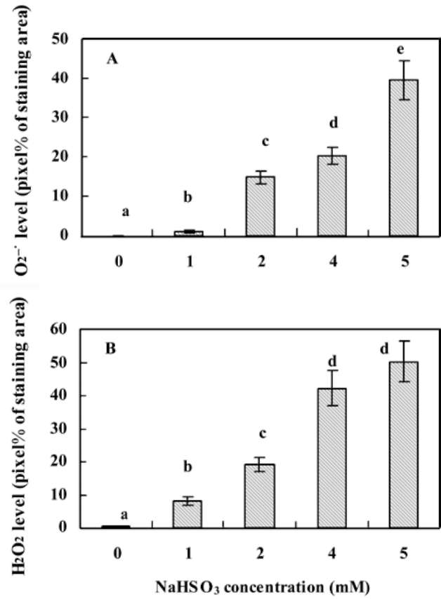
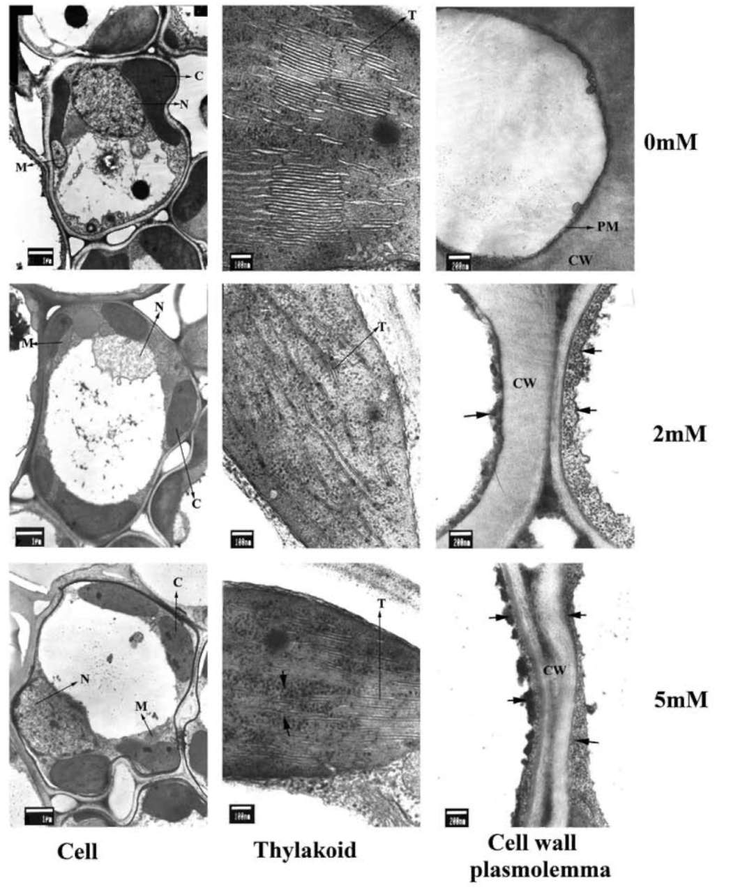
servation of and H2O2 generation in rice leaves in situ. When NBT, a specific probe of , is treated with O;, a dark blue formazan stain appears at the plant tissue's gen­eration site. Increased concentration of NaHSO3 produced significant increases in the quantity of blue formazan spots in rice leaves (Figure 2). Only a trace were detected in the untreated control (leaf segments without NaHSO3), but in samples treated with 5 mM NaHS3, levels increased to 39.6 % pixels (Figure 3A). Generation of H2O2 in leaf seg­ments was determined by the occurrence of reddish-brown staining (DAB-H2O2 polymerization). In the presence of NaHSO3, rice seedlings responded with rapid generation of H2O2 and the appearance of an intensive reddish-brown stain, particularly with NaHSO3 concentrations ranging from 2 mM to 5 mM (Figures 2 and 3B). By 5 mM NaH-SO3 treatment this reached to one half pixel of the entire area of the leaf segment. Microscopic observations of leaf epidermis indicate that H2O2 was predominantly found in the stomata's guard and subsidiary cells and in the cell wall of partial epidermal cells, while was found primar-ily in the epidermal cells (Figure 2).
Cytochemical localization of NaHSO3-induced H2O2 generation in rice leaves
Electron-dense precipitates of cerium perhydroxides, produced from the reaction of H2O2 with CeCl3, were not detectable in the cell wall, plasmalemma or chloroplasts of the control seedlings' mesophyll cells. Treatment with NaHSO3 resulted in the intensive formation of stained pre-
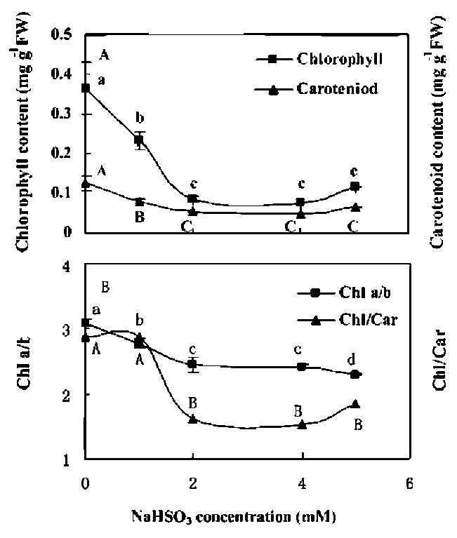
Figure 1. Changes in contents of chlorophyll and carotenoids. (A) ratios of Chl a/b and Chl/Car; (B) in leaves of rice seedlings cultured with sodium bisulfite (NaHS3). Rice seedlings were cultured in hydroponics with 1/2 Hoagland solution and various concentration of NaHSOg for 3 d. Mean values with different letters are significantly different atp < 0.05, n=3.
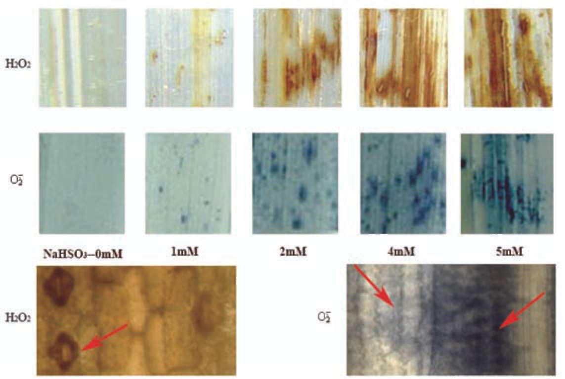
Figure 2. Images of H2O2 (top) and (bottom) generation in leaf tissue induced by NaHS3. Reddish-brown spots are H2O2-DAB re­action product, dark-blue formazan spots are -NBT reaction product. The lower side is microscope pictures of tissue localization of H2O2 and generation sites in leaf epidermis, which show presence of H2O2 in guard and subsidiary cells of stomata and cell wall of epidermal cells, but is found in most epidermal cells. See red arrows in figure.
176
Botanical Studies, Vol. 52, 2011
cipitates spots and the expansion of the generation sites. Staining of precipitates occurred on the plasmalemma and intercellular cleft of the cell wall in samples treated with 2 mM NaHSO3, and expanded to include cell walls and chloroplasts once treated with 5 mM NaHSO3 (Figure 4).
Content of total SO4-sulfur and its allocation in shoots and roots
Treatments with various concentrations of NaHSO3 altered the total sulfur content (determined as sulfate-S after complete oxidation in nitric acid) in rice seedlings' shoots and roots. Whereas shoots'sulfur content increased when NaHSO3 concentrations were elevated, roots' sulfur content decreased when treated with 2-5 mM of NaHS3. When treated with 5 mM NaHSO3, total sulfur content was 2.59 times and 0.56 times that of the corresponding con­trols for shoots and roots, respectively (Figure 5A). Hence, the shoot/root ratio of sulfur increased significantly with increasing NaHSO3 concentrations (Figure 5B). More­over, the generation of sulfate as an oxidative product of NHSO3 in rice shoots was 0.367, 2.311, 3.103 and 3.605 g kg-1DW when treated with 1, 2, 4 and 5 mM NaHSO3, respectively. The results revealed that both the uptake of NaHSO3 and its subsequent conversion to sulfate, and the fraction of sulfur distributed to shoots were enhanced
when exposed to higher concentrations of NaHS3.
Contents of total C, N, P, K, Na, Ca, Mg in shoots and roots
Total N in shoots changed slightly with increased NaHSO3 concentration. N content of sample treated in 5 mM NHSO3 was 83.4% relative to the control rice leaves (Figure 6A). Total C content increased initially in shoots (2 mM NaHSO3) and in roots (1 mM NaHSO3), but declined under higher NaHSO3 concentration treatments (Figure 6B). The total C and N in shoots remained almost un-changed after treatment with 1 mM NaHSO3, likely due to their low concentrations having no effect on metabolism.
In shoots, P content increased after treatment with 1 mM NaHSO3, but higher concentrations of NaHSO3 pro­duced less change. In contrast, a distinct decrease in P content was found in roots induced by NaHSO3 treatment (Figure 6C). When subjected to 5 mM NaHSO3, shoot P content increased by 13.5% but in roots, this decreased by 60%.
The contents of the four metal nutrient elements K, Na, Ca and Mg all increased in shoots and decreased in roots when NaHSO3 concentration was above 2 mM (Figure 6D-F). After treatment with 5 mM NaHSO3 for 3 days, K remained at the same level as the untreated shoot control sample, while Na, Ca and Mg in shoots were higher than the controls by 36%, 24% and 21%, respectively. Never-theless, decrements of 31% (K), 59% (Na), 87% (Ca) and 18% (Mg) were found in roots when compared to the con-trol levels.
Test elements/sulfur ratios in rice shoots
In order to determine the change in stoichiometric equi­librium between sulfur and the other test elements using NaHSO3 , we calculated changes in the shoots for each ele­ment/sulfur ratio. The calculated ratios of normal culture conditions (without NaHSO3) listed in Table 1 showed that N/S ratio was the highest (ca. 12.0) and that Na/S ratio was the lowest (0.49). The presence of NaHSO3, however, led to the reduction of all element/sulfur ratios. The negative correlation coefficient between the calculated ratios and NaHSO3 treatment concentrations was found to be greatest among C/S (-0.9508), P/S (-0.9059) and Na/S (-0.9403), medium among Ca/S (-0.8787) and Mg/S (-0.8952) and smallest among N/S (-0.6472) and K/S (-0.6473). The re-duction in element/S ratio can be attributed to shoot sulfur content dominating over other elements after NaHSO3 treat-ments. The results indicate that the ratios of C/S, P/S and Na/S were considerably modified by excess sulfur in shoots.
Shoot/root ratios of the other five elements
The shoot/root ratio of nutrient element content reflects their distribution in rice seedlings. As shown in Table 2, the shoot/root ratios of five elements including P, K, Na, Ca and Mg were in the range of 0.5410.75, where the shoot/root ratio for K is the highest and lowest for Na. With increasing concentration of NaHSO3, the shoot/root
Figure 3. The quantification of (A) and H2O2 (B) accumula­tion as pixe l% of NBT staining portion/leaf segment area in rice seedlings under hydroponics with NaHSO3. Columns marked with different letters are significantly different at P < 0.05, the vertical line indicates the LSD value, n=3.
LIN et al. ― Bisulfite induced oxidative stress in rice seedlings
177
Figure 4. Subcellular localization of H2O2 by cerium precipitate formation in mesophyll cells of rice seedlings in hydroponics contain­ing NaHSO3. Cerium precipitates are indicated by rough arrows. Bars represent 1 (im (cell), 100 nm (chloroplast) and 200 nm (cell wall and plasmalemma), respectively. Abbreviation: C, chloroplast; M, mitochondrion; N, nucleus; T, thylakoid; PM, plasmalemma; CW, cell wall.
Table 1. Ratios of C, N, P, K, Na, Ca and Mg contents vs. sulfur contents in shoot of rice seedlings under NaHSO; hydroponics. Re­sults represented are from element /S content ratio in shoot calculated from Figure 6 and Figure 5.

Rate
NaHSO3 concentration

0 mM

1 mM

2 mM

4 mM

5 mM

C/S

4.042±

3.499±

3.621±

1.335±

1.364±
P/S
2.401±0.497 a
2.171±0.433 a
1.255±0.277 b
1.117±0.209 b
1.051±0.233 b
N/S
12.088±
10.046±
4.835±
3.670±
3.884±
K/S
3.349±0.299 a
3.069±0.345 a
1.457±0.177 b
1.793±0.243 b
1.279±0.156 c
Na/S
0.491±0.106 a
0.415±0.075 ab
0.324±0.026 b
0.264±0.069 b
0.269±0.025 b
Ca/S
3.027±0.049 a
2.777±0.272 a
1.840±0.188 b
1.726±0.124 b
1.441±0.116 c
Mg/S
1.449±0.105 a
1.256±0.095 a
0.768±0.064 b
0.733±0.075 b
0.678±0.056 b

178
Botanical Studies, Vol. 52, 2011
Table 2. Shoot/root ratios of five elements (P, K, Na, Ca and Mg) in rice seedlings under NaHSO^ hydroponics. Values are obtained from the contents of in five elements in shoot vs. root affected by various NaHSO^ concentrations in Figure 5.

Element
NaHSO3 concentration

0 mM

1 mM

2 mM

4 mM

5 mM
P
1.422±0.294 a
2.839±0.566 b
5.050±1.078 c
4.199±0.787 bc
4.013±0.888 bc
K
2.355±0.210 a
3.074±0.346 b
10.750±0.906 c
8.752±1.188 cd
7.437±0.910 d
Na
0.910±0.195 a
0.536±0.097 b
2.358±0.186 c
1.397±0.364 a
2.187±0.200 c
Ca
3.260±0.053 a
3.545±0.347 a
5.124±0.523 b
4.503±0.323 b
4.635±0.375 b
Mg
1.152±0.084 a
1.710±0.130 b
3.160±0.264 c
7.765±0.782 d
7.935±0.655 d

phorylation and ATP supply in chloroplasts (Wang and Shen, 2002). The present results show that 1 mM NaHSO3 in the culture medium did not have adverse effects on the levels of C, N, K, Ca, Mg, S, H2O2 and the Chl/Car ratio in rice shoots, which also implies that sulfate, from bisulfite conversion in this case, was probably used as the source of sulfur nutrient supplement for rice seedlings.
The damaging effect of higher NaHSO3 concentration on the plants was thought to act directly on some compo­nents of the photosynthetic membrane through reactions involving free radicals or by reacting with disulfide bridges in protein and enzymes (Covello et al., 1989). To confirm that HSO3; might undergo aerobic oxidation to sulfate via a free radical mechanism as previously reported (Peiser and Yang, 1977), the direct proof of oxidative stress produced in leaf cells of rice seedlings caused by bisulfite was ob­served by histochemical and cytochemical detections of and H2O2, and by analyzing the change in photosynthetic pigments. It showed that bleaching of photosynthetic pigments associated with excessive generation of reactive oxygen species (ROS) at uncontrol state was triggered by bisulfite stress. ROS are reported to be produced in several subcellular compartments in the normal metabo­lism process following biotic or abiotic stress (Murphy et al., 1998). generation sites in rice leaves detected by the formation of NBT stained blue famazan and H2O2 accumulation sites, observed by the formation of cerium perhydroxides precipitate or brown DAB-H2O2 polymer, demonstrated that the active sites of ROS generation were localized primarily on epidermal cells, stomatal apparatus and plasmalemma of mesophyll cells. This is consistent with our results finding generation sites in Alocasia macrorrhiza leaves undergoing various stresses (Lin et al., 2009). H2O2 found predominantly in the plasmalemma was also found in cultured tobacco cells induced by cadmium (Olmos et al., 2003), in birch leaf cells induced by O3 (Pel-linen et al., 1999), and in the plasma membrane and cell wall of rice and wheat root tip meristematic tissue induced by anoxic stress (Blokhina et al., 2001). Production of
and H2O2 in plasma membrane and cell wall has been mainly attributed to plasma membrane NADPH oxidase activity (Sagi and Fluhr, 2006). The plasma membrane (PM) bounding NADPH oxidase is associated with super-oxide dismutase (SOD) and peroxidase (Pellinen et al., 1999), which functions as an generation enzyme
Figure 5. Total sulfur content (A) and the shoot/root ratio of sul­fur (B) in rice seedlings under hydroponics with NaHSO;.
ratios for P, K, Na, and Ca initially increased to reach the corresponding maximum value at 2 mM NaHSO3 and then decreased; whereas the ratios for Mg showed a steady increase. This demonstrates that more absorbed elements were distributed to the shoot under NaHSO3 tress.
DISCUSSION
The effect of NaHSO3 on plants was reported to be con­centration-dependent, where NaHSO3 at high concentra­tion (> 1 mM) was toxic to alga, but at low concentration (0-1 mM) it had no adverse influence on growth. Addi­tionally, the sulfate formed from NaHSO3 oxidation could be utilized as a sulfur source (Yang and Wang, 2004). Nevertheless, spraying low concentrations of NaHSO3 on the leaf surface was reported to enhance cyclic photophos-
LIN et al. ― Bisulfite induced oxidative stress in rice seedlings
179
Figure 6. Contents of total nitrogen (A), carbon (B), phosphorus (C), potassium (D), sodium (E), calcium (F) and magnesium (G) in shoot and root of seedlings under hydroponics with NaHSO3.
complex. NADPH oxidase transfers electron from cytoso-lic NADPH to O2 to form O;, which is transformed rapidly to H2O2 by a tight connection of extra-cellular Cu-Zn SOD (Ogawa et al., 1997). Treating living spinach leaf cells with bisulfite led to a remarkable stimulation in the activ­ity of PM NADPH oxidase, while the use of a NADPH oxidase inhibitor, DPI (diphenyleneiodonium), reduced the production of H2O2 (Li et al., 2007). Groom et al. (1996) report that a NADPH oxidase subunit gp91 phox homologue has been identified in rice. Embs and Markakis (2006) infer that NaHSO3 also forms a complex with peroxidase iron. Pretreatment of detached maize leaves with NaHSO3 (4.8 mM) for 24 h increased peroxidase activity (Akhtar and Garraway, 1990). Therefore, ROS generated on the plasma membrane and cell wall of rice leaves in vivo, induced by bisulfite in this study, likely involved the contribution from an enzyme complex consisting of a combination of trans-
membrane synthase, NADPH oxidase and peroxidases.
The excess sulfur accumulated in the shoots of Arabi-dopsis thaliana has been reported as sulfate and organic S in a 3:1 ratio (Van Der Kooij et al., 1997). Rice seedlings cultured with 1-5 mM NaHSO3 resulted in accumulation of different amounts of sulfate-S in shoots, indicating that the bisulfite absorbed by rice roots had oxidized into SO42-. The significant enhancement of sulfate-S content as sodi­um bisulfite concentration was increased demonstrated that up-take of HSO3; and the subsequent conversion to SO42-is concentration- dependent. Meanwhile, the levels for all elements tested (except nitrogen) increased in shoots but decreased in roots under NaHSO3 treatments, resulting in increased shoot/root ratios for each element. This change in element distribution between shoots and roots may re­flect a metabolic regulation that favors the shoot part, the
180
Botanical Studies, Vol. 52, 2011
main photo-assimilation organ of the plants, in response to bisulfite stress. The presence of a much higher quantity of Mg2+ in shoots, when compared to other elements, after treatments with 4-5 mM bisulfate, may indicate a protec­tive response. This response would be to compensate for the loss of the Mg molecule that occurs during the bleach­ing process within chlorophyll structure. Chlorophyll bleaching in conjunction with the increase in sulfate-S content at NaHSO3 concentrations above 2 mM, indicate that destruction of the chlorophyll molecule is closely related to bisulfite oxidation (Peiser and Yang, 1977). Similarly, a higher concentration of Mg and Sulfate-S was found in in situ lichen thalli collected from an industrial town in Israel than from a forest there (Garty et al., 1997).
The linearly negative relation between the fraction ra­tios of C, P, K, Na, Ca, Mg and N contents versus sulfate-S content in shoots with increasing bisulfite concentration indicate that the physiological balance and stoichiometry among the main nutrient elements in rice plants were mod­ified by excess sulfate-S to distinct degree of imbalance. In general, sulfate and nitrate assimilation pathways are well coordinated; where plant tissue contains one part sul­fur for every 15 to 20 parts nitrogen for optimum growth (Kazuki, 2004). The N/S ratio in control rice seedlings was found to be ca. 12.1, but reduced to only 3.7-3.9 under 4-5 mM of bisulfite treatment. The latter is much lower than the requirement of 15-20 parts for optimal growth of most plants. Similar a decrease in the ratio of total N to total sulfate content was reported in Arabidopsis thaliana exposed to SO2 by Van Der Kooij et al. (1997). Hence, the growth of rice seedlings could be limited by the im­balance of N/S ratio induced by NHSO3 treatment. Very little is known about the interactions of sulfur and carbon assimilation (Kopriva et al., 2002). The reduction of total carbon and ratio of C/S in shoots might be due to the in­hibition of photosynthesis by HSO3-. It has been reported that bisulfite treatment resulted in the rapid loss of photo-synthetic oxygen evolution and a marked decline in chlo­rophyll fluorescence in moss (Baxter et al., 1991; Bharali and Bates, 2002); a 70% reduction in PSII activity and the peroxidation of thylakoid membrane lipids in Phaseolus vulgaris leaves (Covell et al., 1989); and an estimated 50% bleaching of chlorophyll in spruce needles (Elstner et al., 1985). An earlier study performed in our laboratory found an increase in chlorophyll fluorescence polarization and a reduction in excited energy transport from PSII to PSI in leaves of five woody plants under simulated SO2 treatment (Liu et al., 2006). These simultaneous occurrence of these events might cause the reduction of carbon accumulation in leaves by inhibiting photosynthesis.
CONCLUSION
In summary, HSO3; at concentrations higher than 1 mM in hydroponics medium induced oxidative stress in rice seedlings. was generated in most epidermal cells, while H2O2 was mainly generated in guard and subsidiary cells and the plasmalemma and cell wall of mesophyll cells.
The uptake of HSO3; increased as NaHSO3 concentra­tion was increased, which subsequently transformed into SO42- -S that was predominantly allocated to shoots. N, C and chlorophyll contents in shoots were reduced under higher HSO3- concentrations. A negative correlation coeffi­cient was observed between HSO3; concentrations and the ratios of all tested elements/S.
Acknowledgement. This research was financially sup­ported by the Knowledge Innovation Program of the Chi­nese Academy of Sciences, Grant No. KSCX2-EW-J-28, National Natural Science Foundation of China (30770173) and Scientific Start-up Foundation of Ministry of Educa­tion for Returned Oversea Scientist.
LITERATURE CITED
Adam, A., T. Farkas, G. Somlyai, M. Hevesi and Z. Kiraly.1989. Consequence of O2 generation during bacterially induced hypersensitive reaction in tobacco: deterioration of mem­brane lipids. Physiol. Mol. Plant Pathol. 34: 13-26.
Akhtar, M. and M.O. Garraway. 1990. Effect of sodium bisulfite on peroxidase activity and electrolyte leakage in maize in relation to sporulation of Bipolaris maydis Race T. Ohio. J. Sci. 90: 71-76.
Baxter, R., M.J. Emes, and J.A. Lee.1991. Short term effects of bisulphate on pollution-tolerant and pollution sensitive populations of Sphagnum cuspidatum Ehrh (ex.Hoffm). New Phytol. 118: 425-431.
Bestwick, C.S., I.R. Brown, M.H.R. Bennet, and J.W. Mansfield. 1997. Localization of hydrogen peroxide accumulation dur­ing the hypersensitive reaction of lettuce cells to Pseudomo-nas syringepvphaseolicola. Plant Cell 9: 209-211.
Bharali, B. and J.W. Bates. 2002. Soil cations influence Bryo-phyte susceptibility to bisulfite. Ann. Bot. 90: 337-243.
Bharali, B. and J.W. Bates. 2006. Detoxification of dissolved SO2 (bisulfite) by terricolous mosses. Ann. Bot. 97: 257-263.
Blokhina, O.B., T.V. Chirkova, and K.V. Fagerstedt. 2001. An-oxic stress leads to hydrogen peroxide formation in plant cells. J. Exp. Bot. 52: 1179-1190.
Covello, P.S., A. Chang, E.B. Dumbroff, and J.E. Thompson. 1989. Inhibition of photosystem ll proceeds thylakoid membrane lipid peroxidation in bisulfite-treated leaves of
Phaseolue vulgaris. Plant Physiol. 90: 1492-1497.
Dittrich, A.P.M., H. Pfang, and U. Heber. 1992. Oxidation and reduction of sulfite by chloroplasts and formation of sulfite addition compound. Plant Physiol. 88: 738-744.
Elstner, E.F., W. Osswald, and R.J. Youngman. 1985. Basic mechanisms of pigment bleaching and loss of structural re­sistance in spruce (Picea abies) needles: advances in phyto-medical diagnostics. Experienta 41: 591-597.
Embs, R.J. and P. Markakis. 2006. Bisulfite effect on the chem­istry and activity of horseradish peroxidase. J. Food Sci. 34:
500-501.
LIN et al. ― Bisulfite induced oxidative stress in rice seedlings
181
Garty, J., N. Kloog, R. Wolfson, Y. Cohen, A. Karnieli, and A. Avni. 1997. The influence of air pollution on the con­centration of mineral elements, on the spectral reflectance response and on the production of stress-ethylene in the lichen Ramalina duriaei. New Phytol. 137: 587-597.
Groom, Q.J., M.A. Torres, A.P. Fordham-Skelton, K.E. Ham-mond-Kosack, N.J. Robinson, and J.D.G. Jones. 1996. rbohA, a rice homologue to mammalian gp91 phox respiratory burst oxidase gene. Plant J. 10: 515-522.
Kazuki, S. 2004. Sulfur assimilatory metabolism. The long and
smelling road. Plant Physiol. 136: 2443-2450.
Kopriva, S., M. Suter, P. von Ballmoos, H. Hesse, U. Krahen-buhl, H. Rennenberg, and C. Brunold. 2002. Interaction of sulfate assimilation with carbon and nitrogen metabolism in Lemna minor. Plant Physiol. 130: 1406-1413.
Li, B, D. Xing, and L. Zhang. 2007. Involvement of NADPH oxidase in sulfur dioxide-induced oxidative stress in plant
cells. Photochem. Photobiol. Sci. 6: 628-634. Lin, Z.F., S.S. Li, G.Z. Lin, G.C. Sun, and J.Y. Guo. 1984.
Superoxide dismutase activity and lipid peroxidation in relation to senescence of rice leaves. Acta Bot. Sin. 26:
605-615.
Lin, Z.F., N. Liu, G.Z. Lin, and C.L. Peng. 2009. In situ local­ization of superoxide generated in leaves of Alocasia mac-rorrhiza (L.) Shott under various stresses. J. Plant Biol. 52:340-347.
Liu, N., C.L. Peng, Z.F. Lin, G.Z. Lin, L.L. Zhang, and X.P.
Pan. 2006. Chang in photosystem ll activity and leaf reflectance features of several subtropical woody plants under simu­lated SO2 treatment. J. Integ. Plant Biol. 48: 1274-1286.
Liu, N., Z.F. Lin, L.L. Guan, G.Z. Lin, and C.L. Peng. 2009.
Light acclimation and HSO3- damage on photosynthetic ap­paratus of three subtropical forest species. Ecotoxicology 18: 929-938.
Murphy, T.M., H. Vu, and T. Nguyen. 1998. The superoxide synthases of rose cells. comparison of assays. Plant Physiol.
117: 1301-1305.
Ogawa, K., S. Kanematsu, and K. Asada. 1997. Generation of superoxide anion and the localization of CuZn-superoxide dismutase in the vascular tissue hypocotyls: Their associa­tion with lignification, Plant Cell Physiol. 38: 1118-1126.
Olmos, E., J.R. Martinez-Solano, A. Piqueras, and E. Hellin. 2003. Early steps in the oxidative burst induced by cad­mium in cultured tobacco cells (BY-2 line). J. Exp. Bot. 54: 291-301.
Pellinen, R., T. Palva, and J. Kangasjravi. 1999. Subcellular lo­calization of ozone-induced hydrogen peroxide production in birch (Betulapendula) leaf cells. Plant J. 20: 349-356.
Peiser, G.D. and S.F. Yang. 1977. Chlorophyll destruction by the bisulfite-oxygen systems. Plant Physiol. 60: 277-281.
Ranieri, A., F. Pieruccetti, A. Panicucci, A. Castagna, G. Lo-reazini, and G.F. Soldatini. 1999. SO2-induced decrease in photosynthetic activity in two barley cultivars. Evidence against specific damage at the protein-pigment complex level. Plant Physiol. Biochem. 37: 919-929.
Sagi, M. and R. Fluhr. 2006. Prodution of reactive oxygen species by plant NADPH oxidases. Plant Physiol. 141: 336-340.
Schrauder, M., W. Moeder, C. Wiese, W. van Camp, D. Inze, C. Langebartels, and Jr. H. Sandermann. 1998. Ozone -induced oxidative burst in the ozone biomonitor plant, to­bacco Bel W3. Plant J. 16: 235-245.
Shimazaki, K-I. and K. Sugahara. 1980. Inhibition site of the electron transport system in lettuce chloroplasts by fumiga­tion of leaves with SO2. Plant Cell Physiol. 21: 125-135.
Van Der Kooij, T.A.W., L.J. De Kok, S. Haneklaue, and E.
Schnug. 1997. Uptake and metabolism of sulphur dioxide by Arabisdopsis thaliana. New Phytol. 135: 101-107.
Wang, H.W. and Y.G. Shen. 2002. How bisulfite enhances pho­tosynthesis. J. Plant Physiol. Mol. Biol. 28: 247-252.
Yang, S., J. Wang, W. Cong, Z. Cai, and F. Ouyang. 2004. Ef­fects of bisulfite and sulfite on the microalga Botryococcus braunii. Enz. Microb. Tech. 35: 46-50.
182
Botanical Studies, Vol. 52, 2011
亞硫酸氫鈉水培誘導水稻幼苗氧化脅迫並影響營養元素的組分
林植芳1 劉楠1 陳少微1 林桂珠1 莫輝1 彭長連2
1中國廣州中國科學院華南植物園,中國科學院返化生態系統植被恢復與管理重點實驗室
2中國廣州華南師範大學生命科學院
為了探明SO2的衍生物 NaHSO3(HSO-3) 誘致植物組織和細胞水準的氧化脅迫狀態及主要營養元
素的吸收與平衡的變化,我們將水稻品種兩優培九的幼苗用加入亞硫酸氫鈉(NaHSO3)1、2、45 mM
Hoagland溶液培養3天。分析其光合色素,S P N C K Na Ca Mg的含量,並以組織化學
和細胞化學方法定位研究活性氧H2O2和的產生位點與數量。結果表明,經HSOs處理後幼苗的莖葉
S顯著積累,P K Na Ca Mg的積累較少,C N和葉綠素含量下降。每種元素與S的比率隨
HSO3—濃度增高而降低,兩者之間呈負相關性。與此相反,根中的大多數營養元素含量均下降導致莖
/根比率升高。葉片中兩種活性氧產生的數量明顯增多,在葉表皮組織中,H2O2主要定位於氣孔的保
衛細胞,附衛細胞和一些表皮細胞中,而則出現於大多數表皮細胞中。葉肉細胞中的H2O2首先出現
於細胞質膜和胞壁之間的連接區,隨後因濃度增高而擴展至胞壁和葉綠體的類囊體上。
關鍵詞:水稻;亞硫酸氫鈉;硫含量;超氧陰離子自由基;過氧化氫;元素/硫比率。