Botanical Studies (2011) 52: 305-312.
MICROBIOLOGY
Characterization of a green microalga UTEX 2219-4: Effects of photosynthesis and osmotic stress on oil body formation
Szu-Ting WANG1, Yi-Ying PAN1, Chiu-Chin LIU1, Lu-Te CHUANG2, and Ching-Nen Nathan
CHEN1,3*
1Institute of Marine Biology, National Sun Yat-sen University, Kaohsiung 804, Taiwan
2Department of Biotechnology, Yuanpei University, Hsinchu 300, Taiwan
(Received September 9, 2010; Accepted December 29, 2010)
ABSTRACT. Microalgae are an important potential feedstock for biodiesel production. Understanding the physiology of lipid biosynthesis in microalgae is pivotal to their management in aquaculture. A freshwater green microalgal strain, UTEX 2219-4, was isolated from UTEX 2219, which was reported to contain two strains. Its ITS sequences were closely related to those in the family Scenedesmaceae. Nitrogen starvation, salt stress and osmotic stress greatly enhanced lipid biosynthesis in this strain, while a combination of nitrogen deficiency and osmotic stress produced the most dramatic effects. Chloroplasts condensed and photosynthe­sis efficiency declined about 50% after 3 days of nitrogen starvation. Chlorophyll degradation followed the same trend but was more severe than the reduction of photosynthesis efficiency. Oil body formation was not observed in the cells kept in the dark under nitrogen starvation, suggesting that photosynthesis, rather than autophagy, is the main player in oil body formation. Under non-saturating light intensity levels coupled with nitrogen starvation, the oil body formation under 200 (imol/m2s light intensity was more efficient than that under 100 (imol/m2s. DCMU blocked both photosynthesis and oil body formation, again suggesting that pho­tosynthesis, rather than autophagy, provides the energy for oil body formation during nitrogen starvation.
Keywords: Autophagy; Microalgae; Oil body; Osmotic stress; Photosynthesis.
INTRODUCTION
Seeking renewable feedstock for bioenergy production is imperative considering the current problems of exces­sive carbon dioxide emission and petroleum depletion. Biodiesel is one of the better choices among varieties of bioenergy, and microalgae are claimed to be the best crop for biodiesel production (Chisti, 2008; Hu et al., 2008). Most green microalgae produce starch under normal conditions. Upon exposure to nutritional stress, such as nitrogen or phosphate starvation, they start to synthesize triacylglycerol which is stored in their oil bodies (Shifrin and Chisholm, 1981). Isolation of new microalgal species that can adapt to tropical or subtropical environments with high growth rates and high lipid contents is pivotal to algal biodiesel production. Understanding the physiology of oil body formation in green microalgae is essential for effec­tive aquaculture management and for the development of the microalgal biodiesel industry.
Oil contents of green microalgae increase significantly in a few days of nitrogen starvation treatment (Wang et al., 2009; Li et al., 2010; Moellering and Benmng, 2010). Although the sensing and signaling process is still not clear, a nitrate transporter is proposed to function as a ni­trate sensor in Arabidopsis (Ho et al., 2009). In nitrogen-deficient conditions, many organisms undergo autophagy to recycle part of the cytoplasm including organelles, which is a self-degrading process common in eukaryotes that provides needed energy and raw materials for cellu­lar repair (Bassham et al., 2006; Yorimitsu and Klionsky, 2005). In Chlamydomonas, autophagy is also induced by nitrogen starvation and other stress (Perez-Perez et al., 2010). Whether the regained energy and raw materials are solely for life support or can be converted to storage lipids is unknown. Under nitrogen starvation, biosynthesis of amino and nucleic acid is arrested, but the light reac­tions of photosynthesis are still active. The cells have to prevent electron accumulation in the thylakoid membrane to minimize free radical production. The primary product of carbon fixation reactions is triose phosphate, which can be used for either starch or lipid biosynthesis. Biosynthesis of the two kinds of macromolecules does not require ni-

*Corresponding author: E-mail: nathanc@mail.nsysu.edu. tw; Tel: 07-525-2000 ext. 5106; Fax: 07-525-5100.
306
Botanical Studies, Vol. 52, 2011
trogen, thus they are good destinations for photosynthetic energy. However, it appears that lipid biosynthesis is more favored by oleaginous green microalga than starch under nitrogen starvation. No conclusive reasons for this phe­nomenon have been identified. According to our present results, there is apparently a mechanism(s) that alters the carbon partitioning of the cells to lipid biosynthesis upon exposure to nitrogen deficient conditions and osmotic stress. This mechanism(s) is useful from the perspective of biodiesel production because this switch(s) may control oil yield in microalgae.
Here we report the isolation and characterization of a green microalga UTEX 2219-4, that was misplaced in the genus Chlorella under the collection number UTEX 2219. UTEX 2219-4 is more closely related to species in the family Scenedesmaceae than to others. We found that the energy for lipid biosynthesis in these cells during ni­trogen starvation came mostly from photosynthesis rather than from autophagy, and that osmotic stress presumably enhanced this lipid biosynthesis by further altering car­bon partitioning towards lipid biosynthesis. The reason why oleaginous green microalgae are in favor of produc­ing lipids rather than starch under nitrogen starvation is discussed.
again under the above conditions. The upper phase was transferred to a new tube, and an equal volume of isopro-panol was mixed into the supernatant. After incubation at room temperature for 10 min, this tube was spun again under the above conditions. The supernatant was discard­ed, and the pellet was rinsed with 70% ethanol twice and dried in the air. The pellet was dissolved in 50 fiL of TE buffer containing RNase.
PCR amplification and sequencing of rDNA
The 18 S rDNA was amplified by PCR using the prim­ers: 18S forward-"TTTCTGCCCTATCAACTTTCGATG" and 18S reverse-"TACAAAGGGCAGGGACGTAAT". The ITS1-5.8 S-ITS2 region rDNA was amplified using the primers: ITS forward2: "CTAGAGGAAGGAGAAGTCG-TAACA" and ITS reverse2: "TGATATGCTTAAGT-TCAGCGG". The PCR products amplified by the two pairs of primers were single bands on the agarose gels. DNA sequencing was carried out by the dideoxy method using the primers above.
Phylogenetic analysis
Thirteen ITS2 sequences were aligned using the software Mega 4.1 (Tamura et al., 2007). Well-aligned regions were selected from each sequence, and used for phylogenetic analysis. There were a total of 178 positions in the final dataset. The Neighbor-Joining Method was used to construct the tree in Figure 1. The percentage of replicate trees in which the associated taxa clustered to­gether in the bootstrap test (500 replicates) is shown next to the branches.
MATERIALS AND METHODS
Microalgae and growth conditions
The freshwater green microalga UTEX 2219, under the registration name Chlorella minutissima, was purchased from the Culture Collection of Algae at the University of Texas at Austin (UTEX). It is noted in the UTEX web that this collection contains two strains (Kessler and Huss, 1992), (http://web.biosci.utexas.edu/utex/algaeDetail. aspx?algaeID=4682). We isolated one strain from this col­lection by growing single colonies and sequencing their rDNAs and internal transcribed spacers (ITS). This strain was designated as UTEX 2219-4. These single species cells were grown in a modified Bold 3N medium contain­ing 4.4 mM NaN3, 0.17 mM CaC^, 0.3 mM MgS4, 0.22 mM K2HPO4, 0.65 mM KH2PO4, 0.43 mM NaCl, and the same levels of metals and vitamins described in Table 2 of Berges et al. (2001) on an orbital shaker at 150 rpm under continuous cool white light at room temperature. Light in­tensities are specified in the Results.
Lipid extraction and oil body staining
Total lipids were extracted based on the methods de­scribed in Bligh and Dyer, 1959; and Chiu et al., 2008.
Cells were harvested by centrifugation and smashed in 1 ml methanol chloroform mix (v/v = 2/1) using 0.5 g glass beads (Sigma G-9268, 425-600 fm) in a mini beadbeater. Cell debris was removed using centrifugation. The super­natant was transferred to a glass tube, and 1 mL chloro­form and 1 mL 1% NaCl solution were added to this tube. These components were mixed well then centrifuged at 10,000 rpm for 5 min to allow phase separation. The chlo­roform phase was transferred to a new glass tube, blow-dried using nitrogen air and weighted. Oil bodies in live cells were stained by the fluorescence dye Nile Red (1 mg/ mL in acetone) and imaged using a Nikon Eclipse 80i fluo­rescence microscope.
DNA extraction
Microalgal cells were harvested by centrifugation at 4000 rpm (Eppendorf 5810 R), 25°C for 5 mm. The cells were broken by a beadbeater in 0.5 mL lysis buf­fer containing 0.3 M NaCl, 50 mM Tris-HCl (pH 8), 20 mM EDTA, 0.34 mM N-Lauroylsarcosine Sodium, and 1.75 M urea. This mixture was incubated at 65°C for 10 min, and spun at 13,200 rpm (Eppendorf 5415 R), 25°C for 5 min. The supernatant was transferred to a new tube, and 0.5 mL of phenol/chloroform (1:1) was added to this tube. After mixing to denature proteins, the tube was spun
Measurement of photosynthesis efficiency and chlorophyll content
Photosynthesis efficiency was determined based on oxygen evolution rates. Ten mL of algal cells at 4x10⁷ cell/ mL density were gently harvested using centrifugation. These cells were resuspended in fresh medium with or without 0.68 M sorbitol as indicated in the results. These cells were kept in the dark for 10 min to deplete oxygen,
WANG et al. ― Physiology of microalgal oil body formation
307
which was measured by a Clark-type oxygen electrode (Hansatech Inc. England). NaHCO3 solution was then added to the sample to reach 3 mM, and the oxygen evo­lution rate of these cells was measured for 10 min under 100 fmol/m2s light intensity at room temperature. DCMU (3-(3,4-dichlorophenyl)-1,1-dimethylurea) was purchased from the Sigma company, USA (Cat. # D-2425). Chloro­phyll of the cells was extracted using 80% acetone. After mixing by vortex for 20 min, the sample was stored at 4°C overnight. The debris was pelleted by centrifuga-tion, and absorbance at 645 and 663 nm of the superna­tant was measured by a spectrophotometer. Chlorophyll content was calculated by the equation: Total chlorophyll (μg/mL) = 20.2(A645)+ 8.02(A663);Chlorophyll a (μg/ mL) = 12.7(A663)- 2.69(A645);Chlorophyll b (μg/mL)=
22.9(A645)- 4.68(A663).
tively), and the ITS sequences were used to BLAST the GenBank. The results showed that the most closely related species to this green alga are members of the Scenedesmaceae family rather than the Chlorellaceae family, which comprises Chlorella. The ITS2 sequences of 12 closely related species representing 12 genera were aligned, and a phylogenetic tree was constructed based on the alignment using the Neighbor-Joining method. As shown in Figure 1, UTEX 2219-4 is most closely related to Neodesmus danubialis, which is a species in the Scenedesmaceae. Of the 12 species, two are mosses (Plagiomnuim and Imbribryum), one is a member of the Haematococcaceae family (Haematococcus) and the re­maining are within Scenedesmaceae.
It is not surprising to find that mosses are closely re­lated to green algae, it has already been well accepted that mosses are descendents of a green algal species. On the other hand, one might wonder how the UTEX 2219-4, that is supposed to be a Chlorella species, can be closely relat­ed to members belonging to Scenedesmaceae and Haema-tococcaceae, rather than to Chlorellaceae. The taxonomy of green algae has been found increasingly inaccurate as more microalgal genomes are sequenced. For example, Chlorella vulgaris C-169 has been renamed Coccomyxa sp. C-169 after the Joint Genome Institute sequenced its genome (http://genome.jgi-psf.org/Chlvu1/Chlvu1.home. html). Therefore, there is a need to regroup microalgal tax­onomy based on molecular markers.
Transmission Electron Microscopy (TEM)
The algal cells were fixed in 2.5% glutaraldehyde and 4% paraformaldehyde in 0.1 M sodium phosphate buffer, pH 7.0 at 4°C for 4 hours. After three 20 min buffer rins­es, the samples were postfixed in 1% OsO^ in the same buffer for 4 hours at room temperature and then rinsed in three 20 min changes of buffer. Samples were dehydrated in an acetone series, embedded in Spurr's resin, and sec­tioned with a Lecia Reichert Ultracut S or Lecia EM UC6 ultramicrotome. The ultra-thin sections (70-90 nm) were stained with uranyl acetate and lead citrate. A Philips CM 100 transmission electron microscope at 80 KV was used for viewing.
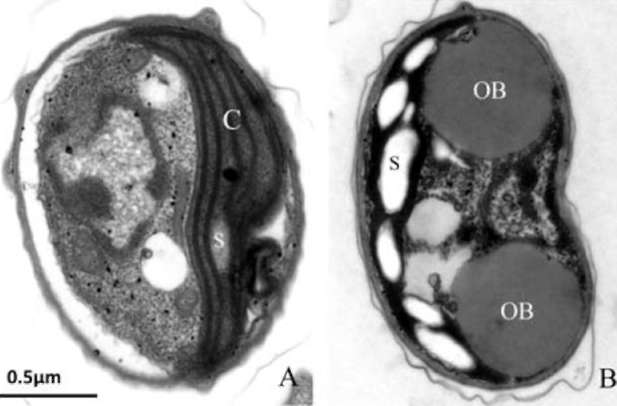
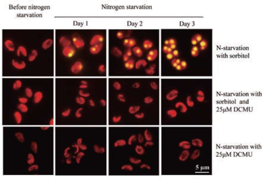
UTEX 2219-4 contained both starch granules and oil bodies after three days of nitrogen star­vation
RESULTS AND DISCUSSION
UTEX 2219-4 is closely related to members in Scenedesmaceae
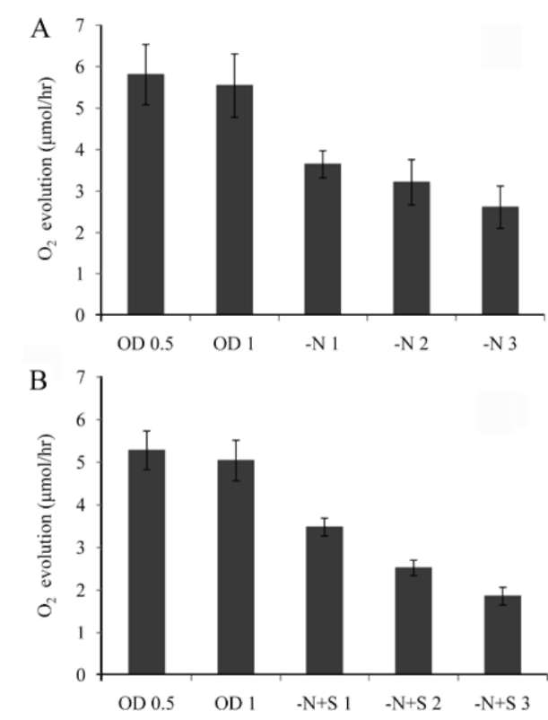
Most green microalgae produce starch as an energy res­ervoir under normal conditions. In nitrogen-deficient con­ditions, they start to accumulate triacylglycerol, which is stored in oil bodies (Moellering and Benning, 2010; Wang et al., 2009; Li et al., 2010). A few possible energy and carbon sources for the triacylglycerol biosynthesis at this
The ITS1-5.8S-ITS2 and the 18S rDNA regions of
UTEX 2219-4 were amplified and sequenced (GenBank accession numbers HQ218939 and HQ218940, respec-
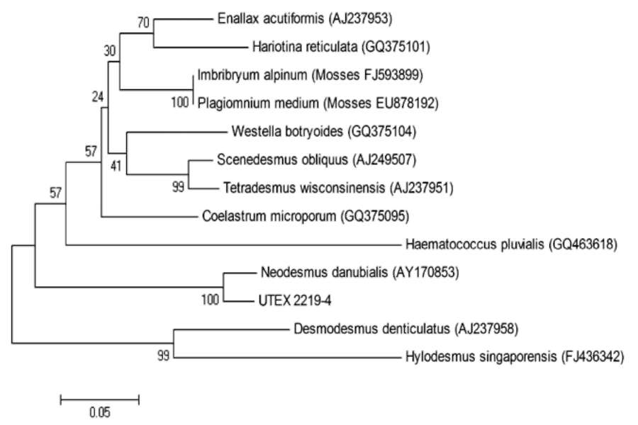
Figure 1. Phylogenetic analysis of UTEX 2219-4 and its closely related species based on ITS2 sequences in GenBank. ITS1 and ITS2 sequences of UTEX 2219-4 were used to BLAST in GenBank, and the 12 most closely related species, representing 12 genera, were selected for phylogenetic analysis. This tree was constructed based on the well-aligned regions in ITS2 se­quences of each species. This tree showed that UTEX 2219-4 is closely related to spe­cies in the family Scenedesmaceae rather than to Chlorellaceae species. The numbers in the parenthesis are the accession num­bers of the respective ITS sequences in GenBank. The scale bar measures the dis­tance between species. The percentage of replicate trees in which the associated taxa clustered together in the bootstrap test (500 replicates) are shown next to the branches.
308
Botanical Studies, Vol. 52, 2011
stage include photosynthesis, autophagy and the degrada­tion of chloroplast constituents such as their membrane, pigments, pyrenoid, and starch granules. Autophagy is a common phenomenon among eukaryotes when they are exposed to nitrogen-deficient conditions. Part of the cyto­plasm, sometimes including organelles, is transported into lysosomes or vacuoles for recycling (Chen et al., 2009). To gain more insight into the physiological changes be­fore and after nitrogen starvation stress, the subcellular structures of UTEX 2219-4 were examined by transmis­sion electron microscopy. As shown in Figure 2, the cell had a relaxed chloroplast with a low quantity of starch prior to nitrogen starvation, compared to the cell after 3 days of nitrogen starvation, which showed a more con­densed chloroplast with oil bodies and significant amount of starch granules. The condensed chloroplast suggests damage due to nitrogen starvation. The increased quantity
of starch granules, and its co-existence with oil bodies in these cells, suggests that carbon sources in these cells during nitrogen starvation were not only allocated to stor­age lipid production but also to starch biosynthesis. This phenomenon leaves room for carbon partitioning manipu­lation in the cells.
It was reported very recently that wild type Chlamy-domonas has the same phenomenon (Li et al., 2010). In wild type Chlamydomonas, a large quantity of starch gran­ules was observed after two days of nitrogen starvation. Upon blocking its starch biosynthesis pathway, the mutant Chlamydomonas produced storage lipids almost exclusive­ly. Thus, it seems whether a green microalga is oleaginous depends on the energy partitioning between starch and lipid biosynthesis when under nitrogen starvation.
Osmotic stress enhances lipid biosynthesis in both with or without nitrogen source in microal-gal cells
An insignificant ratio of UTEX 2219-4 cells contained very low quantities of oil bodies before nitrogen starvation treatment. When these cells were shifted to the nitrogen starvation conditions for 3 days, their lipid content in­creased about 50% as shown in Table 1A, compared to the lipid level before nitrogen starvation. When the cells were treated with nitrogen starvation coupled with 0.34 M (2%) NaCl or KCl salt stress, their lipid contents increased by 80% and 66%, respectively. To our surprise, when the cells were treated with nitrogen starvation coupled with 0.68 M sorbitol, which has the same osmolarity as 0.34M NaCl and KCl, their cell lipid content increased by 150% after 3 days. We wondered whether osmotic stress alone has the same effect. As shown in Table 1B, when non-starved cells were treated in 0.68 M sorbitol for 3 days, their lipid content reached 33% of the biomass. This lipid level was higher than those cells nitrogen-starved for 3 days, and
Figure 2. TEM of UTEX 2219-4 before and after nitrogen star­vation. These algal cells were grown in modified Bold 3N me­dium under continuous 200 jimol/m2 s light intensity until OD682 = 1. (A) Before nitrogen starvation, (B) After three days of nitro­gen starvation. C: Chloroplast; S: Starch granule; OB: Oil body.
Table 1. Lipid contents of UTEX 2219-4 in various conditions. These algal cells were grown in Bold 3N medium under continuous 100 jimol/m2 s light intensity until OD682 = 0.5, and subjected to different treatments.
A. These cells were nitrogen-starved for 3 days, with or without salt or sorbitol stress. Mean SD with three repeats.

Culture condition

Lipid (mg/L)

Biomass (dry weight, mg/L)

Lipid content (% of dry weight)

Before nitrogen starvation

19 1.15

113 5.77

16.53 1.60
Nitrogen starvation for 3 d
33 4.16
133 5.77
24.95 2.04
Nitrogen starvation with 0.34M NaCl for 3 d
40 3.46
133 11.55
30.08 2.55
Nitrogen starvation with 0.34M KCl for 3 d
39 3.06
143 15.28
27.58 2.78
Nitrogen starvation with 0.68M Sorbitol for 3 d
53 3.06
126 11.55
42.30 4.09

B. These cells were grown in normal conditions until OD682 = 0.5. Sorbitol solution was added to reach 0.68 M to create osmotic stress. Equal volume of water was added into the control. The treatment was lasted for 3 days. Mean SD with three repeats.

Culture condition

Lipid (mg/L)

Biomass (dry weight, mg/L)

Lipid content (% of dry weight)

Normal conditions (control)

22 0.53

117 5.57

18.48 0.51
0.68M Sorbitol stress for 3 d
40 1.67
121 6.11
32.88 1.39
WANG et al. ― Physiology of microalgal oil body formation                                                                                                                  309
comparable to the cells under nitrogen starvation coupled with 0.34 M salt stress.
It is well known that nitrogen starvation enhances lipid biosynthesis in green microalgae (Shifrin and Chisholm, 1981; Wang et al., 2009; Li et al., 2010; Moellering and Benning, 2010). The effects of osmotic stress on oil body formation, on the other hand, are less studied. In the case of Dunaliella in which beta-carotene biosynthesis is en-hanced by salt/osmotic stress, the beta-carotene droplets are formed within chloroplasts (Lamerset al., 2008). To our knowledge, this is the first report that osmotic stress enhances oil body formation in microalgae. How the sig-nals of nitrogen starvation or osmotic stress are sensed and executed are still elusive. Nitrogen starvation is one impor-tant cue for terrestrial plants to switch from a vegetative to a reproductive stage. When terrestrial flowering plants face nitrogen deficiency, their leaves turn yellow starting from
the oldest ones, and the nutrients in the leaves are mobi­lized to young leaves, flowers and eventually to seeds, which are best able to survive harsh environments. There are two scenarios that try to explain why green microalgae produce lipids when under nitrogen starvation. The first theorizes that more lipids in the cells increases buoyancy, so that it is easier for the cells to drift to new niches. The second proposes that when microalgal cells are undergoing nitrogen starvation, they are not able to consume ATP and NADPH by synthesizing amino or nucleic acids. One way to quench the electrons in the thylakoid membrane is to synthesize lipids, which do not require nitrogen. However, it is equally true that starch synthesis does not require ni­trogen. But by doing so, cell buoyancy will be decreased. Actually the two hypotheses are not mutually exclusive and can be combined into one. We propose that lipid pro­duction in green microalgae is an adaptation to cope with
Figure 3. Photosynthesis efficiency of UTEX 2219-4 in various conditions. These algal cells were grown in modified Bold 3N medium under continuous 200 jmol/m2 s light intensity until OD682 reached 0.5 or 1, when photosynthesis efficiency was measured. At OD682 = 1, cells were further subjected to nitrogen starvation (-N) or nitrogen starvation plus 0.68 M sorbitol (-N+S) for different days under the same light intensity. (A) Column OD0.5, OD1, -N1, -N2, and -N3 represent photosynthesis ef­ficiency of cells at OD=0.5, 1, and cells at OD682 = 1 stressed by nitrogen starvation for 1, 2, and 3 days, respectively. (B) Col­umn OD0.5, OD1, -N+S1, -N+S2, and -N+S3 represent photo­synthesis efficiency of cells at OD682 = 0.5, 1, and cells at OD682 =1 stressed by nitrogen starvation coupled with 0.68M sorbitol for 1, 2, and 3 days, respectively. The error bars represent stan­dard deviations from three repeats.
Figure 4. Chlorophyll content of UTEX 2219-4 under various conditions. These algal cells were grown in modified Bold 3N medium under continuous 200 jmol/m2 s light intensity until OD682 reached 0.5 or 1 when chlorophyll content was measured. At OD682 = 1, cells were further subjected to nitrogen starvation (-N) or nitrogen starvation plus 0.68 M sorbitol (-N+S) for dif­ferent days under the same light intensity. (A) Column groups OD0.5, OD1, -N1, -N2, and -N3 represent chlorophyll contents of cells at OD682 = 0.5, 1, and cells at OD682 = 1 stressed by ni­trogen starvation for 1, 2, and 3 days, respectively. (B) Column groups OD0.5, OD1, -N+S1, -N+S2, and -N+S3 represent chlo­rophyll contents of cells at OD682 = 0.5, 1, and cells at OD682 = 1 stressed by nitrogen starvation with 0.68M sorbitol for 1, 2, and 3 days, respectively. The error bars represent standard deviations from three repeats.
310
Botanical Studies, Vol. 52, 2011
starvation, because lipid biosynthesis is not only a way to quench electrons in the chloroplast but also to adjust buoy­ancy of the cells that contain starch, so that their probabil­ity of drifting away from a harsh environment is enhanced, especially for those microalga without flagellates.
Osmotic stress imposes a water availability problem within the cells, which impairs physiological and biochem­ical activities. Abilities to escape from or adapt to osmotic stress determine the success of a species. In terrestrial plants, many biochemical modifications are adopted to cope with osmotic stress such as drought and high salt lev­els. One adaptation is the re-enforcement of the wax layer that covers aerial parts and suberin in the roots to prevent water loss. Under osmotic stress, expression of genes in­volved in very-long-chain fatty acids biosynthesis are up-regulated to provide the precursors for wax and suberin biosynthesis (Lee et al., 2009). Apparently, an increase in lipid biosynthesis in microalgal cells under osmotic stress also increases their probability of drifting to new waters. It is interesting to see the differences in physiological func­tions between terrestrial plant and microalgal lipid usage when under osmotic stress.
their photosynthetic efficiency before and during nitrogen starvation. Photosynthetic efficiency and total chlorophyll content of these cells at OD682 = 0.5 or 1 was about the same (Figures 3 and 4, respectively). When these cells at OD682 = 1 were subjected to either nitrogen starvation or nitrogen starvation coupled with 0.68 M osmotic stress, photosynthetic efficiency declined about 35% on the first day. The efficiency declined further, but at a slower pace, on the second and third days, The efficiency of the cells under double stress conditions declined more than that under nitrogen starvation alone. Total chlorophyll content followed a similar trend but its reduction was more severe than that of photosynthetic efficiency.
It is intriguing to consider how, if the energy and car­bon sources for enhanced lipid biosynthesis were from photosynthesis, the double stressed cells could produce more lipids than the cells under nitrogen starvation alone, especially since the photosynthetic efficiency was lower in the double stressed cells. This suggests that there may be an energy source other than photosynthesis involved in lipid biosynthesis, or that carbon partitioning in the downstream of photosynthesis was further shifted in the double stressed cells. As mentioned above, autophagy is a common self-degrading process that occurs in eukaryotic cells undergoing nitrogen starvation. One might assume in this case, that autophagy supplies energy and a carbon source for lipid biosynthesis. However, we did not observe
Photosynthesis efficiency decreased in nitro­gen-starved microalgal cells
Since lipid biosynthesis in the cells was enhanced dur­ing nitrogen starvation, we further investigated changes in
Figure 5. Oil body quantity of UTEX 2219-4 in four conditions stained by Nile Red. The yellow fluorescence is from oil bodies and the red from chlo-roplasts. These algal cells were grown in continuous 100 jmol/m2 s or 200 iimol/m2 s in modified Bold 3N me­dium until OD682 = 0.5, followed by nitrogen starvation for three days with or without 0.68 M sorbitol stress. The higher oil body formation efficiency in both groups under 200 jmol/m2 s compared to their respective 100 iimol/m2 s counterparts suggests that photosynthesis, rather than autophagy, plays the major role in oil formation when algal cells under nitrogen star­vation.
WANG et al. ― Physiology of microalgal oil body formation
311
Figure 6. DCMU blocks oil body forma­tion. The yellow fluorescence is from oil bodies and the red from chloroplasts. These algal cells were grown in modified Bold 3N medium under continuous 200 iimol/m2 s light intensity until OD682 = 1, followed by nitrogen starvation with or without 0.68 M sorbitol and 25 jiM
DCMU for three days. Since DCMU
blocks the electron transport chain in the chloroplasts and no oil body is observed in the treated cells, photosynthesis, rather than autophagy, provides most of the en­ergy for triacylglycerol biosynthesis when algal cells are under nitrogen starvation.
autophagy playing a significant role in the enhanced lipid biosynthesis (see below). The explanation of carbon par­titioning change is thus more favored than is autophagy. We propose that, when nitrogen starvation is coupled with osmotic stress, that the energy and carbon source for biosynthesis is shifted from starch to lipid, resulting in a higher lipid content in the double stressed cells than in the nitrogen starved ones.
based on the minimal concentration necessary to suppress oxygen evolution of the cells. The cells stayed green and looked normal under the microscope through their 3 days of treatment.
Our results show that the energy required for lipid bio­synthesis under nitrogen starvation is mostly derived from photosynthesis and not other processes such as autophagy or chloroplast constituent degradation, and that carbon partitioning downstream photosynthesis is an important factor for efficient oil body formation in green microalgae.
Energy for lipid biosynthesis in green microal-gae under nitrogen starvation is mostly from photosynthesis
Acknowledgments. The authors thank Dr. Wann-Neng Jane of the Institute of Plant and Microbial Biology, Aca-demia Sinica for helping with TEM, and Dr. Keyea Soong of the Marine Biology Institute, National Sun Yat-sen University for critically reviewing this manuscript. This work is supported in part by grants NSC 98-2311-B-110-002 and NSC 98-3114-B-110-001 from National Science Council, Taiwan.
We did not observe oil body formation in nitrogen-starved algal cells kept in the dark (Figure 5), which sug­gests that autophagy does contribute much if any energy to oil body formation. If it is true that the energy for microal-gal oil body formation under nitrogen starvation is derived from photosynthesis, it is expected that, under below satu­ration level of light intensity for photosynthesis, oil body quantity in the cells under nitrogen starvation would have a positive correlation to light intensity. As expected, the oil quantity of cells starved under 100 fmol/m2s was sig­nificantly lower than that of cells starved under 200 fimol/ m2s starting on Day 2, as shown in Figure 5. The same pat­tern was observed for the nitrogen-starved cells under 100 fmol/m2s and 200 fmol/m2s with osmotic stress starting from Day 1. The results support our hypothesis that photo­synthesis supplies most of the energy for oil body forma­tion in green microalgae under nitrogen starvation.
To further verify this hypothesis, DCMU was used to block electron transport in the chloroplast to examine oil body formation. As shown in Figure 6, 25 fM DCMU completely suppressed oil body formation under nitrogen starvation with or without osmotic stress. DCMU com­petes against plastoquinone for a site in the D1 protein of the PSII. Once DCMU binds to this site, electron trans­port is blocked, effectively shutting down photosynthesis
(Trebst, 2007). Twenty-five fM DCMU was selected
LITERATURE CITED
Bassham, D.C., M. Laporte, F. Marty, Y. Moriyasu, Y. Ohsumi, L.J. Olsen, and K. Yoshimoto. 2006. Autophagy in devel­opment and stress responses of plants. Autophagy 2: 2-11.
Epub 2006 Jan 2009.
Berges, J.A., D.J. Franklin, and P.J. Harrison. 2001. Evolution of an artificial seawater medium: Improvements in enriched seawater, artificial water over the last two decades. J. Phy-col. 37: 1138-1145.
Bligh, E.G. and W.J. Dyer. 1959. A rapid method of total lipid
extraction and purification. Canadian J. Biochem. Physiol.
37: 911-917.
Chen, C.N., H.R. Chen, S.Y. Yeh, G. Vittore, and T.H. Ho.
2009. Autophagy is enhanced and floral development is impaired in AtHVA22d RNA interference Arabidopsis. Plant Physiol. 149: 1679-1689.
312
Botanical Studies, Vol. 52, 2011
Chisti, Y. 2008. Biodiesel from microalgae beats bioethanol. Trends Biotechnol. 26: 126-131.
Chiu, S.Y., C.Y. Kao, C.H. Chen, T.C. Kuan, S.C. Ong, and C.S. Lin. 2008. Reduction of CO2 by a high-density culture of Chlorella sp. in a semicontinuous photobioreactor. Biore-
sour. Technol. 99: 3389-3396.
Ho, C.H., S.H. Lin, H.C. Hu, and Y.F. Tsay. 2009. CHL1 func­tions as a nitrate sensor in plants. Cell 138: 1184-1194.
Hu, Q., M. Sommerfeld, E. Jarvis, M. Ghirardi, M. Posewitz, M. Seibert, and A. Darzins. 2008. Microalgal triacylglycerols as feedstocks for biofuel production: perspectives and ad­vances. Plant J. 54: 621-639.
Kessler, E., and V.A.R. Huss. 1992. Comparative physiology and biochenmistry and taxonomic assignment of the Chlorella (Chlorophyceae) strains of the culture collection of the Uni­versity of Texas at Austin. J. Phycol. 28: 550-553.
Lamers, P.P., M. Janssen, R.C. De Vos, R.J. Bino, and R.H. Wi-
jffels. 2008. Exploring and exploiting carotenoid accumula­tion in Dunaliella salina for cell-factory applications. Trends Biotechnol. 26: 631-638.
Lee, S.B., S.J. Jung, Y.S. Go, H.U. Kim, J.K. Kim, H.J. Cho, O.K. Park, and M.C. Suh. 2009. Two Arabidopsis 3-ketoa-cyl CoA synthase genes, KCS20 and KCS2/DAISY, are
functionally redundant in cuticular wax and root suberin biosynthesis, but differentially controlled by osmotic stress. Plant J. 60: 462-475.
Li, Y., D. Han, G. Hu, D. Dauvillee, M. Sommerfeld, S. Ball,
and Q. Hu. 2010. Chlamydomonas starchless mutant defec­tive in ADP-glucose pyrophosphorylase hyper-accumulates triacylglycerol. Metab. Eng. 12: 387-391.
Moellering, E.R. and C. Benning. 2010. RNA interference si­lencing of a major lipid droplet protein affects lipid droplet size in Chlamydomonas reinhardtii. Eukaryot Cell 9: 97­106.
Perez-Perez, M.E., F.J. Florencio, and J.L. Crespo. 2010. Inhibi­tion of target of rapamycin signaling and stress activate au-tophagy in Chlamydomonas reinhardtii. Plant Physiol. 152:1874-1888.
Shifrin, N.S. and S.W. Chisholm. 1981. Phytoplankton lipids:
Interspecific differences and effects of nitrate, silicate and
light-dark cycles. J. Phycol. 17: 374-384. Tamura, K., J. Dudley, M. Nei, and S. Kumar. 2007. MEGA4:
Molecular Evolutionary Genetics Analysis (MEGA) soft­ware version 4.0. Mol. Biol. Evol. 24: 1596-1599.
Trebst, A. 2007. Inhibitors in the functional dissection of the photosynthetic electron transport system. Photosynth Res.
92: 217-224.
Wang, Z.T., N. Ullrich, S. Joo, S. Waffenschmidt, and U. Good-enough. 2009. Algal lipid bodies: stress induction, purifi­cation, and biochemical characterization in wild-type and starchless Chlamydomonas reinhardtii. Eukaryot Cell 8:
1856-1868.
Yorimitsu, T. and D.J. Klionsky. 2005. Autophagy: molecular machinery for self-eating. Cell Death Differ 12 Suppl 2:
綠藻UTEX 2219-4之研究:光合作用與滲透壓逆境
對於油粒體之形成的影響
王思婷1 潘以穎1 呂秋錦1 莊路德2 陳慶能1,2
1國立中山大學海洋生物研究所
2元培科技大學生物技術系
3國立中山大學亞太海洋研究中心
微藻是生質柴油的重要潛在來源之一。瞭解微藻的脂質合成之調控機制有助於生質柴油的生產管
理。我們從 UTEX 2219 中分離出一株淡水綠藻並命名為 UTEX 2219-4 。以它的ITS序列做親緣關係
研究,結果顯示這株綠藻與 Scenedesmaceae 科中物種較為相近。缺氮、高鹽及滲透壓逆境使 UTEX
2219-4
的脂質含量快速上升而以缺氮及滲透壓逆境併用之效果最好。光合作用效率及葉綠素含量在
缺氮下快速下降。在缺氮及黑暗中,UTEX 2219-4 沒有油粒體形成。在缺氮及不飽和照度下,UTEX
2219-4
的油粒體在 200 imol/m2s 之下多於在 100 imol/m2s 下,而DCMU阻斷了油粒體的形成。以上結
果顯示生成油粒體的能源主要來自光合作用,而非從自噬作用的生理調節而來。
關鍵詞:自噬作用;微藻;油粒體;滲透壓逆境;光合作用。