Botanical Studies (2011) 52: 383-391.
MOLECULAR BIOLOGY
Expression and localization of two anther-specific genes in the tapetum and microspore of Lilium longiflorum
Ming-Che LIU, Cheng-Shou YANG, and Co-Shine WANG*
(Received June 18, 2010; Accepted January 14, 2011)
ABSTRACT. Two stage-specific genes have been isolated from a subtractive cDNA library constructed from developing anthers of Lilium longiflorum. Tandem 5'- and 3'-rapid amplifications of cDNA ends with poly­merase chain reaction (5'- and 3'-RACE-PCR) were used to obtain the full length cDNA sequences. The LLA-89 cDNA contained an open reading frame of 303 bp encoding an acidic polypeptide of 100 amino acids with a calculated molecular mass of 10.2 kDa. The LLA-142 cDNA contained an open reading frame of 171 bp encoding a basic polypeptide of 56 amino acids with a calculated molecular mass of 5.7 kDa. The protein en­coded by LLA-89 had a strong hydrophobic region at the N-terminus, indicating the presence of a signal pep-tide (not found in the LLA-142 protein). Sequence alignment revealed that the protein encoded by LLA-142 is novel, while the protein encoded by LLA-89 is identical to a reported LIM4 protein with unknown function. Total RNA blot analysis indicated that the transcripts of LLA-89 and LLA-142 were anther-specific and dif­ferentially detected in the anther wall and in the microspore of developing anthers. The LLA-89 gene could be exogenously induced by gibberellin while the LLA-142 gene could not. In situ hybridization with antisense riboprobes for the two genes in the anther revealed strong signals localized to the tapetal layer of the anther wall. The function of LLA-89 and LLA-142 genes was further discussed.
Keywords: Anther-specific gene; Gibberellin; Lilium longiflorum; Microspore; Tapetum.
Abbreviations: DIG, digoxigenin; GA, gibberellin; LLA, Lilium longiflorum anther; PCR, polymerase chain reaction; RACE, rapid amplification of cDNA ends.
INTRODUCTION
velopment (Huang et al., 2009; Li et al., 2010). Moreover, the tapetal cells provide pollen with adhesive and signaling molecules that are critical for pistil interaction during pol­lination (Suen et al., 2003). Without a differentiated tape-tum, critical nutrient resources are not available for proper microspore/pollen differentiation and maturation (Kapoor et al., 2002; Li et al., 2006; Shi et al., 2009).
Anther/pollen development is controlled primarily by two broad classes of early and late genes. Early gene expression occurs first in microspores but is reduced in mature pollen. Concomitant with the early gene expres­sion in the microspore, a large number of early genes are expressed in tapetal cells (Ma, 2005). These genes might encode proteins needed for cell wall synthesis, starch de­position, and microspore growth. To understand the func­tion of tapetum at the molecular level, several research groups have identified tapetum-specific transcripts from various species (Chen et al., 2006; Grienenberger et al., 2009; Ishiguro et al., 2010; McNeil and Smith, 2010). Gibberellin (GA) is reportedly synthesized in the tape­tum (Kaneko et al., 2003), and tapetal genes regulated by GA and other hormones have been documented (Van den Heuvel et al., 2002; De Grauwe et al., 2007; Hirano et al.,
In higher plants, male gametophyte (pollen) formation occurs in a specialized floral organ, the stamen. The sta­men is the male reproductive organ that consists of anthers and filaments. The young anther is composed primarily of sporogenous tissue surrounded by wall layers. The sporog-enous cells undergo meiosis and give rise to microspores enclosed by callase at the tetrad stage. Subsequently, the enclosed microspores are released into the locule of an­thers by callose that is synthesized and secreted from the tapetum. The microspores further develop into pollen after haploid mitosis and further differentiate into mature and dried pollen (McCormick, 1993; Scott et al., 2004).
The tapetum, an innermost layer of the anther wall, represents a secretory tissue that provides nourishment for microspore development (Ma, 2005). The tapetum synthe­sizes and secretes enzymes and sporopollenin precursors that contribute to the completion of pollen grain wall de-

*Corresponding author: E-mail: cswang2@dragon.nchu.edu. tw; Tel: 886-4-22840328 ext. 771; Fax: 886-4-22853527.
384
Botanical Studies, Vol. 52, 2011
2008; Tzeng et al., 2009). We constructed a subtractive cDNA library for lily anthers at the microspore stage and from it we identified numerous anther-specific genes (Hsu et al., 2008). Of these genes, LLA (LiliumJpngiflorum anther) -89 and LLA-142 were chosen for further investi­gation. The two gene transcripts accumulated specifically at the stage of anther microspore development. The LLA-89 gene could be GA-induced but the LLA-142 gene could not. Gene expression analyses by in situ hybridization and RNA blotting detected mRNAs in the microspore and ta-petum of these two genes.
from other floral and vegetative organs using the Ultra-spec RNA isolation system (Biotecx Laboratories, Inc.). To separate microspores from the anther wall, anthers of young buds were transversely sliced with a scalpel, after which microspores were gently squeezed out and into a 10 mM sodium acetate, 5.2 pH buffer. After 3 min centrifu-gation at 5,000 xg, the pellet (microspores) was ready for total RNA extraction. RNA samples were electrophoresed in 1.0% formaldehyde-3-[A^-morpholino]-propanesulfonic acid gels using standard procedures (Sambrook et al., 1989) and transferred onto nylon membranes (Micron Separation, Inc.). The membranes were prehybridized for 4 h at 42°C in medium containing 5x SSC (750 mM NaCl and 75 mM sodium citrate, pH 7.0), 0.1% polyvinylpyr-rolidone, 0.1% ficoll, 20 mM sodium phosphate, pH 6.5, 0.1% (w/v) sodium dodecyl sulfate (SDS), 1% glycine, 50% formamide and 150 [ig/mLL of denatured salmon sperm DNA. For hybridization, the prehybridization solu­tion was removed and replaced with hybridization buffer that contained the same components as the prehybridiza-tion buffer but with 1% glycine. Denatured salmon sperm DNA (100 |ig/mL), random primed 32P-labeled probe (1.0 x 10⁹ cpm/(ig) of LLA-89 and LLA-142 cDNAs were re­spectively added to the hybridization buffer. Hybridization was carried out at 42°C overnight under constant agitation. The membranes were washed at 42°C in 2x SSC, 0.1% (w/v) SDS twice for 20 min, and at 65°C in 0.1x SSC,
0.1% (w/v) SDS twice for 20 min. The membrane was either visualized using phosphoimager plates or exposed to X-ray film (Konica AX) using one or two intensifying screens (DuPont) for 3 d or less. At least two independent experiments were carried out for the RNA blot analysis.
MATERIALS AND METHODS
Plant material
Lily plants (Lilium longiflorum Thunb. cv. Snow Queen) were grown in the field. The anthers dissected from 10-170 mm buds ranged from 7-23 mm in length. Meiosis occurred in the pollen mother cells when buds were around 20-25 mm, resulting in the formation of tet­rads. After completion of the first microspore mitosis in the 65-70 mm buds, immature pollen formed and later matured and dried. Concomitant with the microspore development, the anther wall tapetum became secretory, after which it degenerated. The anthers and filaments were collected separately in buds longer than 25 mm, but were combined and analyzed together. The first three arrays of young leaves around buds, entire roots (approximately 8 cm from the apex), stems and individual reproductive organs were dissected and immediately frozen in liquid nitrogen. All materials were stored at -80°C until use. For the duration of the GA3 (Sigma-Aldrich, St. Louis, MO, USA) treatment, lily plants with varying bud size were cut around 150 mm from the top and dipped in aqueous solutions containing various concentrations of GA3 for a period of 3-36 h.
RNA in situ hybridization
Anthers dissected from lily buds of various sizes were fixed at 4°C for 16 h under vacuum in 1x PBS buffer (130 mM NaCl, 7 mM Na2HP4 and 3 mM NH2PO4, pH 7.4) containing 4% paraformaldehyde, 0.25% glutaraldehyde, dehydrated and then embedded in Steedman's wax. The embedded tissues were sectioned (7-10 fim) with a mi­crotome and affixed to poly-L-Lys-coated slides. DIG (digoxigenin)-labeled RNA probes were synthesized us­ing a DIG RNA labeling kit (SP6/T7) (Roche Diagnostics GmbH, Penzberg, Germany). In situ hybridization was carried out generally following the DNA in situ hybridiza­tion protocol previously described in Kao et al. (2006) with some modifications. Hybridization sites were immu­nologically detected using an alkaline phosphatase conju­gated anti-DIG antibody diluted 1:500 with 1x TNB buffer [0.1 M Tris-HCl, pH 7.5, 0.15 M NaCl and 0.5% blocking reagent (Roche Diagnostics GmbH, Penzberg, Germany)]. Colorization was for 2 h with 5-bromo-4-chloro-3-indolyl phosphate and nitro blue tetrazolium as substrates of alka­line phosphatase. The hybridization signal viewed under a bright-field microscope was brownish purple. Sections were counterstained with 0.001% Fast Green.
Plasmid isolation and sequence analysis
The LLA-89 and LLA-142 cDNA clones were identified from a subtractive cDNA library at the stage of lily anther microspore development (Hsu et al., 2008). Plasmid DNA was purified from LLA-89 and LLA-142 cDNA clones. The cDNA cloned in the pGEM-T Easy vector (Promega, Mad­ison, WI, USA) was digested with Rsal to determine the insert size. 5'- and 3'- rapid amplification of cDNA ends (RACE) polymerase chain reaction (PCR) was done ac­cording to the SMARTTM RACE cDNA amplification kit user manual (CLONTECH Laboratories, Inc., CA, USA). DNA sequences from both strands of the cloned inserts were obtained using an ABI Model 377 automated se-quencer (Foster City, CA, USA). Sequence alignment was achieved using the Vector NTI Suite 8 software (InforMax, Inc., Bethesda, MD, USA), and the homology search was done using BLAST (Altschul et al., 1997).
RNA blot
Total RNA was extracted from developing anthers and
LIU et al. ― Lily anther-specific genes
385
RESULTS
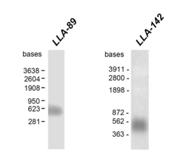
RACE-PCR method was used to obtain the full length 493 bp LLA-89 cDNA (accession no. DQ907930) and the full length 379 bp LLA-142 cDNA (accession no. EF026007), both excluding the poly (A) tail.
The LLA-89 cDNA contains an open reading frame of 303 bp encoding a polypeptide of 100 amino acids with a calculated molecular mass of 10.2 kDa and a pI of 3.2 (Figure 1A). The LLA-89 cDNA insert is close to full-
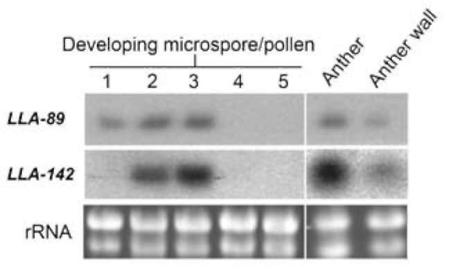
Characterization of the two stage-specific cD­NAs in the anther of L. longiflorum
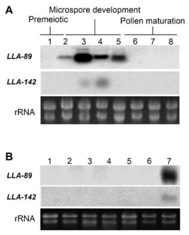
The LLA-89 and LLA-142 cDNAs were identified from a subtractive cDNA library at the L. longiflorum anther microspore development phase (Hsu et al., 2008). Since the insert sizes of both cDNAs were partial, the 5' - and 3'-
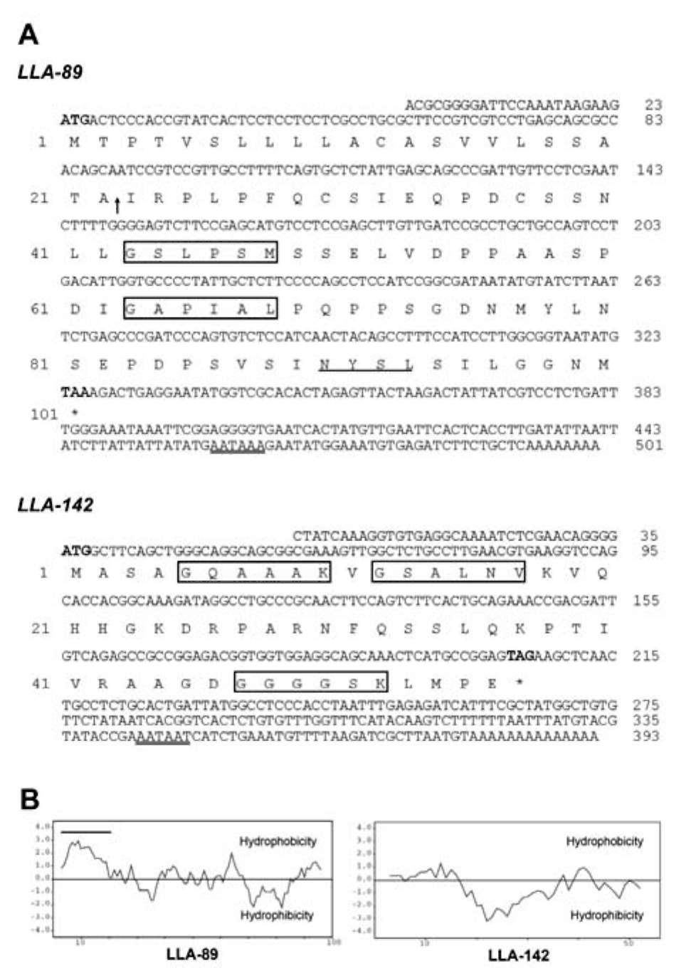
Figure 1. Sequences and hydropathy profiles of LLA-89 and LLA-142 in L. longiflorum anther. (A) Nucleotide and predicted amino acid sequences of LLA-89 and LLA-142 cDNAs. Numbers of nucleotide and amino acid sequences are indicated on the right and left, respectively. Bold letters in the nucleotide sequence indicate start codon and stop codon. The arrow indicates the predicted cleavage site of the signal peptide. The double underlines indicate the putative polyadenylation signal site. The underline indicates the putative N-glycosylation site (N-X-S/T). The putative N-myristoylation sites (G-X-X-X-S/T/A/G/C/N) are boxed; (B) Hydropathy profiles (Kyte and Doolittle, 1982) of LLA-89 and LLA-142 protein sequences. The black line indicates a hydrophobic sequence at the N-terminus of the sequence.
386
Botanical Studies, Vol. 52, 2011
cant resemblance to known proteins, indicating that LLA-142 encodes a novel protein.
Temporal and spatial expression of the two anther-specific genes in L. longiflorum
To determine the expression patterns of the two genes during anther development, blots of total RNA isolated from anthers at different stages, based on lily bud size, were hybridized with 32P-labeled LLA-89 and LLA-142 cDNAs. These two genes were differentially expressed during anther development. The LLA-89 transcripts were first detected in anthers within 20-25 mm-long buds, a stage of anther development concomitant with meiosis of the pollen mother cell. The LLA-89 mRNA accumulated to reach a maximum level in the anther within 34-36 mm-long buds, corresponding to the phase of microspore development and major cytological changes in the wall
Figure 2. An estimation of the full-length of LLA-89 and LLA-142 mRNAs in the anther of L. longiflorum. Total RNA (20 fig) was isolated from anthers within 34-36 mm-long buds. Total RNA was denatured, fractionated on formaldehyde-agarose gels, transferred onto nylon membranes and hybridized with the 32P-labeled LLA-89 and LLA-142 cDNA inserts. The RNA ladder markers are indicated at left.
length compared with its mRNA visualized using RNA blot analysis (Figure 2). In the 3' -untranslated region, a putative AATAAA consensus motif for polyadenylation signals is located 34 bp upstream from the site of poly-adenylation. A putative N-glycosylation site (N-X-S/T) is present at the amino acid position 90. Two putative N-myristoylation sites (G-X-X-X-S/T/A/G/C/N) are pres-ent at the amino acid positions 43 and 63. Assessment of the hydropathy profile (Kyte and Doolittle, 1982) showed that the LLA-89 polypeptide has a strong hydrophobic region near the N-terminus, indicating the presence of a signal peptide (Figure 1B). The proposed cleavage site in LLA-89 is between positions 22 and 23 (Figure 1A). The predicted amino acid sequence of LLA-89 was utilized to search protein databases. Sequence alignment revealed that LLA-89 is identical to LIM4, a protein known to be in-duced during the early prophase of meiosis in microsporo-genesis of L. longiflorum (Kobayashi et al., 1994).
The LLA-142 cDNA contains an open reading frame of 171 bp encoding a polypeptide of 56 amino acids (Figure 1A) with a calculated molecular mass of 5.7 kDa and a pI of 11.1. The LLA-142 cDNA insert is close to full-length compared with its mRNA, visualized using RNA blot analysis (Figure 2). In the 3'-untranslated region, a variant AATAAT consensus motif of polyadenylation is located 36 bp upstream from the site of polyadenylation. Three putative N-myristoylation sites are present at amino acid positions 5, 12, and 47. While the LLA-89 polypeptide contains a signal peptide at the N-terminus, the LLA-142 polypeptide, as shown by hydropathy profile analysis, does not (Kyte and Doolittle, 1982) (Figure 1B). The predicted amino acid sequence of LLA-142 was utilized to search protein databases. Sequence alignment revealed no signifi-
Figure 3. Temporal and organ-specific expression of LLA-89 and LLA-142 genes in the L. longiflorum anther. Total RNA (20 (ig) was isolated (A) from stamen/anthers of different size classes of buds: 1 < 15 mm buds; 2 = 20-25 mm buds; 3 = 34-36 mm buds; 4 = 44-46 mm buds; 5 = 60-65 mm buds; 6 = 90-95 mm buds; 7= 120-125 mm buds; 8 = 150-155 mm buds, and (B) from various vegetative and floral organs: roots (lane 1), stems (lane 2), leaves (lane 3), tepals (lane 4), carpels (lane 5), fila­ments (lane 6) and anthers (lane 7) within 34-36 mm-long buds. Total RNA was denatured, fractionated on formaldehyde-agarose gels, transferred onto nylon membranes, and hybridized with the 32P-labeled LLA-89 and LLA-142 cDNA inserts. Almost equal amounts of total RNA were loaded in each lane, as determined by ethidium bromide staining of the gel.
LIU et al. ― Lily anther-specific genes
387
layers. The LLA-89 mRNA amount decreased signifi­cantly but retained its signal in the 60-65 mm-long bud anthers, during which time the first mitosis occurred in the microspore to form immature pollen. The LLA-142 transcripts, however, were mainly restricted to the anther microspore development phase (Figure 3A). Signals were detected neither in the premeiotic phase nor in the pollen maturation phase. Overall, both LLA-89 and LLA-142 tran­scripts were detected during the microspore development phase.
To determine the tissue-specificity of gene expression in lily plants, total RNA was isolated from vegetative or­gans (roots, stems, and leaves) and floral organs within the 34-46 mm-long buds (tepals, filaments, anthers, and car­pels comprising stigmas, styles and ovaries). The isolated mRNA on a blot was hybridized with 32P-labeled LLA-89 and LLA-142 cDNAs (Figure 3B). With the exception of the anther, no hybridization signal was detected in RNA samples from other lily organs, indicating that the two genes were anther-specific.
Effects of GA on the regulation of the two an­ther-specific genes in developing anthers of L. longiflorum
Figure 4. Transcript induction of LLA-89 and LLA-142 genes by GA3Total RNA (20 (ig) was isolated from anthers (lane 1) within 17-20 mm-long buds. (A) The buds were dissected from lily plants, dipped in solutions containing 1 μM (lane 2), 10 μM (lane 3), and 100 μM (lane 4) GA3for 24 h. (B) The buds were dissected from lily plants dipped in a solution containing 1 μM GA3for 3 h (lane 1), 9 h (lane 2), 18 h (lane 3), 36 h (lane 4), and without GA3 addition for 36 h (lane 5). Total RNA was de-natured, fractionated on formaldehyde-agarose gels, transferred onto nylon membranes and hybridized with the 32P-labeled LLA-89 and LLA-142 cDNA inserts. Almost equal amounts of total RNA were loaded in each lane, as determined by ethidium bro-mide staining of the gel.
pressed in the tapetum when compared to other anther wall layers. However, hybridization signals in the microspores were not discernible due to the structure distortion. As buds grew to 45-50 mm, the tapetal cells in the anther began to disintegrate (Figure 5G-J) and the hybridization signal strength decreased compared to that observed in the tapetum of the 35-40 mm-long buds, suggesting significant down-expression of the LLA-89 and LLA-142 genes (Fig­ure 5G-J).
To investigate the inductive effect of GA on gene ex­pression, we applied GA exogenously. Lily plants with 17-20 mm-long buds were dipped in solutions containing various concentrations of GA3 for 24 h. Total RNA blot analysis revealed that the LLA-89 gene was induced by as little as 1 (μM GA3. In contrast, the LLA-142 gene did not respond to GA3(Figure 4A). The LLA-89 mRNA accumu­lation level increased as the GA3 concentration increased.
In order to determine the kinetics of the GA3-stimulated accumulation of LLA-89 mRNAs in lily anthers, a time-course experiment was conducted. Lily plants with 17-20 mm-long buds were dipped in a solution containing 1 (μM GA3 for various periods of time. Blot analysis showed that the LLA-89 mRNA started to accumulate after 18 h of GA3 treatment (Figure 4B, lane 3) and peaked at 36 h post-treatment (lane 4). As a control, no signal was detected in the anther of lily buds 36 h after treatment without the ad-dition of GA3 (lane 5).
In situ localization of the two anther-specific mRNAs in developing anthers of L. longiflorum
In situ hybridization the two genes with DIG-labeled antisense RNA probes was performed to determine the cellular location of these transcripts. No positive signal was detected in sections of anthers from 35-40 mm-long buds treated with sense probes (Figure 5A and B). When the DIG-labeled antisense riboprobes of LLA-89 and LLA-142 were used, hybridization signals with dark brownish-purple signals were observed in the tapetum of 35-40 mm-long buds (Figure 5C-F). At this time, the tapetal cells became polarized and highly secretory in the anther. The brownish-purple signals observed in the tapetum indicated that transcripts of the two genes were significantly ex-
Accumulation of the two gene transcripts in the developing microspores of L. longiflorum
To verify whether the two genes were expressed in the microspore, anthers of various size classes were dissected into two parts: the anther wall and the microspores. A blot of total RNA isolated from microspores of various devel­opmental stages was hybridized with 32P-labeled LLA-89 and LLA-142 cDNAs. In addition to detection in the anther wall (tapetum), both LLA-89 and LLA-142 transcripts were coordinately detected in the various stages of microspores in buds up to 44-46 mm (Figure 6 lanes 1-3). No signals
388
Botanical Studies, Vol. 52, 2011
Figure 6. Expression patterns of LLA-89 and LLA-142 genes in developing microspores of L. longiflorum. Total RNA (20 (ig) was isolated from the anther and anther wall within 44­46 mm-long buds and from microspores of various size classes of buds: 1 = 20-25 mm-long buds; 2 = 34-36 mm-long buds; 3= 44-46 mm-long buds; 4 = 60-65 mm-long buds; 5 = 70-75 mm-long buds during the microspore development phase. To­tal RNA was denatured, fractionated on formaldehyde-agarose gels, transferred onto nylon membranes, and hybridized with the 32P-labeled LLA-89 and LLA-142 cDNA inserts. Almost equal amounts of total RNA were loaded in each lane, as determined by ethidium bromide staining of the gel.
were detected from blots of mRNA isolated from the mi-crospore/pollen within 60-65 mm-long buds or thereafter (lanes 4 and 5), further indicating the stage-specificity of LLA-89 and LLA-142 gene expression in the anther. The detection of a signal in the anther and anther wall was used as a control.
DISCUSSION
We previously constructed a subtractive cDNA library to identify genes expressed during microspore develop­ment in L. longiflorum. Of those genes identified only in young anthers, the genes LLA-89 and LLA-142 were cho­sen for further investigation. These two genes were anther-specific and differentially expressed at the microspore development stage (Figure 3). Further, analysis by in situ hybridization demonstrated that both gene transcripts accumulated in the tapetum, the innermost layer of the anther wall (Figure 5). In addition to expression in the ta-petum, the two genes were coordinately expressed in the microspore at the microspore development phase, as con­firmed by microspore RNA blot analysis (Figure 6).
The spatial and temporal expression patterns of LLA-89 and LLA-142 correlated with tapetum development and degeneration. Around meiosis, when the buds were 20-25 mm in length, LLA-89 mRNA began to accumulate, reach­ing a maximum level when the buds were 34-36 mm-long. During this time, the tapetum became highly secretory in the anther. Afterwards, the LLA-89 mRNA expression sig­nificantly decreased as tapetal cells began to disintegrate (Figures 3A and 5). When compared with LLA-89, the lev­el of LLA-142 mRNA accumulation was more temporally
Figure 5. In situ hybridization of LLA-89 and LLA-142 tran­scripts in the developing anther of L. longiflorum. Expression of LLA-89 and LLA-142 was analyzed in bright-field illuminated 7-10μm cross-sections of anthers within 35-40 mm-long buds (A-F) or 45-50 mm-long buds (G-J) using DIG-labeled sense (A) and antisense (C, E, G, I) LLA-89 RNA and sense (B) and antisense (D, F, H, J) LLA-142 RNA probes. E, epidermis; En, endothecium; M, middle layer; T, tapetum. Bar = 100 μM.
LIU et al. ― Lily anther-specific genes
389
restricted to a period when the tapetum became highly ac-tive and secretory.
The full-length cDNAs of both genes were obtained by 5'-and 3'-RACE-PCR (Figure 1). Both cDNA inserts were close to full-length based on the sizes of their mR-NAs demonstrated on the blots (Figure 2). The LLA-89 gene encodes an acidic polypeptide, whereas the LLA-142 gene encodes a basic polypeptide. Sequence alignment revealed that LLA-89 was identical to LIM4, a known gene expressed in lily microsporocytes (Kobayashi et al., 1994). It was reported that the LIM4 transcript was first detected at the zygotene stage of meiotic prophase and continued to be expressed throughout meiosis. Although the mRNA is induced during meiosis, it accumulates to a maximum lev­el during the free microspore development phase prior to microspore mitosis (Figure 6). Therefore, the function of the LLA-89 protein is probably not related to the process of meiosis, as implied by Kobayashi et al. (1994). Further­more, we found that the gene was not only expressed in the microspore, but also strongly expressed in the tapetum (Figures 5 and 6). The accumulation of LLA-89 mRNA was associated with major cytological changes in the ta-petum (Figures 3 and 5). The LLA-89 protein contained a signal peptide at the N-terminus, so it is possible that the protein, once synthesized in the tapetum and microspores, is secreted and deposited on the walls of pollen grains. Thus, we propose that the LLA-89 protein may be in­volved in microspore/pollen wall formation. Alternatively, LLA-89 protein may act as a signaling molecule related to pollination or microbial responses based on its small size and the presence of a putative glycosylation and two puta­tive N-myristoylation sites. Protein N-myristoylation plays a vital role in membrane targeting and signal transduction in the plant response to environmental stress (Podell and Gribskov, 2004). It was reported that N-myristoylation of the ZmPtila protein is required for accumulation on the plasma membrane (Herrmann et al., 2006). Ishitani et al. (2000) reported that N-myristoylation was required for the function of the SOS3 salt tolerance gene product in Ara-bidopsis. Although the function of LLA-89 protein is still not clear, the pattern of expression and structure suggests functions different from meiotic regulation (Kobayashi et al., 1994). Functional investigations of the LLA-89 gene using genetic approaches in Arabidopsis might shed light of the function of LLA-89 protein.
While the LLA-89 protein has a signal peptide at the N-terminus, the LLA-142 protein does not. Sequence align­ment revealed no significant resemblance to any known proteins. Since LLA-142 mRNA is localized to the tape-tum, it is reasonable to suggest that the protein remains in the tapetal cells. The tapetum is known for its nutritive role during microspore and pollen development (Ma, 2005; Huang et al., 2009). In addition to a nutritive role, LLA-142 may be involved in one or more of the following developmental events in lily anthers: First, esterases in the tapetum are known to be active during tapetal degenera­tion (Sawhney and Nave, 1986). Peroxidases and other
proteins are associated with the synthesis of sporopollenin or its precursor in the lily tapetum (Reznickova and Wil-lemse, 1980). Finally, proteins are involved in the synthe­sis of carotenoids and flavonoids that, in turn, form the pollenkitt, a substance deposited on the pollen wall with tapetal debris (Hsieh and Huang, 2007).
We have reported two anther-specific genes in L. longi­florum. These two genes were only expressed during the phase of microspore development in the anther and were specifically expressed in the tapetum and microspores. Our analysis indicated that the LLA-89 gene was induced by the exogenous addition of GA3, while LLA-142 was not (Figure 4). In spite of the fact that the LLA-142 gene was not induced by GA3, both genes were indeed regulated by GAs in growing anthers as indicated by the observation that the expressions of LLA-89 and LLA-142 were partially inhibited by treatment with uniconazole, an inhibitor of GA biosynthesis (Hsu et al., 2008). It is possible that the inconsistency of the LLA-142 gene response to exogenous GA is due to an insufficient length of GA3 exposure. Al­ternatively, although the inductive mechanism of gene ac­tivation is not yet clear, it is obvious that the transduction pathway induced by exogenous GA is not the same as that induced by GAs that are naturally produced in the anther. The identification of the two GA-regulated genes is an im­portant step towards understanding the role of GA and its interaction with other hormones in anther development.
Acknowledgments. We thank Mei-Chu Chung from In­stitute of Plant and Microbial Biology, Academia Sinica for preparing those sections of anthers used for RNA in situ hybridization analyses. This work was supported by National Science Council Grant NSC98-2311-B-005-002-MY3 in Taiwan to C.-S. Wang.
LITERATURE CITED
Altschul, S.F., T.L. Madden, A.A. Schaffer, J. Zhang, Z. Zhang, W. Miller, and D.J. Lipman. 1997. Gapped BLAST and PSI-BLAST: a new generation of protein database search programs. Nucleic Acids Res. 25: 3389-3402.
Chen, R.D., E. Zimmermann, S.X. Xu, G.S. Liu, and A.G.
Smith. 2006. Characterization of an anther- and tapetum-specific gene and its highly specific promoter isolated from tomato. Plant Cell Rep. 25: 231-240.
De Grauwe, L., W.H. Vriezen, S. Bertrand, A. Phillips, A.M. Vidal, P. Hedden, and D. Van Der Straeten. 2007. Recipro­cal influence of ethylene and gibberellins on response-gene expression in Arabidopsis thaliana. Planta 226: 485-498.
Grienenberger, E., S. Besseau, P. Geoffroy, D. Debayle, D. Heintz, C. Lapierre, B. Pollet, T. Heitz, and M. Legrand. 2009. A BAHD acyltransferase is expressed in the tapetum of Arabidopsis anthers and is involved in the synthesis of hydroxycinnamoyl spermidines. Plant J. 58: 246-259.
Herrmann, M.M., S. Pinto, J. Kluth1, U. Wienand, and R. Lor-biecke. 2006. The PTI1-like kinase ZmPti1a from maize
390
Botanical Studies, Vol. 52, 2011
(Zea mays L.) co-localizes with callose at the plasma mem­brane of pollen and facilitates a competitive advantage to the male gametophyte. BMC Plant Biol. 6: 22.
Hirano, K., K. Aya, T. Hobo, H. Sakakibara, M. Kojima, R.A. Shim, Y Hasegawa, M. Ueguchi-Tanaka, and M. Matsuoka. 2008.?Comprehensive transcriptome analysis of phytohor-mone biosynthesis and signaling genes in microspore/pollen and tapetum of rice. Plant Cell Physiol. 49: 1429-1450.
Hsieh, K. and A.H. Huang. 2007. Tapetosomes in Brassica tape­tum accumulate endoplasmic reticulum-derived flavonoids and alkanes for delivery to the pollen surface. Plant Cell 19:582-596.
Hsu, Y.F., J.D. Tzeng, M.C. Liu, F.L. Yei, M.C. Chung, and C.S. Wang. 2008. Identification of anther-specific/predominant genes regulated by gibberellin during development of lily anthers. J. Plant Physiol. 165: 553-563. Huang, M.D., F.J.
Wei, C.C. Wu, Y.I. Hsing, and A.H. Huang. 2009.?Analyses of advanced rice anther transcriptomes reveal global tapetum secretory functions and potential pro-teins for lipid exine formation. Plant Physiol. 149: 694-707.
Ishiguro, S., Y. Nishimori, M. Yamada, H. Saito, T. Suzuki, T. Nakagawa, H. Miyake, K. Okada, and K. Nakamura. 2010. The Arabidopsis FLAKY POLLEN1 gene encodes a 3-hy-droxy-3-methylglutaryl-coenzyme A synthase required for development of tapetum-specific organelles and fertility of
pollen grains. Plant Cell Physiol. 51: 896-911. Ishitani, M., J. Liu, U. Halfter, C.S. Kim, W. Shi, and J.K. Zhu.
2000. SOS3 Function in plant salt tolerance requires N-myristoylation and calcium binding. Plant Cell 12: 1667­1677.
Kaneko, M., H. Itoh, Y. Inukai1, T. Sakamoto, M. Ueguchi-Tanaka, M. Ashikari, and M. Matsuoka. 2003. Where do gibberellin biosynthesis and gibberellin signaling occur in rice plants? Plant J. 35: 104-115.
Kao, F.I., Y.Y. Cheng, T.Y. Chow, H.H. Chen, S.M. Liu, C.H.
Cheng, and M.C. Chung. 2006. An integrated map of Oryza sativa L. chromosome 5. Theor. Appl. Genet. 112: 891-902.
Kapoor, S., A. Kobayashi, and H. Takatsuji. 2002. Silencing of the tapetum-specific zinc finger gene TAZ1 causes prema­ture degeneration of tapetum and pollen abortion in petunia. Plant Cell 14: 2353-2367.
Kobayashi, T., E. Kobayashi, S. Sato, Y. Hotta, N. Miyajima, A. Tanaka, and S. Tabata. 1994. Characterization of cDNAs induced in meiotic prophase in lily microsporocytes. DNA Res. 1: 15-26.
Kyte, J. and R.F. Doolittle. 1982. A simple method for display­ing the hydropathic character of a protein. J. Mol. Biol. 157: 105-132.
Li, H., F. Pinot, V. Sauveplane, D. Werck-Reichhart, P. Diehl, L.
Schreiber, R. Franke, P. Zhang, L. Chen, Y. Gao, W. Liang,
and D. Zhang. 2010. Cytochrome P450 family member CYP704B2 catalyzes the co-hydroxylation of fatty acids and is required for anther cutin biosynthesis and pollen exine
Li, N., D.S. Zhang, H.S. Liu, C.S. Yin, X.X. Li, W.Q. Liang, Z. Yuan, B. Xu, H.W. Chu, J. Wang, T.Q. Wen, H. Huang, D. Luo, H. Ma, and D.B. Zhang. 2006. The rice tapetum de­generation retardation gene is required for tapetum degrada­tion and anther development. Plant Cell 18: 2999-3014.
Ma, H. 2005. Molecular genetic analyses of microsporogenesis and microgametogenesis in flowering plants. Annu. Rev. Plant Biol. 56: 393-434.
McCormick, S. 1993. Male gametophyte development. Plant Cell 5: 1265-1275.
McNeil, K.J. and A.G. Smith. 2010. A glycine-rich protein that facilitates exine formation during tomato pollen develop­ment. Planta 231: 793-808.
Podell, S. and M. Gribskov. 2004. Predicting N-terminal myris-toylation sites in plant proteins. BMC Genomics 5: 37.
Reznickova, S.A. and M.T. Willemse. 1980. Formation of pol­len in the anther of Lilium. II. The function of surrounding tissues in the formation of pollen and pollen wall. Acta Bot.
Sambrook, J., E.F. Fritsch, and T. Maniatis. 1989. Molecular cloning: a laboratory manual, 2nd edn. Cold Spring Harbor, New York.
Sawhney, V.K. and E.B. Nave. 1986. Enzymatic changes in postmeiotic anther development in Petunia hybrida. II. His-tochemical localization of esterase, peroxidase, malate- and alcohol dehydrogenase. J. Plant Physiol. 125: 467-473.
Scott, R.J., M. Spielman, and H.G. Dickinsonb. 2004. Stamen
structure and function. Plant Cell 16: 46-60.
Shi, Y., S. Zhao, and J. Yao. 2009. Premature tapetum degenera­tion: a major cause of abortive pollen development in pho-toperiod sensitive genic male sterility in rice. J. Integr. Plant
Biol. 51: 774-781. Suen, D.F., S.S. Wu, H.C. Chang, K.S. Dhugga, and A.H.
Huang. 2003. Cell wall reactive proteins in the coat and wall of maize pollen: potential role in pollen tube growth on the stigma and through the style. J. Biol. Chem. 278:43672-43681.
Tzeng, J.D., S.W. Hsu, M.C. Chung, F.L. Yeh, C.Y. Yang, M.C.
Liu, Y.F. Hsu, and C.S. Wang. 2009. Expression and regula­tion of two novel anther-specific genes in Lilium longiflo-rum. J. Plant Physiol. 166: 417-427.
Van den Heuvel, K.J., R.H. Van Lipzig, G.W. Barendse, and G.J. Wullems. 2002. Regulation of expression of two novel flower-specific genes from tomato (Solanum lycopersicum)
by gibberellin. J. Exp. Bot. 53: 51-59.
LIU et al. — Lily anther-specific genes
391
鐵炮百合兩花藥專一基因LLA-89LLA-142之表現和定位
劉明哲  楊正守  王國祥
國立中興大學生物科技學研究所
利用抑制扣除雜合法(suppression subtractive hybridization)從鐵炮百合(JJlium longiflorum)花藥的
小孢子發育期cDNA集合庫選殖出兩個花藥專一性基因。利用 5'- 3'-RACE-PCR 延伸而得到LLA-89
LLA-142 cDNAs全長序列。LLA-89 cDNA含有一段303 bp可編譯框架(open reading frame),可轉譯
100個胺基酸的酸性蛋白質'其分子量爲10.2 kDa LLA-142 cDNA含有一段171 bp可編譯框架可
轉譯出56個胺基酸的鹼性蛋白質,其分子量爲5.7 kDa 。 LLA-89 蛋白質N端有一段疏水性的訊息胜
(signal peptide)序列,而LLA-142則無。序列比對後顯示LLA-89即是已知的百合LIM4 ,而LLA-
142
則是嶄新的未知蛋白質。利用北方墨漬法分析,得知LLA-89LLA-142 mRNA均在小孢子發育時
期偵測到且具花藥專一性。LLA-89基因會受外加的激勃素(gibberellin)誘導而表現'LLA-142則不會。
利用反式核醣探針(riboprobe)進行原位雜合(in situ hybridization)的實驗證實LLA-89LLA-142 mRNA
在花藥壁的絨氈層中呈現強烈的訊號。兩基因的mRNA也可在花藥發育中的小孢子偵測到。LLA-89
LLA-142兩蛋白質的功能有進一歩加以討論。
關鍵詞:花藥專性基因;激勃素;鐵炮百合;小孢子;絨氈層。