Botanical Studies (2011) 52: 445-454.
PHYSIOLOGY
Ultrastructural studies on actin-like filament in mung bean mitochondria and its potential functional significance
Yih-Shan LO, Lin-June HSIAO, Wann-Neng JANE, Ning CHENG, and Hwa DAI*
Institute of Plant and Microbial Biology, Academia Sinica, Taipei, Tai-wan 11529, ROC
(Received July 9, 2010; Accepted August 3, 2010)
ABSTRACT. This document reports that actin-like filaments, about 5 nm in width, were found in mung bean mitochondria after negative staining of mitoplast. The 5 nm filaments, composed of globular proteins, were visualized beneath the intramembranous particles by freeze-fracture electron microscope. Freeze-fracture im-muno-labeling against anti-actin antibody suggested that actin-like filaments attached to the protoplasmic sur­face of mitochondrial inner membrane. These filaments were also found in the matrix and underneath of outer membrane. The presence of actin-like protein in mitochondria was also substantiated by immunolocalization in situ. Interestingly, some 5 nm filamentous bundles were often found between mitochondrion and its "bud" It is likely that these images represent an undiscovered novel plant mitochondrial budding process which may involve some 5 nm filamentous structure in separating nascent mitochondrion to its mother mitochondrion. Treatment of cultured tobacco cells with F-actin depolymerization reagent, Latrunculin B (Lat B) might affect the shapes of mitochondria. Taken together, this study suggests that some 5 nm filaments, probably F-actin, may be localized in mitochondria and play a role in mitochondrial propagation and mitochondrial shaping.
Keywords: Actin/actin-like filament; Actin dynamics and mitochondrial morphology; Mitochondrial propaga­tion; Vigna radiata.
INTRODUCTION
drion in terms of its DNA segregation, propagation, mor­phological alteration and macromolecular transportation inside mitochondrion.
FtsZ, a progenitor to tubulin, forms the cytoskeletal framework of cytokinetic ring in bacteria and plays a major role in constriction of the furrow at septation of prokary-otes. Arabidopsis chloroplasts were shown to import FstZ in vitro (Osteryoung and Vierling, 1995), which could mediate their division. It appears that most mitochondria studied thus far have replaced FstZ with dynamin-related proteins for fission (Smirnova et al., 1998; Bleazard et al., 1999, Sesaki and Jensen, 1999; Erickson, 2000; Arimura and Tsutsumi, 2002; Logan, 2006), though mitochondria of a few alga still use FstZ for division (Beech et al., 2000). Unlike chloroplasts, mitochondria are dynamic organelles that often form a complex membrane system consisting of interconnected tubular structures, which undergo frequent fusion and fission.
We document in this report that a novel filamentous structure resembling actin filaments was found in mung bean seedling mitochondria. The 5 nm-wide filaments were visualized beneath the intramembranous particles (IMPs) by freeze-fracture electron microscope, thus placing the filaments on the protoplasmic surface of the mitochondrial membrane. This evidence for the presence of actin-like protein in isolated mung bean mitochondria was further
It has been well-investigated recently that actin-like filament in bacteria is responsible for bacterial shape, cell division and DNA segregation (Jones et al., 2001; ven den Ent et al., 2001; Carballido-Lopez, 2006; Graumann, 2007; Pogliano, 2008). In vitro biochemical study indi­cates that MreB can self-assemble into actin-like filaments. The progenitor of mitochondria was an a-proteobacterium and it is rational to assume that mitochondria might carry a MreB/actin-like protein involved in mitochondrial propa­gation and highly dynamic shape keeping. The presence of actin and tubulin in mitochondria and the association of actin with mitochondrial DNA has been reported but has not been further verified before 2011 (Etoh et al., 1990; Carre et al., 2002; Lo et al., 2002; Dai et al., 2005; Wang and Bogenhagen, 2006). The existence of actin inside mi­tochondria was verified by Reyes et al. (2011) and Lo et al. (2011) in human and mung bean mitochondria, respec­tively. After the issue of actin and actin filament's presence in mitochondria was evidenced, a more focused effort in searching for its functional significance will contribute greatly to the largely unknown area on plant mitochon-

*Corresponding author: E-mail: bodaihwa@gate.sinica.edu. tw; Fax:886-2- 2783-8609; Tel: 886-2-27871176.
446
Botanical Studies, Vol. 52, 2011
substantiated by immunolocalization in situ and freeze-fracture immuno-labeling in purified mitochondria. A fila­mentous bundle composed of several 5 nm filaments that circled around between mitochondrion and its "bud" was often found in our freeze fracture analysis. This phenom­enon suggests that these 5 nm filaments that circle around mitochondrion and its "bud" may represent the budding process for propagation, which is different from the con­ventional mitochondrial fission/division process in higher plants. In addition, treatment with F-actin depolymeriza-tion reagent Lantrunculin B (Lat B) can cause mitochon-drial shape and size changes in tobacco culture cells. Taken together, our results suggest that the 5 nm actin-like filaments are present in mitochondria and may be closely involved in mitochondrial propagation and shaping.
then carried out as described by Fujimoto et al. (1996) with minor modification. Anti-actin (C4 clone, Chemicon) an­tibody, followed by goat-anti-mouse antibody conjugated with 20 nm gold particles labelling, was carried out. Phal-loidine (20 faM) was used to stabilize actin filaments before SDS treatment. 1 faM phalloidin was present in all washing solutions along labeling procedures. Fine structure exami­nation was done under a Philips CM 100 electron micro­scope. This study was repeated more than 5 times and the same images were consistently observed.
Fluorescent analysis on mitochondrial mor-phology in tobacco culture cell
Wild-type control tobacco cells were stained with 250 nM MitoTracker Red CM-gXRos (Molecular Probes) for 30 minutes at room temperature followed by 2X washing. Images were collected by a Zeiss LSM510 meta confocal microscope (Carl Zeiss MicroImaging, Inc.) with a C-Aprochromat 63x/1.2 W objective (Carl Zeiss MicroImag-ing, Inc.). The fluorescence of MitoTracker was excited using a 543 nm line of He-Ne laser, and fluorescence was collected by a 565-615 nm band pass filter.
MATERIALS AND METHODS
Mitochondria and mitoplasts preparation
Mung bean mitochondria were prepared from 3-day-old etiolated mung bean (Vigna radiata, TN-5) seedlings and purified by a sucrose gradient, as described by Dai et al. (2005). Mitoplast preparation for electron microscopic examination followed the methods of Kozlowski and Zagorski (1988).
RESULTS
Fine structure and in situ immunolocalization analysis
Freeze-fracture electron microscopic analysis permits a direct visualization of mitochondrial structures on freshly-exposed surfaces beneath the outer membrane of random­ly-fractured (preferentially cleaved along the hydrophobic inner plane of membranous structure). The arrow in Figure 1A/B and 1C points to an ellipsoidal area in which the upper face of the outer membrane appears to have been dislodged, exposing a discrete parallel-aligned filamentous structure and intramembranous particles (IMPs, marker of mitochondrial inner membrane, see Figure 1B). These par­ticles are proteins/lipoproteins embedded in the intra-lipid bilayer of mitochondria (Lang, 1987). The fact that IMPs cast shadows on the surface of the 5 nm filament structure (see Figure 1B) suggests that this filament structure lies beneath the IMPs and presumably attaches to the proto­plasmic surface or possibly to the external surface of the inner membrane. In the magnified image shown in Figure
IB, the width of the filaments measured at stretches with the least metal casting material is approximately 5 nm. This particular filament structure exhibits a distinct organi­zation with an apparent periodicity, uniformly-sized con­stituting units and a possible inter-filament cross linking pattern (Figure 1B). These structural features bear a strik­ing resemblance to an actin filament bundle. The 20 nm immunogold grains seen in Figures 1A and 1B originated from a low intensity background labeling of the COXIII protein as an internal control for the freeze-fracture pro­cedure. A similar filamentous structure is shown in Figure
IC, but with the 5 nm filaments arranged into a meshwork. The same meshwork pattern was also present in the mi-tochondrial matrix (Figure 1D, arrow) but with a looser organization than in the membrane shown in Figure 1C.
The procedure for immunolocalization in tissue sec­tions was performed as described previously (Dai et al., 1998). Tissue sections (100-120 nm) were incubated with anti-actin (C4 clone, Chemicon) antibody followed by goat-anti-mouse antibody conjugated with 12 nm gold par­ticles. The monoclonal antibody PM028 (GTMA) against a yet-unidentified maize mitochondrial protein, with no cross-reactivity to mung bean mitochondria, was used as a control in the immuno-localization study.
EM analysis on whole-mount mitoplasts was carried out by fixing the sample with 1% paraformaldehyde followed by 1% uranyl acetate (pH 4.5) staining for 1-2 min.
For all fine structure analysis, the experiments were repeated 3-6 times and similar results were consistently obtained.
Freeze Fracture and immunogold labeling study
Purified mitochondria were placed on the specimen car­rier and quick-frozen by nitrogen slush. The freeze fractur­ing was carried out in a BAF 400D freeze etch unit (Balzers Union, Liechtenstein) at -105°C. Replicas were made by evaporation of platinum-carbon from an electron-beam gun positioned at a 45° angle, followed by carbon coating from a 90° angle. The replicas were released from the specimen carrier by immersing in 0.1% BSA in PBS buffer, then transferred to 600 fl of 0.05% SDS for 10 minutes at room temperature. Replicas were then washed four times with PBS. The immunogold labeling of anti-actin antibody was
LO et al. ― Structural and functional investigations on actin-like protein filaments in mung bean mitochondria
447
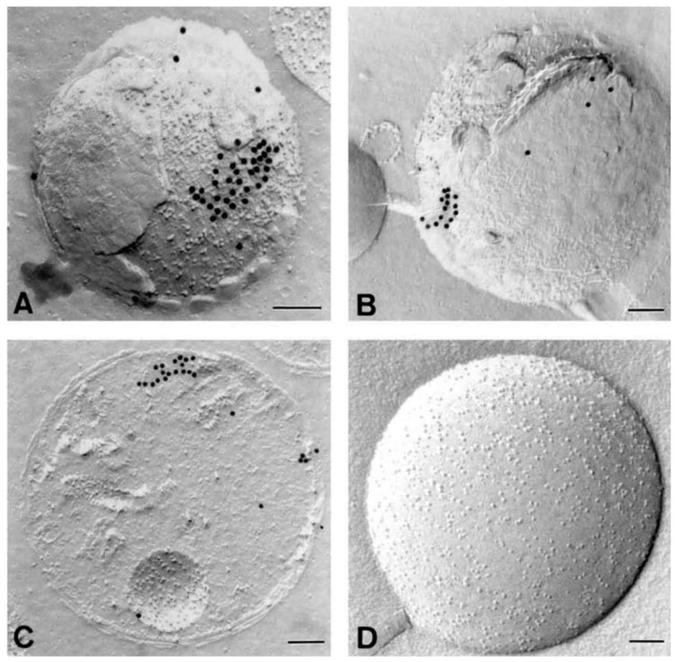
Immunogold labeling of anti-actin antibody in freeze-fractured mitochondria (Figure 2) indicates that a high density of gold particles accumulate as a patch and arrang­ing in lines on protoplasmic surfaces of the mitochondrial membrane (Figure 2A). Since IMPs heavily cover this dislodged and exposed area, actin (or actin-like) filaments are likely attached to the mitochondrial inner membrane (Figure 2A). Figures 2B and 2C show that gold-labeled antibody reacted with actin or actin-like proteins located
under the mitochondrial outer membrane and in the ma­trix, respectively. In both cases, the linear arrangement of gold grains indicates the presence of actin or actin-like filaments with mitochondria. Figure 2D is the control of Figure 2A-C. Almost no cross reaction between the control antibody (PM 028) and the freeze-fractured mitochondria could be detected (Figure 2D and unpublished results).
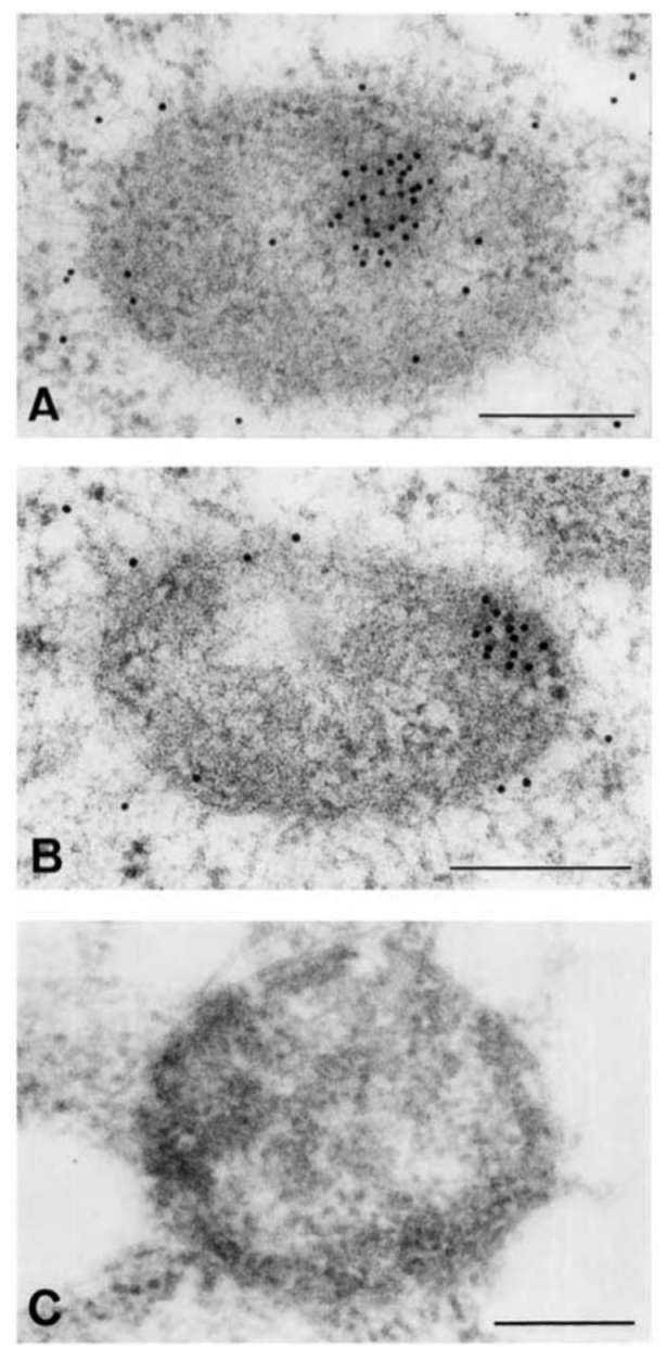
In situ immuno-gold labelling on thin sections of mung bean mitochondria revealed the localization of actin in the
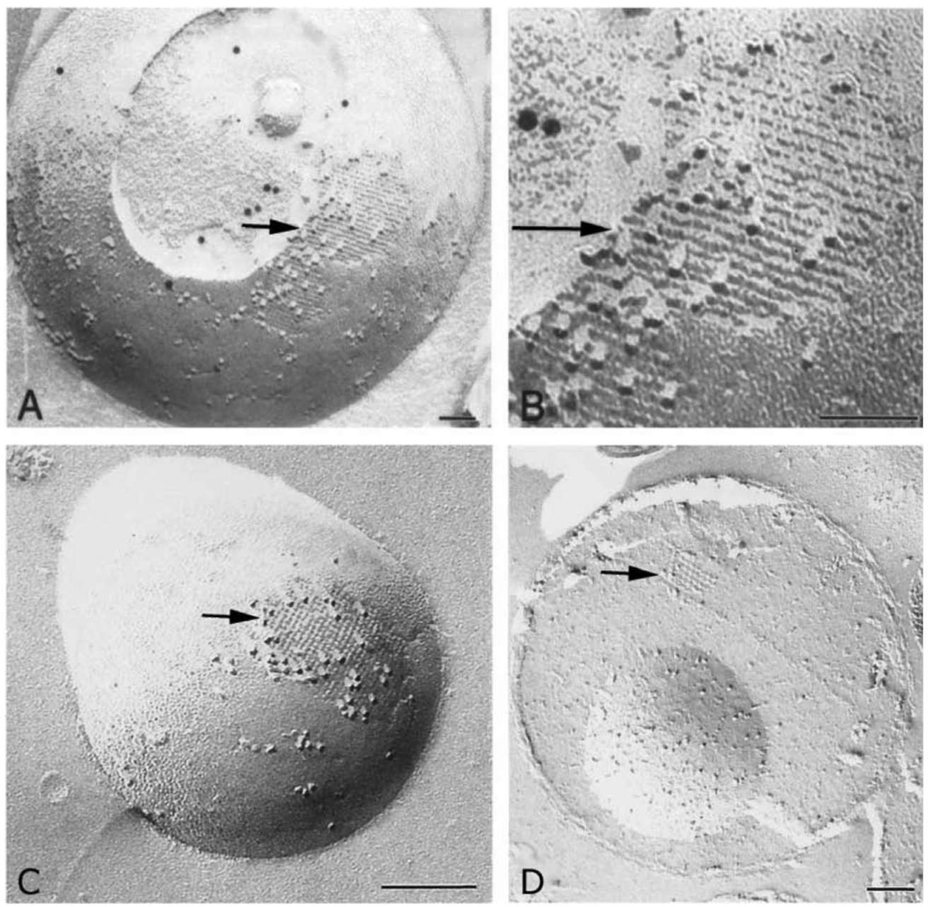
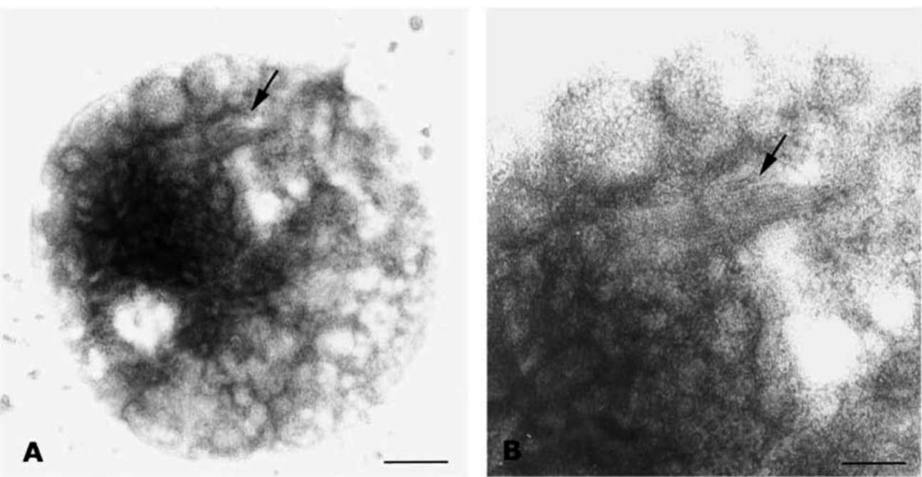
Figure 1. A 5 nm filamentous structure is visualized underneath the surface of mitochondria. Purified mitochondria were subjected to freeze-fracturing, and then immunogold labeling using anti-COX III antibody (for A and B) after laying the metal replica (see Materi­als and Methods). The arrow points to a fractured area in which the mitochondrial outer membrane appears dislodged, exposing the intramembranous particles (IMPs). The 5 nm-wide filaments lie adjacent to and just below these IMPs. Note that the width of the fila­ments and their constituting subunits display a good degree of uniformity and periodicity. The area indicated by the arrow in Panel A is amplified in Panel B. Panels C and D show a 5 nm filamentous structure with cross meshwork arrangements. Panel C shows filaments under the surface of mitochondrion as in Panel A. Panel D indicates cross meshwork and filament structure in mitochondrial matrix. All filaments are composed of globular protein. Bar equals 200 nm.
448
Botanical Studies, Vol. 52, 2011
mitochondrial matrix (Figure 3, Panels A and B). Heavy gold particles representing actin frequently co-localized with the denser area of the mitochondrial matrix under EM examination. Most interesting finding was that nearly all the anti-actin immunogold grains observed inside the mi­tochondria clustered together and co-localized, rather than distributing evenly, in the denser area in the mitochondrial matrix. Some clusters localized in the center of the mito-chondrial domain (Figure 3A) and some localized near the border (Figure 3B). In the same section, only a few im-munogold grains scattered in the cytoplasm and virtually none appeared in the control sections (without the anti-
actin antibody) (Figure 3C, see Materials and Methods).
After staining with 1% uranyl acetate, whole-mount electron microscopic analysis of mitoplasts (with the outer membrane removed by digtonin) revealed a filamentous bundle. Each filament measured about 5 nm wide and was composed of globular proteins, which are associated with mitoplasts (Figure 4, Panel A, arrow; Panel B is the enlargement of the area in Panel A indicated by an arrow). This filamentous bundle was obviously associated with the mitochondrial inner membrane since the mitoplast was repurified after its outer membrane was removed by the appropriate digtonin treatment.
Figure 2. Actin-like protein filaments detected in various locations of mitochondria by freeze-fracture immunogold labeling. A high density of gold grains (linearly-arranged) were accumulated as a patch underneath the protoplasmic surface of mitochondrial inner mem­brane (Panel A). The external face of mitochondrial outer membrane and matrix also show existing of actin-like filaments and hence the gold grains arrangement is mostly linear (Panels B and C, respectively). No gold particles were detected in the control shown in Panel D. Bar equals 200 nm.
LO et al. ― Structural and functional investigations on actin-like protein filaments in mung bean mitochondria
449
brane composition on the very active mitochondrion. No differentiated structure is shown on the smooth lipid mem­brane of the "bud". Panel C presents a negative control of Panel A, the image showing a cleaved surface on the mito-chondrial matrix that exhibits a "pinching out" of the inner membrane (small arrowhead) from an outer membrane (large arrowhead showing surrounding double membrane) and forming a bud-like structure. There is no 5 nm protein structure located at the border region between the "pinch­ing out" inner membrane and the double membrane. This clearly shows that the bud in Panel A does not arise from the "pinched out" inner membrane but arises from its pri­mordial mitochondrion. Panel D represents a possible "na­scent" mitochondrion (small arrowhead) budding out from mother mitochondrion (large arrowhead) and still connect­ed to its mother mitochondrion via a tubular structure (long arrow). It shows clearly that the "mother mitochondrion" encapsulated with a double membrane and the "nascent mitochondrion" shows few IMPs on its membrane.
Treatment of cultured tobacco cells with the F-actin de-polymerization reagent, LatB, enhanced the formation of spherical type of mitochondria from 30% up to 48%, and reduced the population of tubular-shaped mitochondria from 70% down to 51% of the mitochondria investigated (Figure 6).
DISCUSSION
The dependency of mitochondria on actin for their in-tracellular movement and localization is well understood. In Saccharomyces cerevisiae, actin also appears to mediate mitochondrial inheritance (Yaffe, 1999). Though not wide­ly known, actin (or actin-like) protein has been reported inside rat liver mitochondria (Etoh et al., 1990), pea chlo-roplasts (McCurdy and Williamson 1987) and mung bean mitochondria (Lo et al., 2002), respectively. Wang and Bogenhagen (2006) also reported that actin was found as a mitochondrial nucleoproteins as evidenced by proteomics analysis. In some previous studies, proteins attached to the surface of the organelles were not eliminated systemati­cally prior to analysis. Close association of actin with the mitochondrial surface is to be expected. In order to dem­onstrate unambiguously that actin (or actin-like) protein exists inside mitochondria, a proper pretreatment of the sample seems necessary. Our study has shown that mung bean mitochondrial actin is protected by the outer mito-chondrial membrane as well as by the inner membrane and is thus resistant to protease, high salt or a combination of both treatments. We also demonstrated that fluorescent actin without a mitochondrial presequence may import into plant mitochondria (Lo et al., 2011). This study aims to further understand the localization, organization and possible function of actin or actin-like filaments in higher plant mitochondria via a cytological approach.
Direct visualization of the 5 nm-wide actin-like fila­mentous bundles or meshwork beneath the outer mito-chondrial membrane via freeze fracture microscopy and
Figure 3. Immunolocalization of actin in mung bean seedling tissue sections. Three-day-old etiolated mung bean seedling tissues at the hook cortex region were fixed, embedded in Lo-wicryl K4M, sectioned to 100-120 nm thickness, incubated with a monoclonal anti-actin antibody and then with colloidal-gold-conjugated second antibody (Panels A and B). Panel C is negative control using PM028 instead of anti-actin monoclonal antibody for labeling (see Materials and Methods). Bar equals 200 nm.
During our ultra-structural study of freeze-fractured mitochondria, we often found that some filamentous bun-dles formed a belt around the neck between a "mother" mitochondrion and its "bud" (Figure 5, Panels A and B; B is the enlargement of arrow pointed area in A). A filamen­tous bundle composed of several 5 nm filaments circles around between mitochondrion and its "bud". Each fila­ment was about 5 nm in width (see arrow pointed enlarged area in Panel B). That the "mother" mitochondrion was heavily covered with IMPs indicates a typical inner mem-
450
Botanical Studies, Vol. 52, 2011
Figure 4. Whole-mount EM micrograph of mitoplast stained with 1% uranyl acetate. Arrow in Panel B shows filamentous bundle (each filament is about 5 nm in width) inside mitoplast. Panel B is an enlarged portion of Panel A (indicated by arrow). Bars equal 200 nm (A)
and 100 nm (B).
whole mount negative staining was shown in Figures 1 and 4, respectively. Freeze-fracture analysis finds that the actin-like filament structure lies beneath the IMPs and presumably attaches to the inner membrane's protoplasmic surface or possibly to its external surface (Figures 1B and 1C) and that it may also exist in the matrix (Figure 1D). The same meshwork pattern was shown in the mitochon-drial matrix with a much looser organization than in the membrane of Figure 1C. We suspect that the matrix's more loosely-oriented structure may be caused by the hydro-philic environment in the mitochondrial matrix. Based on this hypothesis, we believe that these 5 nm filaments are stable, well organized macromolecules which may move and assembly differently under different environmental conditions. This in vitro evidence is further substantiated by the immunolocalization of actin in the mitochondria (Figure 2A-C). In addition, the filamentous bundles re­vealed by whole-mount negative staining in the mitoplast (Figure 4) further supports the suggestion that the 5 nm filaments stabilize under hydrophilic conditions during mitoplast purification. Since the same meshwork was frequently found on inner mitochondrial membranes (Fig­ures 1 and 4) during our freeze-fracture and whole-mount negative staining EM analysis, we suggest that these 5 nm filaments, composed of globular proteins, may co-localize with the mitochondrial membrane and play some role in its shape.
As a control for the in vitro experiments described above, we used intact tissue for the immunolocalization experiment in order to ascertain whether actin (or actin-like) protein could be be detected in mitochondria in situ. Using the same anti-actin antibody as for immunolocaliza-
tion (shown in Figure 2), immunogold grains were local­ized inside mitochondria in thin tissue sections (Figure 3). Because of the random sectioning angle with respect to the alignment of actin filament bundles, only certain sections were expected to contain significant concentrations of gold grains. On those few sections containing a significant number of grains, they should have been clustered together reflecting the in situ geometry of the filament bundle rela­tive to the section angle. This prediction was borne out in the actual result. Most or nearly all anti-actin immunogold grains observed inside mitochondria did not distribute evenly but were clustered together and co-localized with the denser area in the mitochondrial matrix (Figures 3 A and B). Only a few immunogold grains were scattered in the cytoplasm in the same section and virtually none in control sections, done in the absence of anti-actin anti­body (Figure 3C, see Materials and Methods). This result reveals the presence of actin (or actin-like) protein inside mitochondria in situ in a non-random manner suggestive of a confined distribution of mitochondrial actin or actin-like filament bundles in the organelle.
The findings shown in Figure 5 indicates a possibility that an unknown budding process of mitochondrial propa­gation in mung bean seedlings. The presence of 5 nm fila­ments cable circle around the border area between "bud" and "mother" mitochondria (Figure 5A) may indicate that the "actin" cable is forming a mitochondrion-dividing ring and plays a role similar to that of FtsZ division ring (Beech et al., 2000; Momoyama et al., 2003; Osteryoung and Vierling, 1995; Miyagishima et al., 2001) or bacterial division ring (Lutkenhaus and Addinall, 2003). The "bud" gradually pinches out and forms a nascent mitochondrion
LO et al. ― Structural and functional investigations on actin-like protein filaments in mung bean mitochondria
451
Figure 5. A filamentous bundle composed of several 5 nm filaments circling around and between mitochondrion and its "bud" was found in our freeze- fracture analysis under EM (Panels A and B). Each filament is about 5 nm in width (see arrow pointing to enlarged area in Panel B). "Mother" mitochondrion heavily covered with IMPs indicates a typical mitochondrial inner membrane composition. No differentiated structure is shown on the "bud's"smooth lipid membrane. Panel C presents a negative control image of Panel A. Mitochondrion with a different cleaved surface on mitochondrial matrix. Mother mitochondrion surrounded by a double membrane (large arrowhead) and a connected "pinching out" inner membrane entity (small arrowhead) surrounded by a single membrane extend­ing from the inner membrane of mother mitochondrion. No 5 nm protein structure can be detected at the "pinching out" region. Panel D represents a possible "nascent" mitochondrion (small arrowhead) budding out from its mother mitochondrion (large arrowhead) and still connected to each other via a tubular structure (long arrow). It clearly shows that "mother mitochondrion's" matrix is fractured and surrounded by a double membrane, the nascent mitochondrion shows few IMPs on its membrane. Bars equal 100 nm (Panels A and B), 200 nm (Panels C and D).
452
Botanical Studies, Vol. 52, 2011
Figure 6. Dynamics of mitochondrial actin affect mitochondrial morphology. Wild-type tobacco cell culture treated with actin depo-lymerizing reagent-Lat B followed by MitoTracker (Red CM-H2XRos) staining. Confocal image was captured after removal of stain­ing solution. Panels A and B indicates the mitochondria without Lat B (60 faM) treatment and Panels C and D represent mitochondria with Lat B treatment. The percentage of spherical mitochondria and tubular mitochondria are indicated to the right of images. Total number of mitochondria counted is 273 in non-Lat B treated culture cell and 315 in Lat B treated culture cell.
with few IMPs on its surface and connected to its "mother" mitochondrion by a tubular structure (Figure 5D). It is likely that a budding process from image of Figure 5A to image of Figure 5D may represent the unknown novel way of mitochondrial propagation in higher plant besides con­ventional fission/fussion.
Our findings in Figure 6 show that converting F-actin to G-actin in tobacco cells may increase the percentage of spherical mitochondria populations up to 1.6- fold. This result also suggests that mitochondrial (or cellular) actin dynamics are involved in mitochondrial propagation and in the development of a structure (possibly an actin mesh-work) that controls mitochondrial shape.
This observation also implies that some subclasses of cellular actins in higher plants are translocated from cy­toplasm into mitochondria via unknown mechanism(s). However, we have sequenced 40 individual cDNA clones encoding actin from an unamplified mung bean cDNA bank and have found that, with the exception of a single nucleotide substitution in one clone, all 40 clones were identical to one another in sequences (GenBank accession No. 143208). The sequence of mung bean actin deduced
from the coding region of these cDNAs is 96.8% identical to those of ACT11 and ACT12 of Arabidopsis (Meagher et al., 1999) and contains no mitochondrial targeting se­quence (unpublished data). This could mean that either the cDNA encoding an unique actin with a targeting sequence has not yet been identified or, that an actin (or actin-like) protein without a targeting sequence could be imported into mitochondria by an unknown mechanism. Neverthe­less, our unpublished results demonstrated that a trans-genic fluorescent actin without a targeting sequence can be imported into plant mitochondria (Lo et al., 2011).
The discovery of actin filaments in mitochondria raises additional questions: How are the actin molecules polym­erized in the mitochondria? What is the role of polymer­ized actins or actin filaments inside the mitochondria? Based on the observation of the appearance of actin or actin-like filaments on mitochondrial membrane (Figures 1-5) and changes in mitochondrial morphology with the addition of an actin depolymerization reagent (Figure 6), we speculate that actin filaments play a role in maintaining mitochondrial shape or in its propagatation. Considering the data shown above, we wonder whether our observa-
LO et al. ― Structural and functional investigations on actin-like protein filaments in mung bean mitochondria
453
tion is in any way related to the mitochondria propagation model we proposed earlier, that is based on a "nascent mi­tochondria" population we found in mung bean seedlings (Dai et al., 1998). Taken together, we suggest that some 5 nm filaments surround the mitochondrion and its bud may play a role in mitochondrial shaping and propagation.
yeast mitochondria and mitoplasts synthesizing specific polypeptides. Anal. Biochem. 172: 382-391.
Lang, R.D.A. 1987. Electron microscopy of mitochondria. In V.M. Darley-Usmar, D. Richwood and M.T. Wilson (eds.), Mitochondria-a practical approach. IRL press Oxford, Washington DC, pp. 17-33.
Lo, Y.S., N. Cheng, L.J. Hsiao, A. Annamalai, G.Y. Jauh, T.N. Wen, H. Dai, and K.S. Chiang. 2011. Actin in mung bean mitochondria and implications for its function. The Plant Cell (in print).
Lo, Y.S., Y.T. Wang, W.N. Jane, L.J. Hsiao, O.L.F. Chen, and
H. Dai. 2002. The presence of actin-like filaments in higher plant mitochondria. Bot. Bull. Acad. Sin. 44: 19-24.
Logan, D.C. 2006. Plant mitochondrial dynamics. Biochim. Bio-
phys. Acta 1763: 430-441.
Lutkenhaus, J. and S.G. Addinall. 2003. Bacterial cell division
and the Z ring. Proc. Natl. Acad. Sci. USA 100: 2146-2151. McCurdy, D.W. and R.E. Williamson. 1987. An actin-related
protein inside pea chloroplasts. J. Cell Sci. 87: 449-456
Meagher, R.B., E.C. Mckinney, and A.V. Vitale. 1999. The
evolution of new structures: clues from plant cytoskeletal genes. Trends in Genet. 15: 278-284.
Miyagishima, S., M. Takahara, and T. Kuroiwa. 2001. Novel filaments 5 nm in diameter constitute the cytosolic ring of the plastid division apparatus. Plant Cell 13: 707-721
Momoyama, Y., Y. Miyazawa, S.Y. Miyagishima, T. Mori, O. Misumi, H. Kuroiwa, and K.Tsuneyoshi. 2003. The division of pleomorphic plastids with multiple FtsZ rings in tobacco
BY-2 cells. Eur. J. Cell Biol. 82: 323-332.
Osteryoung, K.W. and E. Vierling. 1995. Conserved cell and or-ganelle division. Nature 376: 473-474.
Pogliano, J. 2008. The bacterial cytoskeleton. Curr. Opin. Cell
Biol. 20: 19-27.
Reyes, A., J. He, C.C. Mao, L.J. Bailey, M. Di Re, H. Sembongi,
L. Kazak, K. Dzionek, J.B. Holmes, T.J. Cluett, M.E. Har-bour, I.M. Fearnley, R.J. Crouch, M.A. Conti, R.S. Adel-stein, J.E. Walker, and I.J. Holt. 2011. Actin and myosin contribute to mammalian mitochondrial DNA maintenance. Nucleic Acids Res. 39: 5098-5108.
Sesaki, H. and R.E. Jensen. 1999. Division versus fusion: Dnm1p and Fzo1p antagonistically regulate mitochondrial shape. J. Cell Biol. 147: 699-706.
Smirnova, E., D.L. Shurland, S.N. Ryazantsev, and A.M. van der Bliek. 1998. A human dynamin-related protein controls the distribution of mitochondria. J. Cell Biol. 143: 351-358.
Van den Ent, F., L.A. Amos, and J. Lowe. 2001. Prokaryotic ori­gin of the actin cytoskeleton. Nature 413: 39-44.
Wang, Y.S. and D. F. Bogenhagen. 2006. Human mitochondrial DNA nucleoids are linked to protein folding machinery and metabolic enzymes at the mitochondrial inner membrane. J.
Biol. Chem. 281: 25791-25802.
Yaffe, M.P. 1999. The machinery of mitochondrial inheritance
and behavior. Science 283: 1493-1497.
Acknowledgments. This research was supported by re­search grants from the National Science Council and from Academia Sinica, ROC.
LITERATURE CITED
Arimura, S.I. and N. Tsutsumi. 2002. A dynamin-like protein (ADL2b) rather than FtsZ, is involved in Arabidopsis mi­tochondrial division. Proc. Nat. Acad. Sci. USA 99: 5727­5731.
Beech, P.L., T. Nheu, T. Schultz, S. Herbert, T. Lithgow, P.R.
Gilson, and G.I. McFadden. 2000. Mitochondrial FtsZ in a Chromophyte Alga. Science 287: 1276-1279.
Bleazard, W., J.M. McCaffery, E.J. King, S. Bale, A. Mozdy, Q. Tieu, J. Nunnari, and J.M. Shaw. 1999. The dynamin-related GTPase Dnm1 regulates mitochondrial fission in yeast Nat.
Cell Biol. 1: 298-304.
Carballido-Lopez, R. 2006. The bacterial actin-like cytoskeleton. Microbiol. Mol. Biol. Rev. 70: 888-909.
Carre, M., N. Andre, G. Carles, H. Borghi, L. Brichese, C. Briand, and D. Braguer. 2002. Tubulin is an inherent com­ponent of mitochondrial membranes that interacts with the voltage-dependent anion channel. J. Biol. Chem. 277:
33664-33669.
Dai, H, Y.S. Lo, W.N. Jane, L.W. Lee, and K.S. Chiang. 1998. Population heterogeneity of higher-plant mitochondria in
structure and function. Euro. J. Cell Biol. 75: 198-209. Dai, H., Y.S. Lo, A. Litvinchuk, Y.T. Wang, W,N. Jane, L.J.
Hsiao, and K.S. Chiang. 2005. Structural and functional characterizations of mung bean mitochondrial nucleoids. Nucleic Acids Res. 33: 4725-4739.
Erickson, H.P. 2000. Dynamin and FtsZ - Missing links in mi-tochondrial and bacterial division. J. Cell Biol. 148: 1103­1105.
Etoh, S., H. Matsui, M. Tokuda, T. Itano, M. Nakamure, and O.
Hatase. 1990. Purification and immunohistochemical study of actin in mitochondrial matrix. Biochem. Inter. 20: 599­606.
Fujimoto, K., M. Umeda, and T. Fujimoto. 1996. Transmem-brane phospholipid distribution revealed by freeze-fracture replica labeling. J. Cell Sci. 109: 2453-2460.
Graumann, P.L. 2007. Cytoskeletal elements in bacteria. Annu.
Rev. Microbiol. 61: 589-618.
Jones, L.J., R. Carballido-Lopez, and J. Errington. 2001. Control of cell shape in bacteria: helical, actin-like filaments in Ba-cillus subtilis. Cell 104: 913-922.
Kozlowski, M. and W. Zagorski. 1988. Stable preparation of
454
Botanical Studies, Vol. 52, 2011
以微細結構分析來研究似肌動蛋白細絲和綠豆粒線體的相關性
以及它潛在的功能性
羅意珊 蕭玲君 簡萬能 成 寧 戴 華
中央研究院植物暨微生物學研究所
我們利用對nntoplastnegative stainning方法,在綠豆線粒體中發現5毫微米似肌動蛋白細
絲的存在。由冷凍裂解電子顯微鏡觀察,球狀蛋白質組成的5毫微米似肌動蛋白細絲和粒線體的
intramembranous particles共存。冷凍裂解的免疫標記法結果,建議似肌動蛋白細絲附在粒線體內膜
protoplasmic內外表面。他們也可能位於外膜下層和基質中。由原位immunolocalization方法,也證實肌
動蛋白或似肌動蛋白出現在線粒體基質。有趣的是,一些5毫微米似肌動蛋白細絲經常圍繞在線粒體和
它的「芽體」之間。這也許代表一種新發現的由5毫微米似肌動蛋白細絲介入的一種新穎的植物粒線體
發芽繁殖的過程。線狀肌動蛋白解聚作用試劑,Latrunculin B處理煙草細胞,可影響粒線體形狀。綜合
而論,這項研究建議一種5毫微米似肌動蛋白細絲,在高等植物粒線體中扮演著和粒線體繁殖和粒線體
形態相關的角色。
關鍵詞:肌動蛋白細絲;肌動蛋白動力學和線粒體形態;線粒體繁殖;Vigna radiata