Botanical Studies (2011) 52: 455-463.
PHYSIOLOGY
In vitro propagation of Platycerium bifurcatum (Cav.) C. Chr. via green globular body initiation
Yue Ken LIAO1* and Yi Hui WU2
1Department of Forestry and Natural Resources, National Chiayi University, No. 300, Syuefu Rd., Chiayi City 60004, Taiwan
2Miaoli District Agricultural Research and Extension Station, Council of Agriculture, Executive Yuan, Miaoli Coimty 36343, Taiwan
(Received September 13, 2010; Accepted March 25, 2011)
ABSTRACT. This study describes a Platycerium bifurcatum (Cav.) C. Chr. propagation method through initiation of green globular bodies (GGBs) from juvenile leaf explants of in vitro grown sporophytes. The GGB initiation was first obtained by incubating the explants on MS medium supplemented with a-naphthalene acetic acid (NAA) and two other cytokinins, 2-isopentenyl adenine (2ip) or 6-benzylaminopruine (BA). NAA at 5.37 (iM was determined a better concentration for the initiation. However, as the cytokinin concentra­tion in the medium increased, it inhibited GGB production. Individually harvested GGBs proliferated well on a medium supplemented with 5.37 [iM NAA and 2.22 [iM 2ip; they displayed less sporophyte formation but a faster increase in diameter. Multiple sporophytes were subsequently regenerated by culturing GGBs on a hormone-free MS medium at the regeneration stage. Non-rooted and separated sporophytes were directly transplanted to four different types of potting media, namely sphagnum, vermiculite, peat, and a mixed medi­um (vermiculite:peat = 1:1). Spontaneous rooting was observed during the first 4-week acclimatization period. Even with the omission of an adventitious root induction step, the survival rate of the plants was 88% or high­er. No significant difference in plant survival was detected among the different media. Using our proposed propagation procedures, it was possible to obtain an estimated production of about 170 sporophytes per initial leaf explant (proximal section; 1/2 of the leaf < 2.0 cm in length) during a 32-week culturing period.
Keywords: Green globular body (GGB); Platycerium bifurcatum; Micropropagation; Staghorn fern.
Abbreviations: 2ip, 2-isopentenyl adenine; BA, 6-benzylaminopruine; FAA, formalin acetic acid alcohol; GGBs, green globular bodies; NAA, a-naphthalene acetic acid; PGRs, plant growth regulators.
INTRODUCTION
1979; Teng and Teng, 1997), entire leaf explants (Camloh and Gogala, 1991; Camloh et al., 1994), rhizomes (Wee et al., 1992) and bud scales (Ambrozic-Dolinsek and Cam-loh, 1997). Instances of more complicated procedures such as the initiation of sporophytic callus (Kwa et al., 1997), the induction of apogamy from gametophytes (Kwa et al., 1995) and apospory from a leaf culture (Ambrozic-Dolinsek et al., 2002) have also been documented over the past decade. Jambor-Benczur et al. (1995) and Huang (2004) have also briefly reported on the possibility of P. bifurcatum propagation via green globular body (GGB) initiation, which is expected to be a rapid and convenient culture method for this genus.
The GGB culturing system has been applied to many fern species, including Nephrolepis spp. (Higuchi et al., 1987; Amaki and Higuchi, 1991), Asplenium nidus L. (Higuchi and Amaki, 1989), Adiantum raddianum C. Presl, Pteris ensiformis Burm. f., Rumohra adiantiformis (G. Forst.) Ching (Amaki and Higuchi, 1991), Blechnum spicant L. (Fernandez et al., 1996), and Polypodium cam-
Platycerium bifurcatum (Cav.) C. Chr. is a common or­namental fern species in Taiwan. It is commercially avail­able in local markets and has become popular at leisure farms and recreational parks where tropical scenes are de­signed to attract tourists. This species can be propagated sexually through spore germination (Camloh, 1993; 1999) and asexually through root bud development (Richards et al., 1983). However, propagation from spores is gener­ally a time-consuming procedure (Thentz and Moncousin, 1984), while ex vitro spore cultivation is vulnerable to at­tack by phytopathological agents (Lane, 1981). A variety of methods for in vitro vegetative propagation of several species in this genus has been more successful, including direct sporophyte regeneration from the shoot tip (Hen-nen and Sheehan, 1978), homogenized leaf tissue (Cooke,

*Corresponding author: E-mail: ykliao@mail.ncyu.edu.tw; Tel: +886-5-2717471; Fax: +886-5-2775852.
456
Botanical Studies, Vol. 52, 2011
bricum L. (Bertrand et al., 1999). The general procedure for GGB initiation depends on using cytokinin and auxin, either alone or in combinations, to stimulate the unique structures grown from various explants. The structure thus initiated functions just like the protocorm-like body in orchids (Amaki and Higuchi, 1991); both have vigor­ous cell division centers (meristem cells) that regenerate into adventitious shoots (Higuchi and Amaki, 1989; Kraus and Monteiro, 1989; Hong et al., 2010; Vyas et al., 2010). GGBs derived from different fern species exhibit distinc­tive features in terms of their growth rate and appearance, and scale- or hair-like structures are occasionally seen on the surface of certain GGB cultures (Amaki and Higuchi, 1991). The most common feature of GGBs is that they contain areas of multiple cell divisions inside the tissue or distributed on the epidermal layer of the GGBs themselves (Higuchi et al., 1987; Amaki and Higuchi, 1991). This characteristic maintains the GGB cultures in a quick divid-ing phase, with high capacity for morphological compe-tency.
Amaki and Higuchi (1991) noted two advantages of using the GGB system for fern propagation: it is easy to control the progress of organogenesis during culturing operations simply by the addition or removal of a single plant growth regulator (PGR), and a high multiplication rate can be expected. Jambor-Benczur et al. (1995) and Huang (2004) have briefly reported the GGB system for P. bifurcatum propagation. Since their protocol is relatively complicated, we have tried to develop a more simplified procedure through GGB propagation for this species, which is described in detail below.
browning rate were determined by examination under a stereomicroscope. The browning rate (%) was calculated as follows: (number of browned explants / total explant number) x 100%.
GGB multiplication
Eight PGR combinations (Table 4) were chosen from the previous GGB initiation experiments to be applied in this stage. These combinations were selected due to their more effective stimulation of GGB production (details giv­en in Table 2). The "control" and Combinations No. 1, 3, 6 and 7 were selected from the NAA + 2ip and NAA + BA experiments (since they produced more GGB in both ex­periments); the others were selected from the NAA + 2ip tests (for the same reason). GGBs of similar sizes (~2.0 mm in diameter), initiated from these eight PGR combi­nations, were isolated from the explants and subcultured back on their originally initiated fresh medium for three more weeks. At the end of this incubation period, the diameter of the enlarged GGBs was measured. The sporo-phyte regeneration (undesired leaf growth of sporophyte from the GGB) was taken into account as a negative indi­cator for GGB multiplication at this stage. The sporophyte regeneration rate (%) = (number of GGBs with sporophyte development / total number of GGBs incubated) x 100%.
Sporophyte regeneration
During the stage of conversion of GGBs into sporo-phytes, the GGBs from 5.37 (iM NAA + 2.22 (iM 2ip incubation were first collected. They were then incubated for five more weeks in the same NAA + 2ip combina­tion to prepare enough material for the experiments. Such prepared GGB cultures (8.0-10.0 mm in diameter) were then transferred to the PGR-free MS basal medium supplemented with different amounts of activated char­coal (0, 0.05, and 0.1%) and maintained for four weeks. Regenerated sporophytes were excised from the surface of the GGB with a scalpel and then examined under a stereomicroscope. Large (> 0.5 cm) and small (< 0.5 cm) leaves of sporophyte were counted individually and evalu­ated as indicators for charcoal influence to determine the best treatment for sporophyte regeneration. Following this evaluation, a second experiment was carried out using the best treatment (MS medium without charcoal) to find the total amount of sporophyte that a GGB culture might produce. Fifteen GGB cultures (prepared as previously described) were incubated on the MS medium minus char­coal. After eight weeks, the regenerated sporophytes were separated and counted under a stereomicroscope. The average production of sporophyte from two repeated tests was reported.
MATERIALS AND METHODS
Plant material and GGB initiation
Juvenile P. bifurcatum plants were grown in vitro and purchased as a commercial product, subcultured on MS basal medium (Murashige and Skoog, 1962) in our labora­tory as stock plants. In preliminary experiments, a serious browning problem was observed on cultured leaf explants larger than 2.0 cm and a dark secreted material accumulat­ed in the medium (data not shown). Therefore, only small leaves (< 2.0 cm) were used in subsequent procedures of our study. Each leaf explant was cut into separate proximal and distal sections across the midpoint of its long axis. Both sections were cultured on GGB initiation media, which were MS medium supplemented with the follow­ing two PGR regimes in various combinations: (1) NAA
(0, 5.37, 26.85, and 53.71 (M) plus 2ip (0, 2.22, 4.44, and 22.2 (M); and (2) NAA (0, 5.37, 26.85, and 53.71 (M) plus BA (0, 2.22, 4.44, and 22.2 (M) (Table 2). Indepen­dent PGR regime experiments were therefore conducted for 16 combinations. For each PGR combination, 15 ex-plant sections from both proximal and distal half leaf were included respectively and five pieces of both sections were incubated in a Petri dish of 90 mm diameter. After being cultured for 4 weeks, the number of GGBs and the explant
Histological examination
Cultured samples from both the GGB multiplication and sporophyte regeneration stages were examined un­der a light microscope. Samples were first fixed in FAA (50% alcohol : glacial acetic acid : formalin, 18:1:1), then
LIAO and WU ― Propagation of staghorn fern via GGB culture
457
dehydrated through a tertiary butyl alcohol series, and embedded in paraffin (Jung et al., 2005). The samples were sectioned to a thickness of 10 (m and stained with safranin-fast green, which allowed developmental events and morphological changes to be observed in detail.
were evaluated in this test. Data on GGB multiplication (diameter increase) and influence of charcoal application on sporophyte regeneration (number of large and small leaves produced), derived from the completely random­ized experimental design, were analyzed the same way except that one-way ANOVA was applied. The percentage of browning on different explants during the GGB initia­tion stage, the ratios of undesired sporophyte formation form GGBs in the multiplication test, and the young plants rooted and their survival rates in the acclimatization stage were analyzed by Chi-squared (x2) test. All experiments were repeated twice and typical results are shown in this report.
In vitro culture conditions
MS medium was used as the basal medium in each tis­sue culture stage. Sucrose (3.0%; w/v) was supplemented as the carbon source and Difco Bacto agar (0.8%; w/v) was added for medium solidification. Activated charcoal and different PGR chemicals (all purchased from Sigma, USA) with their combinations were added as indicated. All media were adjusted to pH 5.8 by the addition of NaOH or HCl prior to autoclaving. Cultures at all in vitro stages were maintained at 21 1°C under a 16 h photope-riod with cold white fluorescent light mixed with incandes­cent light at 55.6 (mol m-2 s-1. An additional water-agar plate was used to cover all the culture dishes to prevent water condensation inside the vessels.
RESULTS
GGB initiation
GGB formation was first observed 2-3 weeks after in­cubation of the explants in GGB initiation medium. They exhibited a fresh green color with a glassy bead-like shape (Figure 1A). Most of the GGBs were initiated from the basal region of the proximal explant sections or from adaxial surface of both sections. Pale brown rhizoids were seen growing abundantly around the GGBs (Figure 1B).
The two factorial designed experiments demonstrated that the NAA and 2ip or BA levels as well as explant ori­gin all had a significant effect on GGB production. There were also significant interactions detected among all these effects and their combinations conducting a complicated relationship while GGB was initiated (Table 1). Bet­ter initiation was obtained using the proximal part of the explants when treated with low NAA + 2ip or BA. NAA at 5.37 (M in both experiments exhibited a higher GGB yield (p<0.05) especially compared with its higher dos­ages. However as cytokinin (2ip or BA) concentration increased, we observed a gradual decrease in GGB pro­duction (Table 1).
A further and detail examination of the GGB yield from all of the PGR combinations were carried out and shown in Table 2. We observed that the GGBs were even initi­ated with the MS PGR-free medium (control). However the best PGR combination was determined, using 5.37 (iM NAA with no cytokinin addition, yielding 7.17 1.23 and 5.37 1.22 GGBs per explant (proximal part), re­spectively. This was significantly greater than the second place combination (4.03 1.19 GGBs from 5.37 (iM NAA + 4.44 (iM 2ip combination or 2.47±0.77 GGBs from 5.37 (M NAA + 2.22 (M BA combination). There was also a significant difference in the browning rate between the dis­tal and proximal explant sections (Table 3), which firmly excludes the possible application of distal sections as cul­ture explants in future studies.
Acclimatization of regenerated plants
Small sporophytes that regenerated from the sporophyte regeneration experiments (MS medium without activated charcoal) were transferred to the same medium in glass jars for further growth. As is consistent with documented processes (Hennen and Sheehan, 1978; Wee et al., 1992), these regenerated but non-rooted leaf entities were col­lected when they developed 3-6 leaves, with individual leaves measuring about 4.0-6.0 cm in length. The basal part was washed to remove the agar residues. The entities were then transferred into four different ex vitro potting media for acclimatization. These media were sphagnum, vermiculite, peat and a mixture (vermiculite: peat = 1:1 by volume). During the first week of acclimatization the plastic pots (top diameter 7.5 cm, bottom diameter 5.3 cm, height 6.0 cm) were filled with these media and then placed in shallow water (1.5 cm deep) in a container covered by transparent cellophane. Each pot contained a single transplanted leaf entity; 30 entities were tested for survival in each medium. The rooted and survival rate (%) was calculated after four weeks as follows: (number of survivals / number of transplanted leaf entities) x 100%. The experiment was carried out in a shaded greenhouse. Light intensity inside the greenhouse was 1.17% that of natural sunlight. The measured photo flux density in the greenhouse on a typical sunny day was 19.75 (mol m-2 s-1. The greenhouse temperature was controlled to be between 20 and 30°C for this experiment.
Statistics and data analysis
Data regarding the number of GGBs initiated during the first experiment (obtained using a completely random­ized factorial design; 4 x 4 x 2) were subjected to a three-way ANOVA analysis followed by Duncan's new multiple range test. The effects of the explant origin, PGR types/ concentrations and their interactions on GGB initiation
GGB multiplication
The GGBs continuously increased in size in all selected PGR combinations as well as in the PGR-free control (Table 4). The multiplication was clearly seen under the
458
Botanical Studies, Vol. 52, 2011
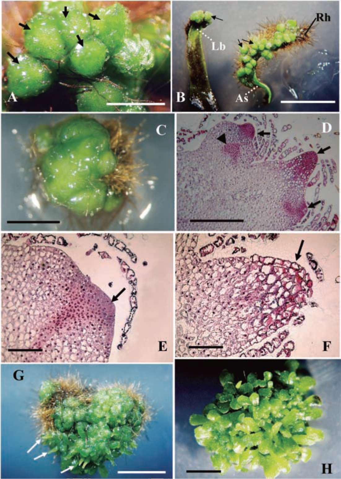
Figure 1. Microscopic examination of tissue-cultured Platycerium bifurcatum via GGB initiation and multiplication (A-D), and subse­quent stages of sporophyte regeneration (E-H). (A) A close-up view of a GGB cluster cultured on MS medium supplemented with 5.37 (M NAA after 4 weeks; each GGB (arrows) exhibits a green glassy bead-like shape (bar = 0.1 cm); (B) GGBs (arrows), surrounded by rhizoids (Rh), mainly initiated from the adaxial surface (As) or leaf base (Lb) of the proximal explant sections (bar = 0.5 cm); (C) Single GGB cultured on MS medium containing 2.22 (M 2ip and 5.37 (M NAA after 3 weeks. Multiplication of small nodule-like structures made them difficult to separate at this stage (bar = 0.25 cm); (D) GGB tissues showing multiple meristematic areas with dividing cells distributed internally (triangular marker) or around the nodule surface (arrows) (bar = 0.5 mm); (E) The initial stage of sporophyte formation occurred due to the high division of cells on the epidermis of the GGB tissue (arrow) (bar = 0.2 mm); (F) Sub­sequent elongation of initial sporophyte (arrow) from the GGB surface (bar = 0.2 mm); (G) Light green elongated sporophyte (arrows) becomes visible. Stereomicroscopic examination shows straightforward growth out of the GGB tissue (bar = 0.5 cm); (H) Regenerated sporophytes with fully extended leaves grown on the GGB tissue (bar = 0.5 cm).
LIAO and WU ― Propagation of staghorn fern via GGB culture
459
Table 1. Factorial experiments showed effects of the NAA + 2ip and NAA + BA combinations on GGB initiation in Platyc­erium bifurcatum from both distal and proximal leaf sections.
microscope by the formation of a solid mass of green tis­sue at the end of this test (Figure 1C). Single GGB was not well distinguished and could not be easily separated at this time, but multiple meristematic areas developed and could be clearly seen on the surface or inner part of the GGB culture (Figure 1D). GGB multiplication was significantly better for the 2.22 (M 2ip, 2.22 (M 2ip + 5.37 (M NAA
Sporophyte regeneration
Sporophytes started to form in the cultured GGBs dur­ing the second week of incubation. The sporophyte for­mation was first seen at the epidermis of the GGB, where a group of small but rapidly dividing cells with dense cytoplasm became activated (Figure 1E). This area further increased in size and elongated toward the outer layer of the GGB surface (Figure 1F). Under the stereomicroscope it could be seen that this elongation was formed of a short rod-like tissue, light green in color (Figure 1G). In the fourth week it finally extended to become a young sporo-phyte (Figure 1H). Intact separable sporophytes were collected after culturing for eight weeks. We recorded an treatments and the PGR-free control (Table 4). A worse diameter growth was obtained using the medium with only NAA supplement either at 5.37 (M or 26.85 (M (without cytokinin), which exhibited the requirement of cytokinin for GGB multiplication. Undesired sporophyte growth was also seen in this multiplication experiment, especially for the "control" and 5.37 or 26.85 (M NAA. However sporophyte growth was not seen in the combination of 2.22 (M BA + 5.37 (M NAA, which only led to an intermedi-ate level of GGB multiplication. This result strongly sug-gests that just a low level of BA may completely inhibit sporophyte development. Among the best group for diam-eter growth was the combination of 2.22 (M 2ip + 5.37 (M NAA that performed the lowest sporophyte regenera-tion (15.56%). This was therefore our determination for routine GGB multiplication procedures in the entire study.

Source factor

Levels

GGB production per NAA + 2ip

leaf section1 NAA + BA

2ip or BA

0.00

1.95±0.30a

1.55±0.25a
(iM)
2.22
1.58±0.23a
0.53±0.13b
4.44
1.50±0.25a
0.07±0.02c
22.2
0.28±0.06b
0.00±0.00c
NAA ((M)
0.00
1.59±0.25a
0.54±0.14b
5.37
2.03±0.85a
1.13±0.23a
26.85
0.76±0.18b
0.36±0.11bc
53.71
0.91±0.18b
0.13±0.05c
Leaf section
Distal
0.23±0.08b
0.16±0.05b
Proximal
2.42±0.20a
0.93±0.14a


Significance2

2ip
*
BA
*
NAA
*
NAA
*
Leaf section (L)
*
Leaf section (L)
*
2ip x NAA
*
BA x NAA
*
2ip x L
*
BA x L
*
NAA x L
*
NAA x L
*
2ip x NAA x L
*
BA x NAA x L
*

1GGB production (mean ± SE) corresponding to different lev­els for each source factor where the mean values followed by different letters in superscript are significantly different as detected by Duncan's new multiple range test atp<0.05. 2Source factor and their interaction effects on GGB initiation where *represents a significant difference detected atp<0.05.
Table 2. GGB production in Platycerium bifurcatum independently initiated from two PGR regimes.

NAA (μM)
0.0
2ip (μM)
2.22
4.44
22.2

0.0

3.87±0.89bc

3.67±0.80bc

3.07±0.78bcd

0.17±0.02e
5.37
7.17±1.23a
3.27±0.62bc
4.03±1.19b
0.67±0.23de
26.85
2.22±1.25bcde
2.37±0.58bcde
0.73±0.38de
0.70±0.35de
53.71
1.50±0.60cde
1.77±0.67bcde
3.07±0.89bcd
0.50±0.28e

NAA (μM)

0.0

BA (μM)
2.22

4.44

22.2

0.0

2.27±0.87b

0.77±0.35c

0.37±0.16c

0.00±0.00c
5.37
5.37±1.22a
2.47±0.77b
0.13±0.10c
0.00±0.00c
26.85
1.60±0.68c
0.55±0.35c
0.08±0.02c
0.00±0.00c
53.71
0.63±0.36c
0.43±0.30c
0.00±0.00c
0.00±0.00c

Note: The results derived from PGR combinations were analyzed by ANOVA and grouped by Duncan's new multiple range test at p < 0.05. The mean values SE followed by different letters in superscript are significantly different, separately presented in this combined table.
460
Botanical Studies, Vol. 52, 2011
Table 3. Comparison of explant browning rate between distal and proximal sections treated with NAA + 2ip and NAA + BA, respectively.

Leaf sections

Browning (%)1

NAA + 2ip NAA + BA

Distal

91.19 95.99
Proximal
39.92 63.14

1Explant browning rate analyzed by Chi-squared tests both showing significant difference in NAA +2ip experiment (χ2 = 277.55 > χ2 (1 001) =6.63) and in NAA + BA experiment (χ2 = 154.79 > χ2 (1, 0.01) =6.63).
Table 4. The PGR combinations specifically selected from initiation stage (Table 2) show various effects on both GGB diameter growth and sporophyte regeneration. The letters in bold indicate that the respective treatments are better for GGB growth or prohibiting sporophyte regeneration.
Figure 2. Surviving plantlet of Platycerium bifurcatum grown through acclimatization in a greenhouse for 8 weeks; this indi­vidual was planted in sphagnum as potting medium. For better examination of the root system, the attached sphagnum was re­moved.
Combination
No.
PGR (iM)
2ip BA NAA
GGB
diameter1 (mm)
Sporophyte rgeneration2 (%)
Control
0.0
0.0
0.0
9.11±0.33a
100
1
0.0
0.0
5.37
6.15±0.45c
95.56
2
2.22
0.0
0.0
9.60±0.32a
66.67
3
2.22
0.0
5.37
9.20±0.24a
15.56
4
4.44
0.0
0.0
7.15±0.22b
31.11
5
4.44
0.0
5.37
7.50±0.27b
27.27
6
0.0
0.0
26.85
4.81±0.40d
71.43
7
0.0
2.22
5.37
7.11±1.11b
0.0
survival rate among the various media (x2 = 2.92 < x2 (3, 05= 7.81). Several randomly selected samples that had sur­vived in each potting medium were removed from their containers and examined. All showed spontaneous root development. The surviving plants were all well-main­tained for another four weeks. Thereafter, they were ready to be transferred and fastened to a tree-fern-fiber plaque for further growth (Figure 2).
DISCUSSION
1Mean values ± SE within column followed by different letters in superscript are significantly different as detected by Dun­can's new multiple range test at p < 0.05. 2Percentage of sporophyte regeneration analyzed by Chi-squared test showing a significant difference (x2 = 169.93 > x2(7,0.01)=18.48).
P. bifurcatum is a plant species with relatively high competency for regeneration. Several different tissue culture methods (as noted in the Introduction) have been successfully applied for its propagation. Tissue brown­ing during culturing operations has rarely been described in these well-documented studies, except for P. stemaria (Hennen and Sheehan, 1978). However, in this study we observed that the explant browning in the GGB initiation stage causes serious and lethal effects, especially when large leaves or the distal sections of the incubated small leaves were used. This problem is greatly improved once GGB growth was initiated. It is no longer observed in the later culturing stages. The proper selection of explants turns out to be necessary and is sufficient to avoid this cul-turing barrier.
No statistically different results were obtained during the initiation stage of GGB when explants received differ­ent low dosages of 2ip (Table 1). But the GGB produc­tion was gradually decreased under increasing levels of 2ip, which implicated a possible inhibitory effect of this cytokinin. Furthermore under the same experimental de­sign, the inclusion of BA in the combinations had a much stronger inhibitory effect, which significantly reduced GGB production even for the lowest level (2.22 (M) of BA application. The addition of synthetic BA during the
average production of 25 ± 2.18 sporophytes from one single GGB (initial size 8-10 mm) through this procedure.
However, medium supplemented with activated char­coal (which we expected to facilitate the removal of carried-over PGR) significantly inhibited sporophyte development. The count of both large and small leaf num­bers showed a highly significant reduction (p < 0.01) in charcoal containing medium (either at 0.05 or 0.1%)(data not shown). Moreover the GGB explants that cultured on charcoal containing medium exhibited serious browning problems which suggested unbeneficial effect caused by addition of activated charcoal.
Acclimatization of regenerants
The sporophyte survival rate was recorded as indicated after transplanting into ex vitro conditions. The percent­ages of survival were 95, 88, 90, and 95% in sphagnum, vermiculite, peat, and a mixture of vermiculite and peat, respectively. We did not detect a significant difference in
LIAO and WU ― Propagation of staghorn fern via GGB culture
461
initiation stage seemed more harmful than the application of 2ip, perhaps because 2ip is a naturally produced PGR in ferns (Auer, 1997). Our recommendation for a better GGB initiation is to use 5.37 (M NAA as the sole PGR without cytokinin (Table 2). Wee et al. (1992) reported that they obtained direct sporophyte regeneration (not via GGB initiation) from young fronds of two Platycerium species using this level of NAA, which verified our conclusion.
Our experiments showed that although the average number of GGBs initially induced per leaf explant was less than 10 and an increase in the PGR concentrations could not improve this number (Table 2). However GGB initiation is not the final object of these tissue culture oper­ations, so the low GGB yield does not become a hindrance in this whole procedure aimed at the mass propagation of P. bifurcatum. After the initiation and separation of GGBs, they could be rapidly turned into a multiplication phase given the specific PGR combination. These GGB cultures will soon become a key and continuously supplied materi­als for propagation purposes.
The goal of our GGB multiplication study is to find a PGR combination which best supports the division and rapid growth of the tissues, not for sporophyte formation as seen in the "control" (MS only) (Table 4). As demon­strated in the figures (Figures 1E, 1F, and 1G), the sporo­phyte first regenerated from GGBs emerged as elongated tissue from the surface. The greater the sporophyte regen­eration, the less surface area the GGB retained for spots of active cell division and multiplication. For example, as in the "control" (Table 4), once the GGB mass was totally covered by regenerated sporophytes, the single GGB was deformed and was no longer available for the next cycle of multiplication. Therefore, to achieve multiplication, we note that a low dosage of BA (2.22 (M) applied to the medium containing 5.37 (M NAA strongly inhibited this undesirable sporophyte development but had less effect on increasing GGB diameter. Although this medium also contained 5.37 (M NAA, this auxin was obviously not the PGR conducive to the inhibition of sporophyte forma­tion, since the medium containing only this same level of NAA showed no evidence of inhibition at all (Table 4; Combination 1). The naturally produced 2ip (2.22 (M) ± NAA (5.37 (M) may also depress sporophyte formation. In contrast, it has a positive effect on GGB multiplication, as seen in Table 4. Obviously, cytokinin is required dur­ing this stage to depress sporophyte formation, as well as to promote GGB multiplication. The choice is that a less effective one in the chemical characteristics of cytokinin seems to be more suitable for this purpose. This is similar to the findings reported by Higuchi et al. (1987) for Neph-rolepis cordifolia Presl and by Higuchi and Amaki (1989) for Asplenium nidus. We found 2.22 (M 2ip with 5.37 (M NAA to be a better combination to accomplish these two goals (sporophyte depressing and diameter increas­ing) than could be expected from the addition of BA to the medium (Table 4). However, for long-term maintenance or subculturing of pure GGB tissues without sporophyte regeneration, BA might be the best choice.
Rooting of tissue culture propagules derived from adventitious shoot cultures or callus organogenesis is an essential step for the establishment of independently ex vitro grown plants. Usually a greenhouse environment with high moisture and low light intensity is preferred for the growth of in vitro pre-rooted plants (with root primor-dia or initially grown roots). Therefore, a programmed mist-spray system plus shedding materials constructed in the greenhouse is needed and a root induction treatment for the propagules is also required. As search through the references shows that Camloh et al. (1994), Jambor-Benczur et al. (1995) and Huang (2004) all used an auxin-stimulated rooting treatment to achieve better rooting performance in P. bifurcatum, but this step is not necessary for some other Platycerium species. Wee et al. (1992) concluded that, for P. coronarium and P. ridleyi, rooting and acclimatization abilities are species dependent, so rooting treatment was not applied in their experiments. We therefore omitted the procedure for rooting of regener-ants in this study and carried out testing to determine its necessity. We observed spontaneous rooting displayed by the in vitro grown non-rooted propagules of P. bifurcatum, which again proved that this fern species is relatively high in competency. Plants received acclimatization achieved acceptable rooting percentages and could survive after being directly transplanted into the potting media in a shaded greenhouse, which allowed for additional savings of equipment, labor and time. We did not observe any significant difference in survival rates among the tested media; however we recommend using sphagnum as the potting material, because with this water-holding material we easily obtained an integrated root system grown deeply into the medium. The tightly formed root system as plants grown in sphagnum was observed after an 8-week growth period (data not shown). This type of structure makes the plant easier to handle when fastened to the tree-fern-fiber plaque for further growth or exhibition.
Based on our study, we could obtain about 170 sporo-phytes per initial leaf explant suitable for transplantation from pot container to plaque after a 32-week culturing period. This amount of sporophyte production could be further increased by transferring the GGB cultures into cyclic multiplication conditions. It is also safe to assume that GGB cultures of P. bifurcatum are also easily manipu­lated as a stock material due to their firm structure and di­viding characteristics while using cyclic multiplication as a strategy for in vitro maintenance. All these advantages observed during this study are essential to developing a commercial tissue culture protocol for mass propagating of a plant species. The proposed propagation procedure presented here for P. bifurcatum might also be applied to other Platycerium species available in our local markets.
LITERATURE CITED
Amaki, W. and H. Higuchi. 1991. A possible propagation system of Nephrolepis, Asplenium, Pteris, Adiantum and Rumohra (Arachniodes) through tissue culture. Acta Hortic. 300:
462
Botanical Studies, Vol. 52, 2011
237-243.
Ambrozic-Dolinsek, J. and M. Camloh. 1997. Gametophytic and sporophytic regeneration from bud scales of the fern Platyc-erium bifurcatum (Cav.) C. Chr. in vitro. Ann. Bot. 80: 23­28.
Ambrozic-Dolinsek, J., M. Camloh, B. Bohanec, and J. Zel. 2002. Apospory in leaf culture of staghorn fern (Platcerium
bifurcatum). Plant Cell Rep. 20: 791-796.
Auer, C.A. 1997. Cytokinin conjugation: recent advances and patterns in plant evolution. Plant Growth Regul. 23: 17-23.
Bertrand, A.M., M.A. Albuerne, H. Fernandez, A. Gonzalez, and R. Sanchez-Tames. 1999. In vitro organogenesis of Polypo-dium cambricum. Plant Cell Tissue Org. Cult. 57: 65-69.
Camloh, M. 1993. Spore germination and early gametophyte de­velopment of Platycerium bifurcatum. Am. Fern J. 83: 79­85.
Camloh, M. 1999. Spore age and sterilization affects germina­tion and early gametophyte development of Platycerium bifurcatum. Am. Fern J. 89: 124-132.
Camloh, M. and N. Gogala. 1991. Platycerium bifurcatum ~Adventitious bud and shoot formation without growth regulators in vitro. Acta Hortic. 289: 89-90.
Camloh, M., N. Gogala, and J. Rode. 1994. Plant regeneration from leaf explants of the fern Platycerium bifurcatum in vitro. Sci. Hortic. 56: 257-266.
Cooke, R.C. 1979. Homogenization as an aid in tissue culture propagation of Platycerium and Davallia. HortSci. 14: 21­22.
Fernandez, H., A.M. Bertrand, and R. Sachez-Tames. 1996. Mi-cropropagation and phase change in Blechnum spicant and Pteris ensiformis. Plant Cell Tissue Org. Cult. 44: 261-265.
Hennen, G.R. and T.J. Sheehan. 1978. In vitro propagation of Platycerium stemaria (Beauvois) Desv. HortSci. 13: 245.
Higuchi, H. and W. Amaki. 1989. Effects of 6-benzylaminopu-rine on the organogenesis of Asplenium nidus L. through in vitro propagation. Sci. Hortic. 37: 351-359.
Higuchi, H., W. Amaki, and S. Suzuki. 1987. In vitro propaga­tion of Nephrolepis cordifolia Prsel. Sci. Hortic. 32: 105­113.
Hong, P.-I., J.-T. Chen, and W.-C. Chang. 2010. Shoot develop­ment and plant regeneration from protocorm-like bodies of Zygopetalum mackayi. In Vitro Cell. Dev. Biol. Plant 46:306-311.
Huang, Z.Y. 2004. Study on tissue culture of Platycerium bifur­catum. J. Biol. 21: 22-24. [in Chinese with English abstract]
Jambor-Benczur, E., A. Csillag, A. Marta-Riffer, and G. Csizma-dia. 1995. In vitro regeneration and propagation of Platyc-erium bifurcatum. Acta Agron. Hung. 43: 59-66.
Jung, M.-J., G.-I. Liao, and C.-S. Kuoh. 2005. Phryma leptos-tachya (Phrymaceae), a new family record in Taiwan. Bot.
Bull. Acad. Sin. 46: 239-244.
Kraus, J.E. and W.R. Monteiro. 1989. Formation of protocorm-like bodies from root apices of Catasetum pileatum (Orchi-daceae) cultivated in vitro. I. morphological aspects. Ann.
Bot. 64:491-498.
Kwa, S.-H., Y.-C. Wee, T.-M. Lim, and P.P. Kumar. 1995. IAA- induced apogamy in Platycerium coronarium (Koenig) Desv. gametophytes cultured in vitro. Plant Cell Rep. 14:
598-602.
Kwa, S.-H., Y.-C. Wee, T.-M. Lim, and P.P. Kumar. 1997. Mor-
phogenetic plasticity of callus reinitiation from cell suspen­sion cultures of the fern Platycerium coronarium. Plant Cell Tissue Org. Cult. 48: 37-44.
Lane, B.C. 1981. A procedure for propagation ferns from spores using a nutrient-agar solution. Comb. Proc. Int. Plant Prop.
Soc. 30: 94-97.
Murashige, T. and F. Skoog. 1962. A revised medium for rapid growth and bioassays with tobacco tissue cultures. Physio.
Plant. 15: 473-497.
Richards, J.H., J.Z. Beck, and A.M. Hirsch. 1983. Structural in­vestigation of asexual reproduction in Nephrolepis exaltata and Platycerium bifurcatum. Am. J. Bot. 70: 993-1001.
Teng, W.-L. and M.-C. Teng. 1997. In vitro regeneration patterns of Platycerium bifurcatum leaf cell suspension culture. Plant Cell Rep. 16: 820-824.
Thentz, M. and C. Moncousin. 1984. Micropropagation in vitro de Platycerium bifurcatum (Cav.) C. Chr. Rev. Horticole.
Suisse. 57: 293-297.
Wee, Y.-C., S.-H. Kwa, and C.-S. Loh. 1992. Production of sporophytes from Platycerium coronarium and P. ridleyi frond strips and rhizome pieces cultured in vitro. Am. Fern
J. 82: 75-79.
Vyas, S., S. Guha, P. Kapoor, and I.U. Rao. 2010. Micropropaga-tion of Cymbidium Sleeping Nymph through protocorm-like bodies production by thin cell layer culture. Sci. Hortic.
123: 551-557.
LIAO and WU ― Propagation of staghorn fern via GGB culture
463
建立二歧鹿角蕨組織培養綠球體之繁殖系統
廖宇賡1   吳怡慧2
1國立嘉義大學森林暨自然資源學系
2行政院農業委員會苗栗區農業改良場
本硏究描述取用二歧鹿角蕨(Platycerium bifurcatum (Cav.) C. Chr.)之幼葉培殖體'經由誘導綠球體
(GGB)的方式進行組織培養繁殖方法。在添加a-naphthalene acetic acid (NAA)2-isopentenyl adenine
(2ip)
6-benzylaminopurine (BA)MS培養基中可誘導GGB發生,NAA的濃度以維持在5.37 μM
佳'而增加細胞分裂素(cytokinin)的濃度則抑制GGB的發生。單一 GGB可以在添加5.37 μM NAA
2.22 μM 2ip之培養基中進行增殖,此時GGB展現較少的孢子體增生而有較快的直徑生長。增殖的
GGB接著可以在不添加植物生長調節劑(PGR)MS培養基中形成多數孢子體。個別分離且無根的孢
子體可以直接轉移到試管外的四種栽培介質(濕水苔、蛭石、泥炭土以及等體積混合蛭石及泥炭土 )中
生長,在健化的前四週,植物體出現自發性的發根現象,在沒有使用不定根誘導的處理之下,植物體健
化成活率高於88% 。在不同介質之間植物存活率並無顯著差異。採用本硏究建議的繁殖歩驟,推估在
32週之內可以從一個葉片培殖體(2.0 cm的葉子;取近葉基1/2之部分;)產生170個孢子體。
關鍵詞:綠球體;二歧鹿角蕨;微體繁殖;鹿角蕨。