Botanical Studies (2011) 52: 465-470.
PHYSIOLOGY
Two soybean (Glycine max L.) GmPM proteins reduce liposome leakage during desiccation
Shu-Yi YANG1'2'3'4, Ming-Der SHIH 2,4, Tsan-Piao LIN1, and Yue-Ie C. HSING2 *
1Institute of Plant Biology, National Taiwan University, Taipei, 10617, Taiwan
2Institute of Plant and Microbial Biology, Academia Sinica, No. 128, Sec. 2, Academia Rd., Nangang District, Taipei City 115, Taiiwan
(Received February 7, 2011; Accepted May 9, 2011)
ABSTRACT. Immature seeds can acquire the traits of desiccation tolerance and germination after precocious maturation. This process is associated with the accumulation of non-reducing sugars and late embryogenesis abundant (LEA) proteins. LEA proteins were first identified from late developmental seeds and were proposed to be associated with desiccation tolerance. In the present study, we examined whether purified LEA fusion proteins from soybean seeds can maintain the integrity of artificial liposomes under desiccation. We also stud­ied the relationship between the protective effects of LEA protein-sucrose glasses and desiccated membrane. GmPM6 (LEA II) but not GmPM16 (LEA IV) provided significant protection to dehydrated liposomes. How­ever, neither GmPM6- nor GmPM16-sucrose glasses could protect these liposomes. We discuss the functions of cellular glass in mature orthodox seeds.
Keywords: Dehydration; Glass; GmPM; LEA proteins; Liposome; Soybean.
INTRODUCTION
shell around membranes and thus prevent their structural damage when water is depleted. This situation results in the depression of the liquid crystalline to gel phase transi­tion temperature (Tm) in the dry phospholipids (Crowe et al., 1997). Sugars also facilitate vitrification and thus avoid the damage caused by crystallization when water is with­drawn (Williams and Leopold, 1989).
LEA proteins are a subset of osmotic responsive pro­teins; their accumulation has been observed during em­bryo maturation in nearly all vascular plants (Dure et al., 1989; Shih et al., 2008). Hundreds of LEA proteins have been identified from plants, fungi, bacteria, and even animals (Tunnacliffe and Wise, 2007; Shih et al., 2008). Most LEA proteins are hydrophilic and remain soluble at high temperature (Dure, 1993). Five groups of LEA proteins have been identified from common amino acid sequence domains and have been proposed to contribute to desiccation tolerance in the embryo (Shih et al., 2008). The accumulation of LEA proteins is proposed to be in­creased in drought-stressed plants, and these proteins may play a protective role against desiccation-induced cellular damage (Dure et al., 1989; Dure, 1993). For instance, the ectopic expression of LEA proteins in model plants re­sulted in increased tolerance to water stress (e.g., Xu et al., 1996; Borrell et al., 2002; NDong et al., 2002). Moreover, recombinant yeast or bacteria expressing LEA proteins were reported to be less susceptible to growth inhibition of growth in media of high osmolarity (e.g. Imai et al., 1996; Swire-Clark and Marcotte, 1999; Zhang et al., 2000; Lan et al., 2005). Several in vitro experiments have involved
Immature seeds of certain plants, when detached and slowly dried, acquire traits that mimic those of mature seeds, such as germination and desiccation tolerance (Rosenberg and Rinne, 1986). Thirty-five days after flow­ering, soybean seeds may germinate at a very slow rate and may not grow into seedlings. They are also not desic­cation tolerant. If moisture is gradually withdrawn over at least 4 days, seeds become functionally similar to mature seeds in at least five respects: 1) accelerated germination rate, 2) seedling growth, 3) desiccation tolerance acquisi­tion, 4) soluble sugars accumulation, and 5) late embryo-genesis abundant (LEA) proteins accumulation. Soluble sugar levels are markedly different in axes of seeds under­going slow drying and high relative humidity control treat­ment (Blackman et al., 1992). The accumulation of soluble non-reducing sugars, a characteristic of most mature seeds, appears to be important in the development of desiccation tolerance (Koster and Leopold, 1988). The hydroxyl con­stituents of the sugars are believed to replace the hydration

3Current address: Department of Plant Molecular Biology, University of Lausanne, Quartier Unil-Sorge, CH-1015 Lausanne, Switzerland.
4These two authors contributed equally to this work. *Corresponding author: E-mail: bohsing@gate.sinica.edu. tw; Tel: +886-2-27871049; Fax: +886-2-27827954.
466
Botanical Studies, Vol. 52, 2011
the function of LEA proteins. Cryoprotection or desicca­tion protection assays showed that several LEA proteins maintained the function of stress-sensitive enzymes after freezing or dehydration (e.g., Kazuoka and Oeda, 1994; Honjoh et al., 2000; Goyal et al., 2005; Grelet et al., 2005; Nakayama et al., 2008). FTIR analysis results indicated that LEA proteins increased the glass transition tempera­ture (Tg) of non-reducing sugars and reduced the phase transition temperature (Tm) of phospholipids in the dehy­drating condition (Wolkers et al., 2001; Shih et al., 2004;
2010a; 2010c).
Using 4-day pod-dried (PD) soybean seeds at 35 days post-flowering as material, we prepared a cDNA library (Hsing et al., 1990; Hsmg and Wu, 1992) and identified 41 cDNA clones designated Glycine max physiologically ma­ture (GmPM). Twenty-two of them belonged to five groups of LEA genes. Molecular biology, expression profiling, or structural biology was used to reveal the functions of these proteins (e.g., Hsing et al., 1995; Lee et al., 2000; Shih et al., 2004; 2010a; 2010b; 2010c; Tsai et al., 2008). Most of the soybean LEA proteins studied accumulated in precociously or naturally matured seeds. Hydrophilic LEA proteins (groups I to IV) were members of natively un­folded proteins or intrinsically disordered proteins, and the proteins can change their conformation and interact with macromolecules, such as non-reducing sugars, phospho-lipids, or polypeptides.
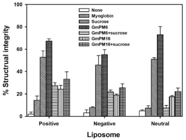
Here, we present the results of desiccation protection assay of two hydrophilic soybean LEA proteins, GmPM6 (LEA II, 23.7 kDa, pI value 6.1) (Soulages et al., 2003; Shih et al., 2010c) and GmPM16 (LEA IV, 14.7, pI value 9.7) (Shih et al., 2004) by monitoring the integrity of lipo-somes after desiccation. We discuss the possible role of the LEA proteins in desiccation and dry storage tolerance.
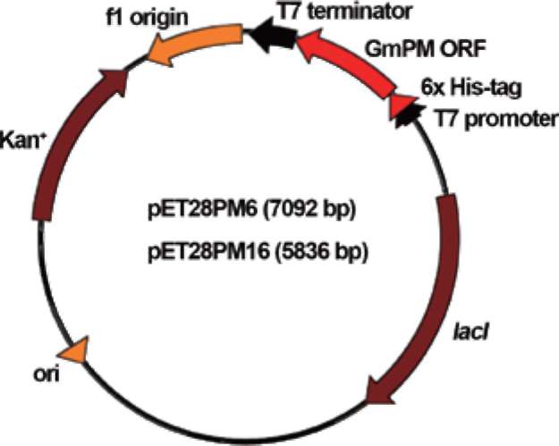
Figure 1. Scheme of construction of the GmPM6 and GmPM16 fusion plasmids.
1 min, with a final extension at 72°C for 5 min. The am­plified PCR products were separated and purified by 1% (w/v) agarose gel electrophoresis. Products were digested with the restriction enzymes Ndel and Xhol, then ligated into the pET28a T7 expression vector (Novagen), which had been pre-cut with the same enzymes (Figure 1). The recombinant plasmids were introduced into E. coli cells, and the expression of the recombinant genes was enhanced by 1 mM IPTG induction. Recombinant proteins were purified by an immobilized-metal affinity column as per the manufacturer's protocol (pET system manual ver. 5.0, Novagen, Germany). Purified recombinant proteins were examined by one-dimensional 12.5% SDS-polyacrylamide gel electrophoresis, followed by Coomassie blue staining.
MATERIALS AND METHODS
Liposome preparation
Bacterial strains and media
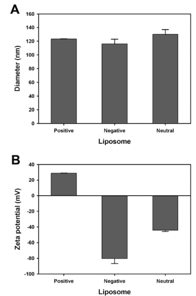
Myoglobin (17.2 kDa, pI value 7.1), 5(6)-carboxy-fluorescein (CF), and sucrose were from Sigma (USA). 1-palmitoyl-2-oleoyl-3-sn-phosphatidylcholine (POPC, 760 kDa) was from Avanti Polar Lipids (USA). Liposomes containing CF in Tris buffer were prepared by reverse-phase evaporation (Szoka and Papahadjopoulos, 1978). Five micrograms of POPC was dissolved in organic sol­vent into which 5 mol% dicetyl phosphate or 5 mol% stearylamine was added to give an overall negative or pos­itive charge. Neutral liposomes were made from POPC. The solvent was removed under reduced pressure at 35°C on a rotary evaporator. The dried lipids were dissolved in 1 ml of 1 mM Tris, pH 8.0, containing 150 mM CF. The tube was sonicated in a bath-type sonicator for 10 min at 30°C until a uniform emulsion formed. Liposome suspensions (multi-lamellar vesicles) were sonicated by use of a probe-type sonicator. Sonication was carried out intermittently at 5°C for 5 min. After sonication, unincorporated mate-rial was removed by passing the large unilamellar vesicles through a Sephadex G-50 column equilibrated with 1 mM Tris, pH 8.0, to stabilize the LEA proteins.
Escherichia coli strains XL1-Blue and BL21(DE3) were used as the host. XLl-Blue was used for cloning plasmids: and BL21(DE3) was used for expressing recombinant pro­teins and growth analysis. All cultures were grown in LB-medium in the presence of 50 fig/ml kanamycin at 37°C.
Recombinant plasmid construction, expression, and purification of its products
Recombinant DNA techniques were performed es-sentially as described (Sambrook and Russell, 2001). The NdeI-XhoI fragment of GmPM6 or GmPM16 containing the coding region was amplified by specific forward and reverse primers (GmPM6: 5'-ggttgacgCATATGgcaacgt-tg-3' and 5'-catgcatg-
CTCGAGgcacgatgc-3'; GmPM16: 5'-gaagaaca-
CATATGcaatcctc-3' and 5'-gacgtactCTCGA-Gaaacacag-3')
from maintenance plasmids harboring the full-length cDNA of the GmPM clones. The PCR protocol was an initial denaturation at 94°C for 5 min, followed by 30 cycles at 94°C for 45 sec, 55°C for 30 sec and 72°C for
YANG et al. ― Two soybean GmPM proteins reduce liposome leakage during desiccation
467
Determination of particle-size distribution and zeta potential of vesicles
Analysis of liposome membrane integrity after desiccation
Mean vesicle sizes of the liposome were measured at 25°C using a Zetasizer 3000HS apparatus (Malvern Instruments, UK). Red light at 632 nm was used as the light source. Particles were measured by photon correla­tion spectroscopy and were in constant random thermal or Brownian motion, which caused the intensity of light scattered from the particles to form a moving speckle pat­tern. Large particles moved more slowly than did small particles, so their scattered light fluctuation rate was also slower. Photon correlation spectroscopy was used to mea­sure the rate of change of these light fluctuations to deter­mine the size distribution of the light scattering particles. The zeta potential of the liposomes was determined from electrophoretic mobility measurements at 25°C. At some distance from the surface, the "shear plane," the ions were no longer dragged along with a moving particle but re­mained in the bulk solution. The potential at this distance was, by definition, the zeta potential Z. A Zetasizer and la­ser Doppler electrophoresis were used to measure particle movement when they were placed in an electric field. The measurement determined the charges on the particles.
Liposomes with different zeta potential and encapsulat­ing the fluorescent dye CF were diluted into Tris buffer containing various concentrations of protein and subjected to desiccation before rehydration. Eight-microliter drop­lets containing liposome and sugar or protein (ratio 1:10) were dried in a stream of dry nitrogen gas for 3 hr. One milliliter of Tris buffer (1 mM) was then added to the dry liposome preparation. CF fluorescence was determined us­ing a spectrofluorometer (excitation wavelength 490 nm, emission wavelength 515 nm). Membrane integrity after desiccation was calculated from the fluorescence of the sample relative to a non-desiccated control sample and the total CF released from the sample after the addition of Tri­ton X-100 to 0.1%. Liposome experiments were repeated 3 times with different preparations. Data are presented as mean±S.D.
RESULTS
Particle size and zeta potential of liposomes
Figure 2A indicates that the three kinds of liposomes prepared by the reverse-phase evaporation method had similar particle size, about 120 nm. Negative and positive liposomes clearly showed negative or positive zeta poten­tial (Figure 2B). The neutral liposome with negative zeta potential may be due to measurement under 1 mM of Tris buffer, which may also cause underestimation of the posi­tive charge of positive vesicles.
Maintenance of liposome membrane integrity after desiccation and rehydration
Figure 3 illustrates that the liposome membrane integ­rity after desiccation and rehydration could be maintained at different levels while being incubated with GmPM6 or GmPM16. Structural integrity was assessed by determin­ing the release of the encapsulated fluorescent dye CF, with total release equivalent to complete loss of integrity. Three types of liposomes desiccated alone showed loss of membrane integrity with rehydration, whereas the li-posomes desiccated in the presence of GmPM6 proteins maintained up to 67%, 52%, and 72% of membrane in­tegrity for stearylamine (positive liposome), dicetyl phos­phate (negative liposome) and POPC (neutral liposome), respectively. The liposomes desiccated in the presence of GmPM16 proteins maintained up to 24%, 19%, and 18% of membrane integrity of positive, negative and neutral li-posomes, respectively. Myoglobin was chosen as a control protein because of its similar molecular size to GmPM6 and pGmPM16 (see MATERIALS AND METHODS) and its having no known function in membrane protection. The protection assay indicated that percentage membrane integrity on co-incubation with myoglobin was 14%, 8%, and 7% for positive, negative and neutral liposomes, re­spectively. Sucrose was also chosen as a control because non-reducing sugars had been proposed to protect mem-
Figure 2. Properties of phospholipid intermediate unilamellar vesicle. A, mean vesicle size; B, zeta potential.
468
Botanical Studies, Vol. 52, 2011
of sugars may substitute for water molecules and provide the required hydrophilic interactions for membrane stabili­zation (Crowe et al., 1995). In the present study, GmPM6 and GmPM16 proteins were used to test the function of membrane stabilization during dehydration. Both pro­teins contained similar molecular mass and shared similar amino acid contents with high amounts of charged amino acid residues (Shih et al., 2008). However, they showed very different results: GmPM6 proteins strongly prevented liposome leakage after drying, whereas GmPM16 only had a weak effect (Figure 3). LEA II proteins were proposed to stabilize plasma membranes during osmotic stresses. A lo­calization study revealed that freezing-induced WCS120, a wheat LEA II protein, accumulated near the plasma mem­brane but did not integrate into the lipid bilayer (Danyluk et al., 1998). However, LEA IV proteins accumulated mainly in the cytosome, and no evidence indicates that the proteins accumulate near or inside membrane (Hsing, unpublished data). Therefore, GmPM6 proteins might function as membrane stabilizers. Although the protective effect of sucrose on the liposome results in the depression of Tm, the degree of depression of Tm for various protective materials disagrees with the protective order we found. For example, sucrose contained the lowest Tm (not datable until -50°C, Hoekstra et al., 2001) to natural liposome, whereas GmPM6 and GmPM16 slightly lowered the Tm values from 56°C to about 45°C (Shih et al., 2010c; Shih and Hoekstra, unpublished data). The order of protective effect in the current study was GmPM6, sucrose, and GmPM16. No Tm value for sucrose, GmPM6, or GmPM16-positive liposome matrix could be detected until -50°C. However, the order of protective effect was still GmPM6, sucrose, and GmPM16. Thus, the protective effect of LEA proteins on phospholipids might not be caused solely by the inter­action between hydroxyl groups of proteins and hydro-philic heads of phospholipids, although such interaction indeed existed.
The results of the liposomes and protein-sucrose glasses interaction (Figure 3) illustrate the different is­sues in glass function. Our results indicate that protein-sugar glasses have little or no protective addictivity. The protective effect of GmPM6-sucrose glasses was markedly less than that of GmPM6 or sucrose alone. However, the LEA protein-sugar glass stabilized protein structures under osmotic stress conditions. For instance, kinetic analysis of protein aggregation suggested that the glass forming from Aphelenchus avenae LEA III proteins and trehalose efficiently prevented the heat- or dehydration-induced ag­gregation of citrate synthase or lactate dehydrogenase, and the glass also revealed significant addictivity (Goyal et al., 2005). These dehydration-sensitive enzymes were embed­ded into glass, and thus the combined effects of LEA pro­teins and non-reducing sugars could be detected. However, in the present study, even if the liposomes were possibly embedded into glass, the interaction between liposomes and glass did not prevent the formation of a solid gel phase during dehydration. The current model suggested that non-reducing sugars could integrate into hydrophilic heads
Figure 3. Effect of the presence of various concentrations of myoglobin, sucrose, GmPM6, GmPM6-sucrose, GmPM16, and GmPM 16-sucrose glass on the structural integrity of positive, negative, and neutral liposomes after desiccation.
brane during dehydration (Hoekstra et al., 2001). Percent­age membrane integrity with sucrose was 53%, 46%, and 51% for positive, negative and neutral liposomes, respec­tively. Hence, GmPM6 is more efficient in protecting membrane integrity during dehydration than other proteins or sucrose.
We then investigated whether the combination of sugar and LEA proteins could improve the protection of lipo-somes against desiccation. Three kinds of liposomes en­capsulating CF containing various concentrations of LEA proteins, sucrose, and combinations of sugar and protein (in 1:1 mass ratio) were desiccated. Compared with the protective efficiency of GmPM6 alone, the efficiency of the GmPM6-sucrose matrix was significantly decreased. The membrane integrity percentage decreased to 27%, 22%, and 8% for positive, negative and neutral liposomes, respectively. By contrast, the protective efficiency of the GmPM16-sucrose matrix was slightly increased: 33%, 25%, and 22% for positive, negative and neutral lipo-somes, respectively. The protective efficiency of sucrose alone for liposomes was at least two-fold that of GmPM protein-sucrose matrixes.
DISCUSSION
Using liposomes as a model membrane system, Crowe et al. (1988) proposed that adjacent lipid bilayers in li-posomes are held apart by the water associated with the phosphate head group. Removal and replacement of these water molecules may alter the bilayer configuration. With the loss of water molecules, hydrogen bonding to each polar head group and the lateral spacing between the phos-pholipids is reduced. This situation leads to increased van der Waals interactions between the hydrocarbon chains of the phospholipid molecules and thus the formation of a solid "gel" phase and an increase in the phase transition temperature (Hoekstra et al., 1989). The hydroxyl groups
YANG et al. ― Two soybean GmPM proteins reduce liposome leakage during desiccation
469
of plasma membrane, and then maintain the liquid-crys­talline phase (Crowe et al., 1992; Hoekstra et al., 2001). However, at the first hour after imbibition, the seeds still contain approximately 10% leakage of cytoplasmic sol­utes (McKersie and Stinson, 1980). The removal of the seed coat greatly increases the leakage to more than five­fold (Duke and Kakefuda, 1981). Of note, the detectable
leakage of cytosol-organelle and mitochondrial marker enzymes suggests that the plasma membrane is damaged and loses semi-permeability at seed maturation (McKersie and Stinson, 1980). The membrane leakage might not lead to cell death; the instant repair system should be critical. Numerous enzymes and transcription factors for early ger­mination are synthesized during maturation and stored in mature seed. These proteins are desiccation-sensitive and should be protected because they have to function immedi­ately during early imbibition. Hence, from our results, the cellular glass may function ahead of the protein or cellular structure protection, whereas the rest of the non-reducing sugars may provide protection for plasma membranes. Further analysis is ongoing.
and E.A. Bray (ed.), Plant responses to cellular dehydra­tion during environmental stress. American Society of Plant Physiologists. Vol. 10. The National Academies Press, Washington, DC, pp. 91-103.
Dure, L., M. Crouch, J. Harada, T.H.D. Ho, J. Mundy, R. Qua-trano, T. Thomas, and Z.R. Sung. 1989. Common amino acid sequence domains among the LEA proteins of higher plants. Plant Mol. Biol. 12: 475-486.
Goyal, K., L.J. Walton, and A. Tunnacliffe. 2005. LEA proteins prevent protein aggregation due to water stress. Biochem. J. 388: 151-157.
Grelet, J., A. Benamar, E. Teyssier, M.H. Avelange-Macherel, D. Grunwald, and D. Macherel. 2005. Identification in pea seed mitochondria of a late-embryogenesis abundant protein able to protect enzymes from drying. Plant Physiol. 137:
157-167.
Hoekstra, F.A., E.A. Golovina, and J. Buitink. 2001. Mecha­nisms of plant desiccation tolerance. Trends Plant Sci. 6: 431-438.
Hoekstra, F.A., J.H. Crowe, and L.M. Crowe. 1989. Membrane behavior in drought and its physiological significance. In R.B. Taylorson (ed.), Plenum Press, New York, pp. 71-88.
Honjoh, K.I., H. Matsumoto, H. Shimizu, K. Ooyama, K. Tana-ka, Y. Oda, R. Takata, T. Joh, K. Suga, T. Miyamoto, M. Iio, and S. Hatano. 2000. Cryoprotective activities of group 3 late embryogenesis abundant proteins from Chlorella vul­garis C-27. Biosci. Biotechnol. Biochem. 64: 1656-1663.
Hsing, Y.C., R.W. Rinne, A.G. Hepburn, and R.E. Zielinski.
1990. Expression of maturation-specific genes in soybean seeds. Crop Sci. 30: 1343-1350.
Hsing, Y.C., Z.Y. Chen, M.D. Shih, J.S. Hsieh, and T.Y. Chow.
1995. Unusual sequences of group 3 LEA mRNA inducible by maturation or drying in soybean seeds. Plant Mol. Biol. 29: 863-868.
Hsing, Y.I.C. and S. Wu. 1992. Cloning and characterization of cDNA clones encoding soybean seed maturation polypep-tides. Bot. Bull. Acad. Sin. 33: 191-199.
Imai, R., L. Chang, A. Ohta, E.A. Bray, and M. Takagi. 1996. A lea-class gene of tomato confers salt and freezing tolerance when expressed in Saccharomyces cerevisiae. Gene 170: 243-248.
Kazuoka, T. and K. Oeda. 1994. Purification and characteriza­tion of COR85-oligomeric complex from cold-acclimated spinach. Plant Cell Physiol. 35: 601-611.
Koster, K.L. and A.C. Leopold. 1988. Sugars and Desiccation
Tolerance in Seeds. Plant Physiol. 88: 829-832.
Lan, Y., D. Cai, and Y.Z. Zheng. 2005. Expression in Escherichia coli of three different soybean late embryogenesis abundant (LEA) genes to investigate enhanced stress tolerance. J. Int.
Plant Biol. 47: 613-621.
Lee, P.F., Y.I.C. Hsing, and T.Y. Chow. 2000. Promoter activ­ity of a soybean gene encoding a seed maturation protein,
GmPM9. Bot. Bull. Acad. Sin. 41: 175-182. McKersie, B.D. and R.H. Stinson. 1980. Effect of Dehydration
on Leakage and Membrane Structure in Lotus corniculatus
Acknowledgements. We thank L.-H. Wu for technical support, and Laura Smales for English editing. This work was supported by the National Science Council, Taiwan, ROC, to T.P. Lin and Y.I.C. Hsing.
LITERATURE CITED
Blackman, S.A., R.L. Obendorf, and A.C. Leopold. 1992. Matu­ration proteins and sugars in desiccation tolerance of devel­oping soybean seeds. Plant Physiol. 100: 225-230.
Borrell, A., M.C. Cutanda, V Lumbreras, J. Pujal, A. Goday, F.A. Culianez-Macia, and M. Pages. 2002. Arabidopsis thaliana atrab28: a nuclear targeted protein related to germination and toxic cation tolerance. Plant Mol. Biol. 50: 249-259.
Crowe, J.H., A.E. Oliver, F.A. Hoekstra, and L.M. Crowe. 1997. Stabilization of dry membranes by mixtures of hydroxyeth-yl starch and glucose: the role of vitrification. Cryobiology
35: 20-30.
Crowe, J.H., F.A. Hoekstra, and L.M. Crowe. 1992. Anhydro-biosis. Annu. Rev. Physiol. 54: 579-599.
Crowe, J.H., L.M. Crowe, J.F. Carpenter, A.S. Rudolph, C.A. Wistrom, B.J. Spargo, and T.J. Anchordoguy. 1988. Interac­tions of sugars with membranes. Biochim. Biophys. Acta 947: 367-384.
Crowe, L.M. and J.H. Crowe. 1995. Freeze-dried liposomes. In P. Puisieux (ed.), Editions de Sante, Paris, pp. 237-272.
Danyluk, J., A. Perron, M. Houde, A. Limin, B. Fowler, N. Ben-hamou, and F. Sarhan. 1998. Accumulation of an acidic de-hydrin in the vicinity of the plasma membrane during cold acclimation of wheat. Plant Cell 10: 623-638.
Duke, S.H. and G. Kakefuda. 1981. Role of the testa in prevent­ing cellular rupture during imbibition of legume seeds. Plant Physiol. 67: 449-456.
Dure III, L. 1993. Structural motifs in Lea proteins. In T.J. Close
470
Botanical Studies, Vol. 52, 2011
L. Seeds. Plant Physiol. 66: 316-320.
Nakayama, K., K. Okawa, T. Kakizaki, and T. Inaba. 2008. Eval­uation of the protective activities of a late embryogenesis abundant (LEA) related protein, Cor15am, during various stresses in vitro. Biosci. Biotechnol. Biochem. 72: 1642­1645.
NDong, C., J. Danyluk, K.E. Wilson, T. Pocock, N.P. Huner,
and F. Sarhan. 2002. Cold-regulated cereal chloroplast late embryogenesis abundant-like proteins. Molecular character­ization and functional analyses. Plant Physiol. 129: 1368­1381.
Rosenberg, L.A. and R.W. Rinne. 1986. Moisture loss as a pre­requisite for seedling growth in soybean seeds (Glycine max
L. Merr). J. Exp. Bot. 37: 1663-1674.
Sambrook, J. and D.W. Russell. 2001. Molecular cloning: A laboratory manual. 3rd Ed. Cold Spring Harbor Laboratory Press, New York.
Shih, M.D., F.A. Hoekstra, and Y.I.C. Hsing. 2008. Late embryo-
genesis abundant proteins. Adv. Bot. Res. 48: 211-255.
Shih, M.D., L.T. Huang, F.J. Wei, M.T. Wu, F.A. Hoekstra, and
Y.I.C. Hisng. 2010a. OsLEA1a, a new Em-like protein of cereal plants. Plant Cell Physiol. in press.
Shih, M.D., M.H. Kao, S.C. Lin, T.F. Shu, K.L. Hsieh, M.C.
Chung, T.H. Tsou, Y.I.C. Hsing, and J.S. Hsieh. 2010b. Tis­sue- and cellular localization of soybean (Glycine max L.) seed maturation protein transcripts. Bot. Stud. 51: 183-194.
Shih, M.D., S.C. Lin, J.S. Hsieh, C.H. Tsou, T.Y. Chow, T.P. Lin,
and Y.I. Hsing. 2004. Gene cloning and characterization of a soybean (Glycine max L.) LEA protein, GmPM16. Plant
Mol. Biol. 56: 689-703. Shih, M.D., T.Y. Hsieh, T.P. Lin, Y.I. Hsing, and F.A. Hoekstra.
2010c. Characterization of two soybean (Glycine max L.) LEA IV proteins by circular dichroism and Fourier trans-
form infrared spectrometry. Plant Cell Physiol. 51: 395-407.
Soulages, J.L., K. Kim, E.L. Arrese, C. Walters, and J.C. Cush-man. 2003. Conformation of a group 2 late embryogenesis abundant protein from soybean. Evidence of poly (l-proline)-type II structure. Plant Physiol. 131: 963-975.
Swire-Clark, G.A. and W.R. Marcotte, Jr. 1999. The wheat LEA protein Em functions as an osmoprotective molecule in Sac-charomyces cerevisiae. Plant Mol. Biol. 39: 117-128.
Szoka, F. Jr. and D. Papahadjopoulos. 1978. Procedure for preparation of liposomes with large internal aqueous space and high capture by reverse-phase evaporation. Proc. Natl.
Acad. Sci. USA 75: 4194-4198. Tsai, Y.C., M.T. Wu, J.S. Hsieh, Y.I.C. Hsing, and M.D. Shih.
2008. Expression of glycine max physiologically mature genes in soybean (Glycine max L.) tissues. J. Genet Mol
Biol. 19: 168-181.
Tunnacliffe, A. and M.J. Wise. 2007. The continuing conundrum of the LEA proteins. Naturwissenschaften. 94: 791-812.
Williams, R.J. and A.C. Leopold. 1989. The glassy state in corn
embryos. Plant Physiol. 89: 977-981. Wolkers, W.F., S. McCready, W.F. Brandt, G.G. Lindsey, and
F.A. Hoekstra. 2001. Isolation and characterization of a D-7 LEA protein from pollen that stabilizes glasses in vitro.
Biochim. Biophys. Acta 1544: 196-206.
Xu, D., X. Duan, B. Wang, B. Hong, T. Ho, and R. Wu. 1996. Expression of a late embryogenesis abundant protein gene, HVA1, from barley confers tolerance to water deficit and salt stress in transgenic rice. Plant Physiol. 110: 249-257.
Zhang, L., A. Ohta, M. Takagi, and R. Imai. 2000. Expression of plant group 2 and group 3 lea genes in Saccharomyces cer-evisiae revealed functional divergence among LEA proteins.
J. Biochem. 127: 611-616.
兩種大豆GmPM蛋白能在乾燥情況下降低微脂體的滲漏
楊淑怡1,2 施明德2 林讚標1 邢禹依2
1國立台灣大學植物科學研究所
2中央研究院植物暨微生物學研究所
本研究係針對大豆種子的晚胚蛋白在乾燥的環境下,對人造微脂體的保護效果。同時,我們也以
由晚胚蛋白與蔗糖所組成的玻璃質體,進行微脂體保護效果的研究。結果顯示,屬於第二群晚胚蛋白的
GmPM6對於乾燥的微脂體具有明顯的保護效果,但第四群的晚胚蛋白GmPM16則較低。另一方面,無
GmPM6GmPM16與蔗糖所組成的玻璃質體皆無法對乾燥的微脂體產生足夠的保護能力。因此,
雖然晚胚蛋白與微脂體之間能夠產生靜電吸引力與氫鍵,但維持微脂體的完整性可能是由於其它的作用
力。此外,關於成熟的正儲型種子的細胞玻離質體的功能也加以討論
關鍵詞:脫水;玻璃質體;GmPM ;晚胚蛋白;微脂體大豆。