Botanical Studies (2012) 53: 9-17.
MOLECULAR BIOLOGY
Lemon ascorbate peroxidase: cDNA cloning and biochemical characterization
Ya-Han DAI1,3,4, Chich-Yu HUANG1,4, Lisa WEN2,4, Dey-Chyi SHEU3, and Chi-Tsai LIN1*
1Institute of Bioscience and Biotechnology and Center for Marine Bioenvironment and Biotechnology, National TaiwanOcean University, Keelung 202, Taiwan
2Department of Chemistry, Western Illinois University, 1 University Circle, Macomb, IL 61455-1390 , USA
3Department of Bioengineering, Tatung University, Taipei 104, Taiwan
(Received October 27, 2010; Accepted June 24, 2011)
ABSTRACT. Ascorbate peroxidase (Apx) plays important roles both as a reductant and as a H2O2 scavenger via ascorbate (AsA). In this paper, we discuss how a ClApx cDNA (1,068 bp, GQ465430) encoding a putative Apx was cloned from lemon (Citrus limon). The deduced amino acid sequence is similar to the Apxes from other plant species. A 3-D structural model of ClApx was constructed based on the crystal structure of Pisum sativum Apx (PDB code 1APX). To characterize the ClApx protein, the coding region was subcloned into an expression vector pYEX-S1 and transformed into Saccharomycgs ce~revisiae. The recombinant His6-tagged ClApx was overexpressed and purified by Ni2+-nitrilotriacetic acid Sepharose. The purified enzyme showed two prominent bands on 15% sodium dodecyl sulfate-polyacrylamide gel electrophoresis. The Michaelis con­stant (KM) values of the recombinant enzyme for AsA and H2O2 were 0.40 and 0.11 mM, respectively. The enzyme was active from pH range 6 to 8. The thermal inactivation of the enzyme showed a half-life of 6.5 min at 45°C, and its inactivation rate constant Kwas 1.1 x 10⁻¹ min-1. The enzyme retained 35% activity after chymotrypsin digestion at pH 8 and 37°C for 40 min.
Keywords: Ascorbate peroxidase; Citrus limon; Saccharom^yces cerevisiae; Three-dimension structural model.
Abbreviations: AsA, ascorbate; Apx, ascorbate peroxidase; IPTG, isopropyl p-D-thiogalactopyranoside; SDS-PAGE, sodium dodecyl sulfate-polyacrylamide gel electrophoresis; MDHA, monodehydroascorbate; PBS, phosphate buffer saline.
INTRODUCTION
crucial component of receptor signaling that serves as an intracellular messenger (Rhee, 1999; Thannickal and Fanburg, 2000; Wahid et al., 2007; Chen et al., 2009). Receptor-mediated production of H2O2 has been studied mostly in phagocytic leukocytes (Babior, 1999). In these cells, one electron reduction of O2 by a multicomponent NADPH oxidase generates superoxides that are then en-zymatically converted to H2O2. Recently, the intracellular generation of superoxides and H2O2 has also been detected in various nonphagocytic cells (Lambeth, 2004). Timely elimination of messengers after completion of their func-tions has been shown to be critical for cellular signaling (Rhee et al., 2005). H2O2 may be eliminated by catalase, glutathione peroxidase, and peroxiredoxin. The Apxes can scavenge H2O2 by oxidizing the abundant AsA in plant cells (Nakano and Asada, 1981). Crystal structures of two known Apxes, PsApx (Pisum sativum, PDB code 1APX; Patterson and Poulos, 1995) and GmApx (Glycine max, PDB code 1OAG; Sharp et al., 2003) have been deter-mined. Thus, Apxes play important roles in eliminating/ detoxifying peroxides. Our aim is to study various lemon enzymes involved in modulating H2O2. Here, we report the cloning of an Apx (designated as ClApx) from lemon. The
The antioxidant properties of ascorbate (AsA) are a major focus in both plant and animal metabolism research. AsA can protect plants and mammals against oxidative stress. In most cases, monodehydroascorbate (MDHA) is the primary oxidation product of AsA. In plants, the reac­tion catalyzed by AsA peroxidase (Apx) is a major source of MDHA, which scavenges hydrogen peroxide (H2O2) (Hossain et al., 1984; Barrows and Poulos, 2005; Lu et al., 2009), and thus aids catalase (Ken et al., 2008) and perox-iredoxin (Wen et al., 2007; Liau et al., 2010) in modulat­ing hydrogen peroxide levels. Some people use lemons to eliminate excess melanin in their skin. The cosmetic effect may be attributed to AsA and superoxide dismutase (SOD), among others found in lemon.
H2O2 has been recognized as a cytotoxic agent and a

4Footnote: The first and the second authors contributed equally to this paper.
*Corresponding author: E-mail: B0220@mail.ntou.edu.tw; Phone: 886-2-24622192 ext. 5513; Fax: 886-2-24622320.
10
Botanical Studies, Vol. 53, 2012
coding region of the ClApx cDNA was introduced into a yeast expression system and the active enzyme was puri­fied and characterized.
sequences, a phylogenetic analysis was performed using Phylip-3.69 program (http://evolution.genetics.washing-
ton.edu/phylip.html) via the Maximum Likelihood (ML) phylogenetic tree.
MATERIALS AND METHODS
ClApx cDNA subcloning into an expression vector
Total RNA preparation from lemon and cDNA synthesis
The coding region of the ClApx cDNA was amplified using gene specific flanking primers. The 5' upstream primer contains the EcoRI recognition site (5' GAA TTC GAT GAC GAA GAA TTA CCC CAC 3') and the 3' downstream primer contains the XhoI recognition site (5'CTCGAG GGC TTC AGC AAA TCC TAG CT 3'). Us-ing 0.2 fig of 5'-RACE-ready cDNA as a template, and 10 pmole of each 5' upstream and 3' downstream primers, a 0.75 kb fragment was amplified by PCR. The fragment was ligated into pCR4 and transformed into E. coli. The recombinant plasmid was isolated and digested with EcoRI and XhoI after which the digestion products were sepa-rated on a 1% agarose gel. The 0.75 kb insert DNA was gel purified and subcloned into the EcoRI and Xhol sites of pET-20b(+) expression vector (Novagen). The recombi-nant DNA (pET-20b(+)-ClApx) was then transformed into E. coli C41(DE3). The recombinant protein was not over-expressed in the E. coli expression system. We decided to subclone the gene into a yeast expression system. The cod-ing region of the ClApx cDNA was re-amplified by using two gene-specific primers: the 5' upstream primer was the same whereas the 3' downstream primer contained a His6-tag and EcoRI recognition site (5' CGT CTC GAA TTC TCA GTG GTG GTG GTG GTG GTG 3'). Using the 0.1 fig recombinant DNA of pET-20b(+)-ClApx as a template, and 10 pmole of each 5' upstream and 3' downstream primers, a 0.75 kb fragment was amplified by PCR. The fragment was ligated into pCR4 and transformed into E. coli. The recombinant plasmid was isolated and digested with EcoRI and the digestion products were separated on an 0.8% agarose gel. The 0.75 kb insert DNA was gel pu-rified and subcloned into the EcoRI site of the pYEX-S1 expression vector (Clontech) and introduced into Saccha-romyces cerevisiae (trp- ura-). The transformed yeast cells were selected by YNBDT (0.17% yeast nitrogen base, 0.5% ammonium sulfate, and 2% glucose) agar plates containing 20 [ig Trp/mL. The presence of ClApx cDNA in the selected transformants was verified by PCR using gene-specific flanking primers. The recombinant ClApx protein was expressed in yeast in YPD medium (1% yeast extract, 2% peptone, 2% glucose). Active recombinant ClApx production was shown using an enzyme assay.
A fresh unpeeled lemon (2.3 g) was obtained from a lo­cal market, frozen in liquid nitrogen, and ground to powder in a ceramic mortar. PolyA mRNA (30 μg) was prepared using Straight A's mRNA Isolation System (Novagen, USA). Four fig of the mRNA were used in the 5'-RACE-Ready cDNA and 3'-RACE-Ready cDNA syntheses with Clontech's SMART RACE cDNA Amplification Kit.
Apx cDNA isolation
A 0.65 kb fragment was amplified by PCR using the lemon 3'-RACE-Ready cDNA as a template and a UPM (universal primer A mix, purchased from BD Biosciences) primer & degenerate primer (5' GAR GGY CGT CTT CCT GAT GC 3'), The degenerate primer was designed based on the conserved Apx sequences from CpApx (Car-ica papaya, EF512304), ZmApx (Zea mays, FJ890983), PpApx (Pinus pinaster, AY485994), AtApx (Arabidop-sis thaliana, D14442), OsApx (Oryza sativa, D45423), CaApx (Capsicum annuum, AF442387), and AmApx (Avicennia marina, EU025130). The 0.65 kb fragment was subcloned and sequenced. Based on this DNA sequence, we synthesized an Apx-3R primer (5' CCA TCC TTC TCA CCA GTC 3'). The primer allowed sequence exten­sion from the 5' end of the 0.65 kb fragment. A PCR was carried out using 0.2 fig of the 5'-RACE-Ready cDNA as a template. The primer pairs were UPM and Apx-3R primer. A 0.7 kb fragment (5'-RACE; 5'-DNA end) was amplified. The 0.7 kb DNA fragment was subcloned into pCR4 vec­tor and transformed into Escherichia coli TOPO10. The nucleotide sequence of this insert was determined in both strands. Sequence analysis revealed that the combined sequences covered an open reading frame of a putative ClApx cDNA (1068 bp, GQ465430).
ClApx sequence bioinformatics analysis
The identity of the ClApx cDNA clone was verified by comparing the DNA sequence and the inferred amino acid sequence with various data banks using the basic lo­cal alignment search tool (BLAST). Multiple alignments were constructed using the ClustalW2 program. Protein secondary structure was predicted by the SWISS-MODEL program and represented as a helices and p strands. A 3-D structural model of ClApx was constructed by SWISS-MODEL (Arnold et al., 2006) (http://swissmodel.expasy.org/SWISS-MODEL.html) based on the known crystal structure of PsApx (Pisum sativum, PDB ID: 1APX). The model was then superimposed onto GmApx (Glycine max, PDB ID: 1OAG) via the SPDBV_4 program. To study the ClApx evolutionary relationships among plant Apx
Recombinant ClApx expression and purification
The transformed yeast containing the ClApx was grown at 30°C in 250 mL of YPD medium for 5 days. The cells were harvested and soluble proteins extracted in PBS with glass beads as previously described (Huang et al., 2010a, b). The recombinant ClApx was purified by Ni-NTA affinity chromatography (elution buffer: 30% PBS
DAI et al. ― Lemon ascorbate peroxidase
11
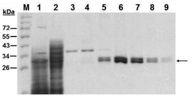
containing 100 mM imidazole) according to the manu­facture's instruction (Qiagen). The purified protein was checked by 15% SDS-PAGE. Proteins on the gel were detected by Coomassie Brilliant Blue R-250 staining. Protein concentration was determined using a Bio-Rad Protein Assay Kit (Richmond, CA) using bovine serum albumin as a standard.
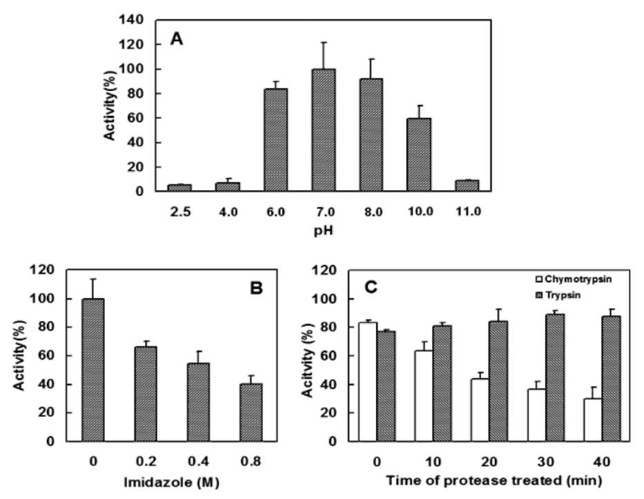
added to the enzyme sample (0.2 [g/2 [L enzyme in 3% PBS containing 5% glycerol per reaction) to the final lev­els (each final volume is 20 iL) of 0.2, 0.4, or 0.8 M and incubated at 37°C for 1 h. (4) Proteolytic susceptibility. The enzyme (1.0 [g/10 [L in 1.5 % PBS containing 2.5% glycerol, additional 10 mM CaCl2 for chymotrypsin) was incubated with one-tenth of trypsin or chymotrypsin (w/w) at pH 8.0, 37°C for a period of 5, 10, 20 or 40 min. Ali-quots (0.2 [g/2 [L) were removed at various time intervals for analysis. After each treatment, the residue Apx activity was tested as described above.
Electrospray ionization quadrupole-time-of-flight (ESI Q-TOF) molecular mass analysis
The purified recombinant ClApx (0.1 mg/mL) was dissolved in 3% PBS containing 0.05 mM imidazole and 0.5% glycerol. The sample (5 [L) was used for molecular mass determination using an ESI Q-TOF mass spectrom­eter (Micromass, Manchester, England).
Apx activity assay and kinetic studies
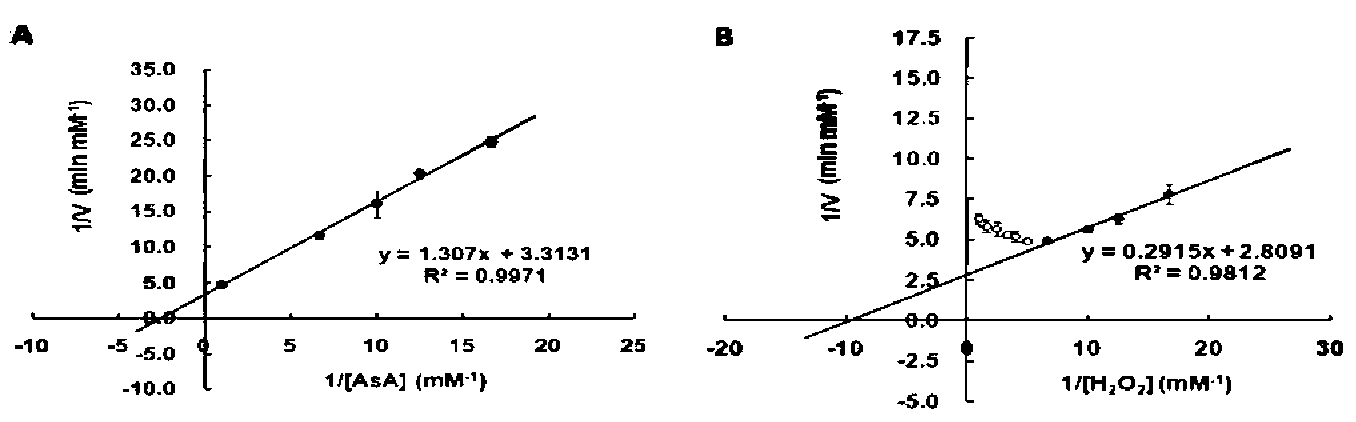
Apx activity was determined by measuring AsA oxi­dation (Nakano and Asada, 1981). The reaction mixture (100 [L) contained 20 mM potassium phosphate (pH 7.4), 1 mM H2O2, 1 mM AsA and 0.2 [g ClApx. The reaction started upon the addition of ClApx. The reaction was fol­lowed by a decrease in A290 due to AsA oxidation.
ClApx (0.2 [g) kinetic properties were determined by varying the concentrations of AsA (0.06 to 1.0 mM) with the fixed amount of 1 mM H2O2 or varying concentrations of H2O2 (0.06 to 1.0 mM) with the fixed amount of 1 mM AsA. The change in absorbance at 290 nm was recorded between 10 sec and 20 sec. The molar absorption coef­ficient of AsA at 290 nm was 2.8 mM-1 cm-1. The Km, Vmax and kcat were calculated from Lineweaver-Burk plots.
Biochemical characterization
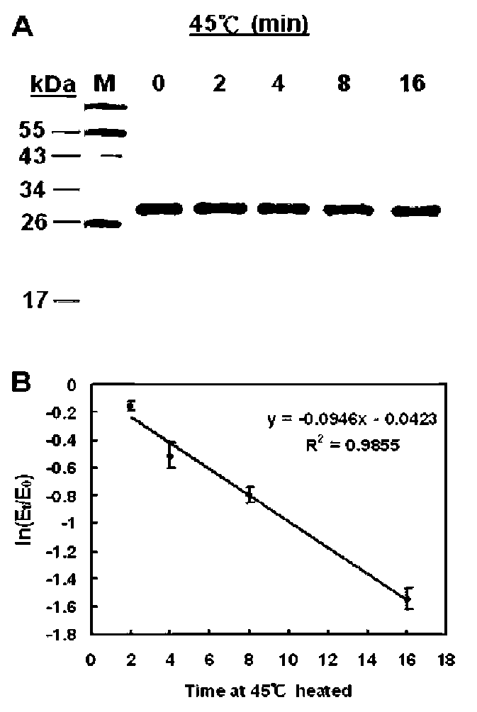
The stability of ClApx under various conditions was studied by assaying its peroxidase activity. Aliquots of the ClApx sample were tested for: (1) Thermal effect. Enzyme sample (0.2 [g/2 [L enzyme in 3% PBS containing 5% glycerol per reaction) was heated to 45°C for 2, 4, 8 or 16 min. (2) pH effect. Enzyme sample (0.2 [g/2 [L enzyme in 3% PBS containing 5% glycerol per reaction) was ad­justed to the desired pH by adding a volume of buffer with
different pHs: 0.2 M citrate buffer (pH 2.5, 4.0), 0.2 M
potassium phosphate buffer (pH 6.0, 7.0 or 8.0) or 0.2 M CAPS buffer (pH 10.4, or 11.0). Each sample was incu­bated at 37°C for 1 h. (3) Imidazole effect. During protein purification, the ClApx enzyme was eluted with imidazole and its effect on activity was examined. Imidazole was

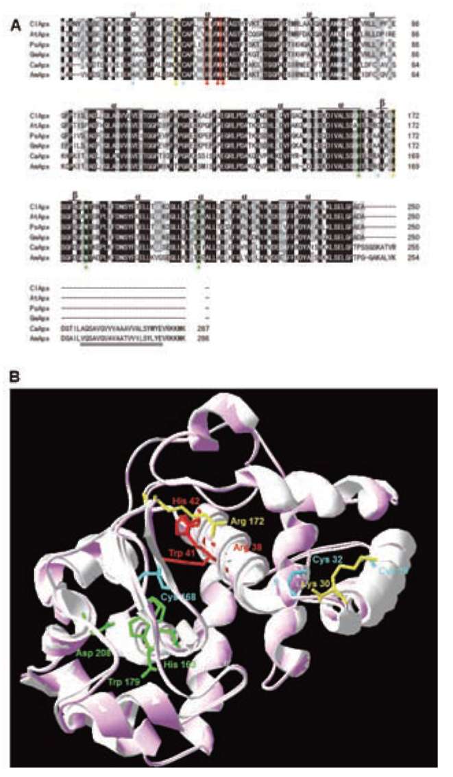
Figure 1. Alignment of the ClApx amino acid sequences with other organism's Apxes and 3-D structural model. (A) Sequence align­ment: ClApx (this study), AtApx (Arabidopsis thaliana), PsApx (Pisum sativumf), GmApx (Glycine max), CaApx (Capsicum annuum) and AmApx (Avicennia marina). Conservative replacements are shaded gray. Protein secondary structure was predicted by the SWISS-MODEL program and predicted a helices and p strands are indicated; (B) A 3-D structural model of ClApx was modeled based on the known X-ray structure of PsApx (Pisum sativum) via the SWISS-MODEL program and was superimposed to obtain a better structure via the SPDBV_4 program. Superimposition of ClApx (pink) and GmApx (Glycine max) (white) was shown using protein solid rib­bons. Blue stars denote the 3 Cys residues in the ClApx protein. The yellow triangles indicate the putative AsA binding sites. The red triangles indicate the putative active sites. The green triangles denote the conserved residues of the putative proximal hydrogen bonded triad. Double underline denote the C-terminal membrane-spanning segment belonging to cm1 type.
12
Botanical Studies, Vol. 53, 2012
RESULTS
with calculated molecular mass of 29 kDa (EMBL acces­sion no. GQ465430). Theoretical pI/Mw was 5.6/27570. Figure 1 shows the optimal alignment of the ClApx amino acid sequences with five related Apx sequences from other sources. ClApx is most similar (80%) to AtApx (Arabidop-sis thaliana, BAA03334), PsApx (Pisum sativum, P48534) and GmApx (Glycine max, Q43758), and 60% similar to CaApx (Capsicum annuum, AF442387) and AmApx (Avi-
Cloning and characterization of a cDNA encod­ing ClApx
A putative ClApx cDNA clone was identified based on its sequence homology to the published Apxes in the NCBI data bank. The coding region of ClApx cDNA was 750 bp that encodes a protein of 250 amino acid residues
Figure 2. Phylogenetic tree showing the relationships among plant Apxes. The sources of the ascorbate peroxidase sequences includ­ing species, abbreviations, accession numbers, enzyme types, and number of amino acids are shown in Table 1. The tree was made us­ing the Phylip-3.69 program (http://evolution.genetics.washington.edu/phylip.html) via Maximum Likelihood (ML) phylogenetic tree. The ML method is a statistical method and a p value is provided for each branch. The right branches of the tree, cht (thylakoid-bound), chs (stroma-bound), cm (cytosol membrane-bound)1, cm2, and cm3 ascorbate peroxidases, represent the evolution of these 14 plant species. The upper left branch of the tree, the cytosol-souble (csl) ascorbate peroxidases, represents the evolution of these 24 plant spe­cies. The lower left branch of the tree, the cytosol-souble (cs2) ascorbate peroxidases, represents the evolution of these 14 plant species including this study, Citrus limon (double underlines). ** denotes significantly different, P < 0.01.
Table 1. Sources of the ascorbate peroxidase sequences.
DAI et al. ― Lemon ascorbate peroxidase
13
cennia marina, EU025130). Like all other Apx sequences, the ClApx sequence contained no signal peptides and ap­peared to be cytosolic. We predicted the secondary struc­ture (Figure 1A, represented as a helices and p strands) and a 3-D structural model (Figure 1B, represented as sol­id ribbon) using SWISS-MODEL program. The 3-D struc­tural model was constructed based on the known crystal structure of PsApx (Pisum sativum). The model (pink) was superimposed onto GmApx (Glycine max) (white) via the SPDBV_4 program. Figure 1A and B show several color coded conserved residues presumably involved in various important functions. Blue stars denote the 3 Cys residues (Cys19, 32, 168) in the ClApx protein. The Cys32 is totally con­served and is located near the AsA binding site based on the 3-D structural model (Figure 1B). The yellow triangles denote the putative AsA binding sites (Lys30, Arg172) (Mac-donald et al., 2006). The red triangles denote the putative active sites (Arg38, Trp41, His42) (Efimov et al., 2007). The green triangles denote the conserved residues of the puta-
tive proximal hydrogen bonded triad (His163, Asp208, Trp179) (Barrows and Poulos, 2005), and the ClApx belongs to heme peroxidase. Its Trp179 in the proximal heme pocket may form the more traditional porphyrin d-cation radical (Barrows and Poulos, 2005). The heme propionates play a role in stabilization of porphyrin d-cation radicals. Using density functional theory, Guallar et al. (2003) reported that protecting the propionate groups by forming hydrogen bonds with the nearby residues is responsible for stabiliza­tion of a porphyrin d-cation radical.
Through sequence analysis, Jespersen et al. (1997) clas­sified plant Apxes (sequences from 20 species) into seven types based on differences in their cellular locations and their structural characteristics: cs1 (cytosol soluble 1), cs2, cml (cytosol membrane-bound 1), cm2, cm3, chs (chlo-roplast stroma), and cht (chloroplast thylakoid-bound). The analysis was expanded in this study to include all new plant Apx sequences published from 1997 to the present, including the ClApx (Table 1). A phylogenetic tree was generated (Figure 2) using the same classification and the ML phylogenetic analysis of the 59 plant species of the Apx sequences. The p values at each branch donated by ** were less than 0.01, and were thus significantly different. This ClApx appeared to belong to the cs2 type.
Recombinant ClApx expression and purification
The ClApx (0.75 kb) coding region was amplified by PCR and subcloned into a yeast expression vector, pYEX-S1, as described in the Materials and Methods section. Positive clones were verified by DNA sequence analysis. The recombinant ClApx was expressed, and the proteins were analyzed on a 15% SDS-PAGE in the absence a re-ducing agent without boiling (Figure 3). The recombinant ClApx was expressed as a His6-tagged fusion protein and was purified by affinity chromatography with nickel-chelating Sepharose. The purified ClApx protein appeared as double bands on SDS-PAGE and had a molecular mass of ~29 kDa (expected size of ClApx) (Figure 3, lanes 5-9). Analysis of the ClApx by ESI Q-TOF confirmed the pres-
Figure 3. Expression and purification of recombinant ClApx in yeast. Fifteen fiL (loading buffer without p-mercaptoethanol and without boiling) of each fraction was loaded into each lane of the 15% SDS-PAGE. Lane 1, crude extract from yeast express­ing ClApx; 2, flow-through proteins from the Ni-NTA column (2 mL); 3-4, washed from Ni-NTA column; 5-9, ClApx (each fraction was 1.5 mL) eluted from Ni-NTA column. Molecular masses (in kDa) of standards are shown at left. Arrow indicated the target protein.
Figure 4. Double-reciprocal plots of varying AsA and H2O2 concentrations on ClApx activity. The initial rate of the enzymatic reac­tion was measured at 1 mM H2O2 with the AsA concentration varied from 0.06 to 1.0 mM (A). The activities were also measured at 1.0 mM AsA with the H2O2 concentration varied from 0.06 to 1.0 mM (B). The KM, Vmaxand kcatwere calculated from the Lineweaver-Burk plots.
14
Botanical Studies, Vol. 53, 2012
ence of a major protein of 28.1 kDa. This indicated that the enzyme was predominantly monomeric in solution. The Ni-NTA-eluted fractions were pooled and character­ized further. The yield of the purified His6-tagged ClApx was 930 μg from 150 mL of culture. Functional ClApx was detected by an activity assay as describe below.
Recombinant ClApx characterization and kinetic studies
The recombinant ClApx was used to catalyze the conversion of AsA to MDHA. Figure 4 shows the AsA consumption in the presence of H2O2 (1 mM) and puri­fied ClApx (0.2 μg/0.1 mL). The electrons produced in the reaction were used to reduce H2O2 into H2O. As shown in Figure 4A-B, the Lineweaver-Burk plot of the velocity (1/V) against 1/[AsA] gave the Km = 0.40 mM, Vmax=0.29 mM/min, and kcat = 4.2 x 10³ min-1. The plot of the veloc­ity (1/V) against 1/ [H2O2] gave the Km = 0.11 mM, Vmax=0.36 mM/min, and kcat = 5.1 x 10³ min-1 for the ClApx, respectively.
ClApx thermal stability and imidazole effects were tested because the information was considered useful for developing enzyme purification protocols. To examine the effect of high temperature on the ClApx activity, the en­zyme was treated as described in the Materials and Meth­ods section and then analyzed for Apx activity residue. The ClApx's half-life of inactivation at 45°C was 6.5 min, and its thermal inactivation rate constant Ki was 1.1 x 10⁻¹ min-1 (Figure 5). The ClApx has an optimal Apx activity at pH 7.0, and the enzyme retained significant activity at pHs 6.0-8.0 (Figure 6A). The enzyme showed a decrease in its activity with increasing imidazole concentration from 0-0.8 M (Figure 6B). Approximately 50% activity was lost in the presence of 0.8 M imidazole. The enzyme lost 65% activity after 40 min of incubation at 37°C with one-tenth its weight of chymotrypsin (Figure 6C). However, the en­zyme retained all the activity after 40 min of incubation at 37°C with one-tenth its weight of trypsin (Figure 6C).
Figure 5. Effect of temperature on the purified ClApx. The enzyme sample was heated at 45oC for various time intervals. Aliquots of the sample were taken at 0, 2, 4, 8 or 16 min and analyzed by SDS-PAGE (A) and assayed for Apx activity. The thermal inactivation kinetics of ClApx activity was plotted (B). E0 and Et are original activity and residual activity after being heated for different time intervals. Data are means of three ex­periments.
Table 2. Kinetic analyses of ClApx and the other two Apxes (from P. tomentosa and C. pea). The kinetic parameters were deter­mined as described in the Materials and Methods. The Km value for ascorbate (AsA) was determined at 0.06-1.0 mM AsA and 1.0 mM H2O2. The Km value for H2O2 was determined at 0.06-1.0 mM H2O2 and 1.0 mM AsA. Data represent the mean ±SE) of three separate experiments.

C. limon
P. tomentosa
C. pea

AsA

KM(mM)

0.40 ±0.03

0.53 ±0.04

0.30 ±0.03
kcat(min-1)
4.29 x 10³
1.04 x 10²
4.80 x 10³
kcat/ KM (min-1 mM-1)
1.07 x 10⁴
1.97 x 10²
1.60 x 10⁴
H2O2
KM(mM)
0.11 0.03
0.12 0.01
kcat (min-1)
5.07 x 10³
1.22 x 10²
kcat/ KM(min-1 mM-1)
4.61 x 10⁴
1.02 x 10³
Values are from this work [C. limon (ClApx)] or from the literature: P. tomentosa Apx (Lu et al., 2009) and C. pea Apx (Macdonald
et al., 2006).
DAI et al. ― Lemon ascorbate peroxidase
15
Figure 6. Effect of pH, imidazole, and chymotrypsin or trypsin on the purified ClApx. A, The enzyme samples were incubated with different pH buffer at 37°C for 1 h and then assayed for Apx activity; B, The enzyme samples were incubated with various concentra­tion of imidazole at 37°C for 1 h and then checked for Apx activity; C, The enzyme samples were incubated with chymotrypsin or trypsin at 37°C for various time and then checked for Apx activity. Data are means of three experiments.
The protease test results will help us understand the effect of digestive enzymes on the ClApx and its suitability as a health food.
purified enzyme existed only in its monomeric form. This is similar to those of the stromal and thylakoid membrane-bound monomeric Apxs from most plants (Ishikawa et al., 1996), but different from the dimeric Apx from Populus tomentosa (Lu et al., 2009).
DISCUSSION
In the present study, we cloned, expressed, purified, and characterized a lemon Apx that plays important roles in scavenging H2O2. Based on the classification of plant Apxes (Jespersen et al., 1997) and the phylogenetic tree (Figure 2), the ClApx belongs to the cs2 type. Many oxi-doreductase enzymes such as monodehydroascorbate re-ductase (Huang et al., 2010a, b), dithiol glutaredoxin (Ken et al., 2009), dehydroascorbate reductase (Jiang et al., 2008), and peroxiredoxins (Wen et al., 2007; Liau et al., 2010) possess Cys residues to catalyze its redox reaction. The ClApx enzyme (Figure 1) contains three Cys residues at the positions 19, 32 and 168. Cys32 is totally conserved in all six Apx enzymes as shown in Figure 1. It is possible that the Cys32 residue (near Lys30 which presumably binds AsA) participates in the transfer of electrons from AsA to catalyze the conversion of AsA to one electron oxidized form of the substrate (MDHA) and H2O2 to H2O. Based on the structural model shown in Figure 1B, Cys19, Cys32 and Cys168 may form an intramolecular disulfide bond between any two of these Cys residues due to their close proximity. It is possible that the lower band seen in the SDS-PAGE (Figure 3, lanes 5-9) represents the formation of a more compact ClApx with an intramolecular disulfide bond.
Based on the PAGE analysis and the ESI Q-TOF data, the
Furthermore, Macdonald et al. reported (2006) that the Apx catalytic mechanism involved formation of an oxidized Compound I intermediate that was subsequently reduced by the substrate (AsA) in two, successive single electron transfer steps [eqs 1-3, where HS (AsA) denotes substrate and S' denotes one electron oxidized form of the substrate]
Apx + H2O2 ― Compound I + H2O (1) Compound I + HS ― Compound II + S' (2) Compound II + HS— Apx + S'+ H2O (3)
Residues Lys30 and Arg172 of Apx enzymes are binding sites of the AsA. Apxes show high specificity for L-AsA but will also oxidize other nonphysiological substrates (Macdonald et al., 2006). ClApx also contains the same Lys30 and Arg172 residues and is expected to possess a simi­lar mechanism of scavenging H2O2.
The ClApx appears to show substrate inhibition at high H2O2 concentration. As shown in Figure 4B, the Lineweaver-Burk plot showed the effects of substrate in­hibition when the H2O2 concentration was over 0.2 mM. Further investigation is necessary to confirm whether H2O2 really can inhibit its own conversion to product at very high concentrations.
16
Botanical Studies, Vol. 53, 2012
We compared the KM and kcat values of the ClApx for AsA with those of P. tomentosa Apx. As shown in Table 2, Lu et al. (2009) reported that P. tomentosa Apx had KM and kcat values of 0.53 mM and 1.22 x 10² min-1 for AsA. The lemon Apx has lower KM (0.40 mM) and higher kcat (4.2 x 10³ min-1) for AsA. Therefore, our results suggest that ClApx can scavenge H2O2 efficiently at a lower AsA concentration.
ture, function and evolution of seven types of ascorbate peroxidase. Biochem. J. 326: 305-310.
Jiang, Y.C., C.Y. Huang, L. Wen, and C.T. Lin. 2008. Dehy-
droascorbate reductase cDNA from sweet potato (Ipomoea batatas [L.] Lam): expression, enzyme properties, and ki­netic studies. J. Agric. Food Chem. 56: 3623-3627.
Ken, C.F., H.T. Chen, R.C. Chang, and C.T. Lin. 2008. Bio­chemical characterization of a catalase from Antrodia camphorata: expression in Escherichia coli and enzyme
properties. Bot. Stud. 49: 119-125. Ken, C.F., C.Y. Lin, Y.C. Jiang, L. Wen, and C.T. Lin. 2009.
Cloning, expression and characterization of an enzyme pos­sesses both glutaredoxin and dehydroascorbate reductase activity from Taiwanofungus camphorata. J. Agric. Food Chem. 57: 10357-10362.
Lambeth, J.D. 2004. NOX enzymes and the biology of reactive oxygen. Nat. Rev. Immunol. 4: 181-189.
Liau, Y.J., Y.T. Chen, C.Y. Lin, J.K. Huang, and C.T. Lin. 2010.
Characterization of 2-Cys peroxiredoxin isozyme (Prx1) from Taiwanofungus camphorata (Niu-chang-chih): expres­sion and enzyme properties. Food. Chem. 119: 154-160.
Lu, H.R., L. Han, and X.N. Jiang. 2009. Heterologous expres­sion and characterization of a proxidomal ascorbate peroxi-dase from Populus tomentosa. Mol. Biol. Rep. 36: 21-27.
Macdonald, I.K., S.K. Badyal, L. Ghamsari, P.C.E. Moody, and E.L. Raven. 2006. Interaction of ascorbate peroxidase with substrates: A mechanistic and structural analysis. Biochem­istry 45: 7808-7817.
Nakano, Y. and K. Asada. 1981. Hydrogen peroxide is scavenged by ascorbate-specific peroxidase in spinach chloroplasts.
Plant Cell Physiol. 22: 867-880.
Patterson, W.R. and T.L. Poulos. 1995. Crystal structure of re-combinant pea cytosolic ascorbate peroxidase. Biochemis­try 34: 4331-4341.
Rhee, S.G. 1999. Redox signaling: Hydrogen peroxide as intrac-ellular messenger. Exp. Mol. Med. 31: 53-59.
Rhee, S.G., K.S. Yang, S.W. Kang, H.A. Woo, and T.S. Chang.
2005. Controlled elimination of intracellular H2O2: regula­tion of peroxiredoxin, catalase, and glutathione peroxidase via post-translational modification. Antioxid. & Redox Sig­naling. 7: 619-626.
Sharp, K.H., M. Mewies, P.C. Moody, and E.L. Raven. 2003.
Crystal structure of the ascorbate peroxidase-ascorbate
complex. Nat. Struct. Biol. 10: 303-307.
Thannickal, V.J. and B.L. Fanburg. 2000. Reactive oxygen species in cell signaling. Am. J. Physiol. Lung Cell Mol.
Physiol. 279: L1005-L1028. Wahid, A., M. Perveen, S. Gelani, and S.M. Basra. 2007. Pre-
treatment of seed with H2O2 improves salt tolerance of wheat seedlings by alleviation of oxidative damage and ex­pression of stress proteins. J. Plant Physiol. 164: 283-294.
Wen, L., H.M. Huang, R.H. Juang, and C.T. Lin. 2007. Bio­chemical characterization of 1-Cys peroxiredoxin from An-trodia camphorata. Appl. Microbiol. Biotechnol. 73: 1314­1322.
Acknowledgement. This work was supported by the Na­tional Science Council of the Republic of China, Taiwan under grant NSC 97-2313-B-019-001-MY3 to C-T. L.
LITERATURE CITED
Arnold, K., L. Bordoli, J. Kopp, and T. Schwede. 2006. The SWISS-MODEL workspace: a web-based environment for protein structure homology modeling. Bioinformatics 22: 195-201.
Babior, B.M. 1999. NADPH oxidase: an update. Blood 93:
1464-1476.
Barrows, T.P. and T.L. Poulos. 2005. Role of electrostatics and salt bridges in stabilizing the compound I radical in ascor-bate peroxidase. Biochemistry 44: 14062-14068.
Chen, X.Y., X. Ding, S. Xu, R. Wang, W. Xuan, Z.Y. Cao, J. Chen, H.H. Wu, M.B. Ye, and W.B. Shen. 2009. Endog­enous hydrogen peroxide plays a positive role in the up-regulation of heme oxygenase and acclimation to oxidative stress in wheat seedling leaves. J. Integrat. Plant Biol. 51:
951-960.
Efimov, I., N.D. Papadopoulou, K.J. McLean, S.K. Badyal, I.K. Macdonald, A.W. Munro, P.C. E. Moody, and E.L. Raven. 2007. The redox properties of ascorbate peroxidase. Bio­chemistry 46: 8017-8023.
Guallar, V., M. H. Baik, S.J. Lippard, and R.A. Friesner. 2003.
Peripheral heme substituents control the hydrogen-atom ab­straction chemistry in cytochromes P450. Proc. Natl. Acad. Sci. USA 100: 6998-7002.
Hossain, M.A., Y. Nakano, and K. Asada. 1984. Monodehy-droascorbate reductase in spinach chloroplasts and its participation in regeneration of ascorbate for scavenging hydrogen peroxide. Plant Cell Physiol. 25: 385-395.
Huang, C.Y., C.F. Ken, H.H. Chi, L. Wen, and C.T. Lin. 2010a.
Cloning, expression and characterization of a thioredoxin reductase cDNA from Taiwanofungus camphorate. J. Agric. Food Chem. 58: 4825-4830.
Huang, C.Y., L. Wen, R.H. Juang, D.C. Sheu, and C.T. Lin.
2010b. Monodehydroascorbate reductase cDNA from sweet potato: expression and kinetic studies. Bot. Stud. 51: 37-44.
Ishikawa, T.S., K. Yoshimura, and K. Takeda. 1996. cDNAs encoding spinach stromal and thylakoid-bound ascorbate peroxidase, differing in the presence or absence of their 3'-
coding regions. FEBS Lett. 384: 289-293.
Jespersen, H.M., I.V.H. Kjaersgaird, L. 0stergaard and K.G. Welinder. 1997. From sequence analysis of three novel ascorbate peroxidases from Arabidopsis thaliana to struc-
DAI et al. ― Lemon ascorbate peroxidase
17
檸檬抗壞血酸過氧化酶選殖及生化特性研究
戴亞涵1,3 黃智郁1 溫麗莎2 許垤棊3 林棋財1
1國立台灣海洋大學生物科技研究所
2美國Western Illinois大學化學系
3大同大學生物工程學系
抗壞血酸過氧化酶(Ascorbate peroxidase, Apx)扮演氧化抗壞血酸(AsA)以清除過氧化氧(H2O2)。從
檸檬(Citrus limon) cDNA庫選殖出Apx cDNA序列(1,068 bp, GQ465430)'全長共1,068個核苷酸'可
轉譯出250個胺基酸。經序列比較ClApx與其他物種的序列有很高的相似性,依據已知結構,建立一
模擬立體結構(3-D structural model)。在演化樹上屬於cs2 (cytosol soluble)。進一歩將其轉譯區選殖入表
現載體pYEX-S1 '以酵母菌Saccharomyces cerevisiae作爲表現宿主,經親和性管柱純化可得到具有活性
ClApx '對AsAH2O2Km値分別爲0.400.11 mM 。其特性在45°C加熱活性降低一半的時間爲
6.5分鐘,pH 6.0 - 8.0仍然具有相當的活性。
關鍵詞:檸檬;抗壞血酸過氧化酶;模擬立體結構;酵母菌。