Botanical Studies (2012) 53: 67-73.
PHYSIOLOGY
Morphological andBot531-1 cellular changes in rice roots (Oryza sativa L.) caused by Al stress
Idioleidys ALVAREZ1*, Ofelia SAM, Ines REYNALDO1, Pilar TESTILLANO2, Maria del CARMEN RISUENO2, and Marta ARIAS3 *
1Instituto Nacional de Ciencias Agricolas. Ctra. Tapaste, Km 31/2. Gaveta Postal No.l, 32700, San Jose de Las Lajas, La Habana, Cuba
2Laboratorio de Organizacion y Desarrollo Nuclear de plantas del Centro de Investigaciones Biologicas del CSIC. Ramiro de Maestzu, 109, 28040, Madrid, Spain
3Cdtedra de Anatomia Vegetal, Facultad de Cs. Naturales e IML. Tucumdn, Argentina
(Received February 24, 2010; Accepted August 30, 2011)
ABSTRACT. Aluminium toxicity significantly limits crop productivity in acid soils and its effects are pri­marily root-related. The aim of this paper was to study the effects of aluminium on the morphology and cell structure of rice roots. Different AlCl; levels were employed to impose stress conditions. Apical root segments were processed using a Progressive Low Temperature Method (PLT) and Lowycril resin, so that the samples could be cut. The stained sections were viewed and photographed using a Zeiss Optic microscope with an at­tached digital camera. Root elongation was reduced in seedlings exposed to Al, accompanied by deformed roots. Some structural changes were detected in epidermal and cortical cells. Reduced length accompanied by radial expansion contributed to cell elongation inhibition in different cellular types. Thickened cell walls and a vacuole size increase were also observed. These results provide evidence of the structural changes provoked in rice root cells by toxic levels of aluminium, as well as the morphological changes observed in Al-stressed rice roots in our laboratory.
Keywords: Aluminium; Rice; Root cells; Structural changes.
INTRODUCTION
The extent of root growth inhibition always depends on Al concentration, length of plant exposure, and genetic­ally-fixed tolerance or sensitivity of the tested plants. It is largely recognized that root tips are the primary site of Al-induced injury (Delhaize and Ryan, 1995; Sivaguru and Horst, 1995).
Aluminum is the most abundant metal and the third most abundant element in the Earth's crust. A large pro­portion of Al is incorporated into soil minerals like alumi-nosilicate, with much smaller quantities appearing in the soluble forms that are capable of influencing biological systems (Guo et al., 2004). In acid soils, however, the release of Al from Al-containing minerals is accelerated, which increases the concentration of phytotoxic forms of Al in the soil and in some farming practices (Zheng and Yang, 2005)
Several methods have been developed for evaluating Al tolerance, and these have also contributed to the elucida­tion of physiological processes (Polle et al., 1978; Ruiz-Torres and Carver, 1992; Moustakas et al., 1993; Zhang et al., 1994). Among these methods, hematoxylin staining has been used as a precocious, non-destructive way to study Al sensitivity in plant species (Polle et al., 1978; Carver et al., 1988; Rincon and Gonzalez 1992; Delhaize et al., 1993; Wagatsuma et al., 1995), including maize (Guevara et al., 1992; Ryan et al., 1993; Jorge and Arruda, 1997), where it has also been used as a selection phenotypic index for Al resistance (Cancado et al., 1999; Eticha et al., 2005).
Al has been shown to inhibit both primary root and root hair growth, resulting in poor nutrient acquisition, and consequently leading to shoot nutrient deficiencies and poor crop yields (Taylor et al., 2000; Kochian et al., 2004). The commonly observable symptom of aluminium injury is the inhibition of root elongation resulting from the interactions between aluminium and root cells and their components (Wang and Kao, 2007; Eticha et al., 2005; Ma et al., 2004).
Root system architecture can also change after long-term Al application. Seedling roots treated with Al may appear stunted and may undergo a color change. Toxic concentrations of aluminium induced cracking on the root surfaces of pea, soybean, maize and wheat (Wagatsuma et al., 1987; Delhaize and Ryan, 1995; De Lima and Coope-land, 1994; Budikova et al., 1998). Increased Al concen­trations in root medium induced severe damage to the root

*Corresponding authors: E-mail: arias@csnat.unt.edu.ar (Marta ARIASP); E-mail: ialvarez@inca.edu.cu (Idioleidys ALVAREZ).
68
Botanical Studies, Vol. 53, 2012
structure of maize (Ciamporova, 2000). At the cellular level, the possible sites of Al effects occurring are in the cell wall, on the plasma membrane or in the cytoplasm. Possible mechanisms of Al toxicity involve Al interactions with the cell wall constituents and the plasma membrane (Delhaize and Ryan, 1995; Horst, 1995).
iments. The experimental results were submitted to ANOVA of simple classification. The statistical package STAT-GRAPHICS (version 4.1 for Windows) was used to calculate both the SE and to compare the means (Tukey's test).
RESULTS
During the past few decades, physiologists have contributed to our understanding of the mechanisms of Al toxicity and tolerance in some of the cereals (Matsumoto, 2000; Kochian et al., 2005; Zheng and Yang, 2005; Rubia et al., 2011). Although rice is generally considered the most Al-tolerant species among small grain cereal crops, the mechanisms responsible for the high Al tolerance of rice are not yet understood (Macedo and Jan, 2008).
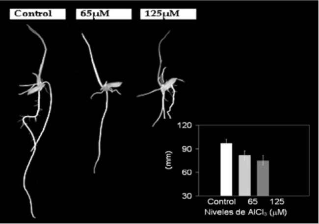
Root elongation was significantly inhibited in rice seedlings exposed to Al (Figure 1). However, growth inhibition was more severe in seedlings exposed to 125 μM than in seedlings exposed to 65 μM AlCl3. In the most stressed roots, elongation was reduced 24% compared to the control plants, while those exposed to 65 μM AlCl3 exhibited a reduction of 15.5% (Figure 1).
The aim of this paper is to study the responses to Al to-xicity in a Cuban rice cultivar (INCA LP-7). The effects of Al on root morphology and on some cellular level changes are also investigated.
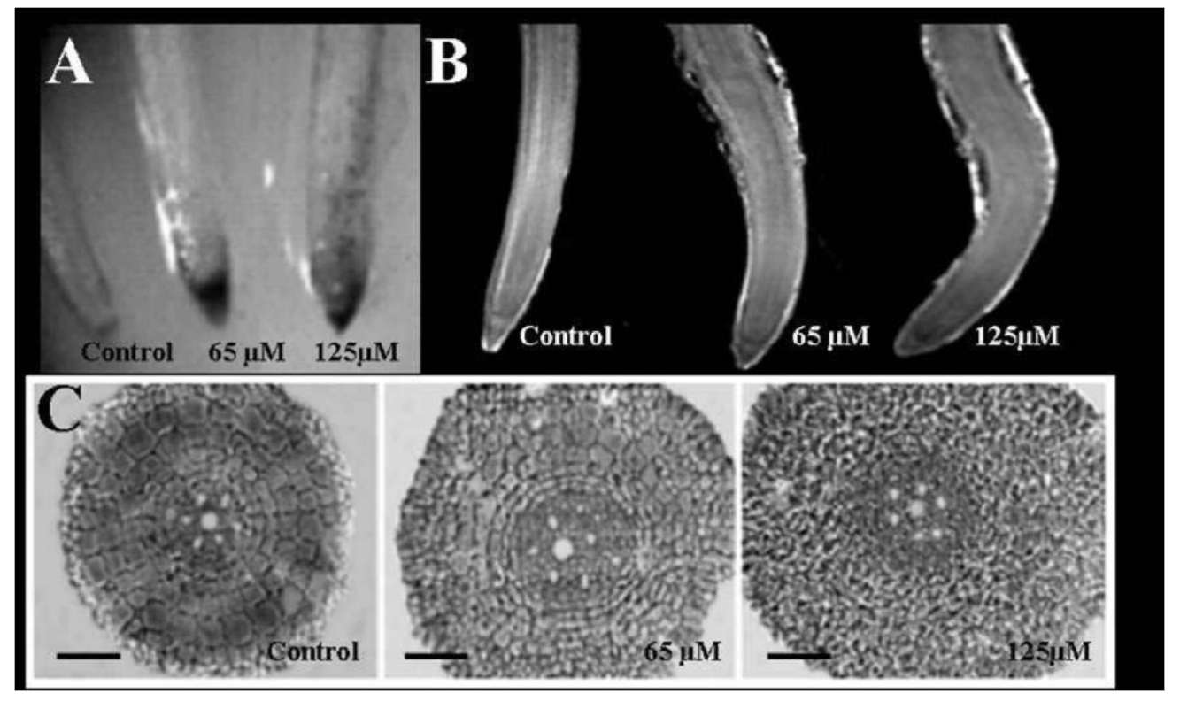
Seminal roots stained with hematoxylin are shown in Figure 2A. In seedlings exposed to Al, the root tips exhibited a red-brown staining, which was more intensive in roots exposed to 125 than to 65 μM Al. Seedlings exposed to 125 μM also exhibited stained spotting. No staining was observed in control plants (Figure 2A).
MATERIALS AND METHODS
Plant material and growth conditions
Roots of stressed seedlings showed symptoms of Al in­jury at both Al concentrations. Root-apices appeared swol­len and irregularly curved in Al-treated seedlings (Figure 2B). A localized thickened zone was observed between 20 and 40 mm from the rice root apex, corresponding to an increased diameter of cross-sections (Figure 2C)
Seeds of Oryza sativa L., cv INCA-LP7 were sterilized with 5% sodium hypochlorite and germinated on filter pa­per for seven days. Two Al concentrations were employed to impose the stress; 0, 65 and 125 (iM AlClg (pH= 4). The seedlings were under 16 h of light/8 h of darkness at 25°C.
To evaluate root elongation, the primary roots of seed­lings (n > 25) were photographed and their length mea­sured with a ruler before microscopic sampling.
Cross-sections of roots exposed to both Al concentrations were observed as more disordered (Figure 2C) than 
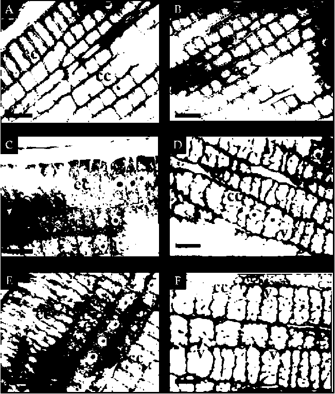
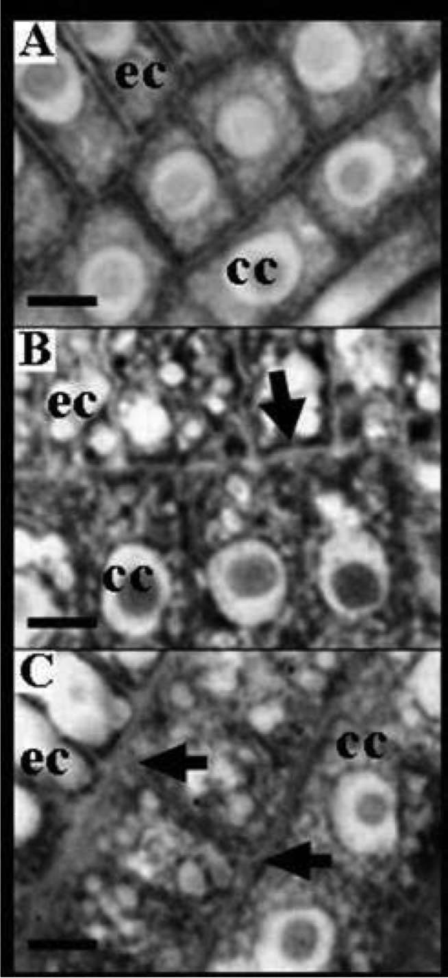
those of the control roots. Some alterations in the size of epidermal and cortical cells provoked by Al were responsible for this change. The epidermal cells were of typical size in longitudinal sections of the control roots (Figure 3), but lost their tissue features and appeared shorter and wider than the cells in control roots, when exposed to 65 and 125 μM AlCl3. These changes were also observed in cortical cells (Figure 3).
Hematoxylin staining
Staining protocol was based on Polle et al. (1978). The roots of seedlings cultivated for seven days, in the presence or absence of Al, were gently shaken in 200 ml distilled water for 15 min. The water was then replaced by 200 ml of aqueous hematoxylin solution [0.2% hematoxy­lin (Merck) and 0.02% potassium iodide, w/v] and left at the same slow agitation for 20 min. The solution was then replaced with 200 ml of water. The root apices were ex­cised and photographed under stereoscopic microscope.
Light microscopy
Apical segments obtained from primary roots (3 mm) were fixed with 4% paraformaldehyde. After three washes in distilled water, the roots were dehydrated by Progres­sive Lowering Methods (PLT) and embedded in Lowicryl resin (Risueno, 2000).
Semi-thin longitudinal and cross-sections were made on a LKB Ultramicrotome and stained with toluidine blue. The sections were viewed with a Zeiss optic microscope and photographed with a digital camera coupled to the mi­croscope.
Figure 1. Effects of 65 and 125 (iM AICI3 on root elongation of rice cv INCA LP-7. Values are means of five independent repli­cates. Means followed by different letters differ significantly at P < 0.05 (Tukey's Test).
Statistical Analysis
Randomized complete design was employed for the exper-
ALVAREZ et al. ― Changes in rice by Al stress
69
Figure 2. Different manifestations of Al stress symptoms in roots. The rice cv INCA LP-7 was cultivated at increasing levels of Al for 7 d. Stereoscopic images (A and B) A, Al injury at apices of adventitious roots, visualized by hematoxylin staining; B, Deformed roots after Al exposure; C, Root transition zone cross-sections exhibiting increased root diameter after Al treatment. Scale bars represent 100 (m.
Increases in vacuolar volume were induced by Al (Figure 3). Intensively vacuolated cells were observed in the cortex and the epidermis in longitudinal sections of stressed roots. This increase was more intense in cortex cells than in epidermal cells (Figure 3).
Structural modifications in the cell walls of root-tip cells were observed in seedlings exposed to Al (Figure 4). A thickening of tangential cell walls occurred in the exter­nal cortical cells (first-third layer). Seedlings exposed to the highest Al concentration (125 μM AlCl3) showed the greatest increase in cell wall thickening (Figure 4).
DISCUSSION
Aluminium toxicity limits plant growth mainly through its adverse effects on root growth and development. Under acidic soil conditions, active, phytotoxic forms of Al are released into the soil solution at levels that can inhibit root growth and damage roots (Liao et al., 2006)
Root inhibition in seedlings exposed to Al (65 and 125 (M AlCl3) was modest, causing no more than 22% inhibition in the most stressed roots, compared to the Al control. A similar inhibitory effect of Al on root elonga­tion has been confirmed in most of the experimental work (reviewed by Ciamporova, 2002) and in previous results obtained using different rice cultivars (Oryza sativa L.) (Alvarez et al., 2005).This is a major consequence of Al
Figure 3. Light-microscopy images from longitudinal section of rice root apices exposed to 0 (A, B), 65 (C, D), 125 μM AlCl3 (E, F). Control roots show organized epidermal cell lines, while Alexposed roots exhibited disorganization and increased vacuolation. Scale bars represent 10 μm.
70
Botanical Studies, Vol. 53, 2012
toxicity, which subsequently affects nutrients and water
uptake (Horst et al., 2010).
These results established that the INCA-LP7 rice cul-tivar is not only more Al tolerant than other rice cultivars studied (Alvarez et al., 2005), but also more Al tolerant than other crop species reported in the literature (Wenzl et al., 2001; Pineros et al., 2002; Amenos et al., 2009).
This degree of Al tolerance was also in accord with a low level of Al accumulation (as indicated by the low degree of Al-specific hematoxylin staining in Figure 2) that was localized, to the first millimeters of the root tip. Differential Al accumulation observed between both cul-tivars is probably related to Al adsorption and desorption kinetics of the root tip cell walls, where polysaccharides are thought to play an important role in excluding Al spe­cifically from the rice root apex (Yang et al., 2008, Silva et al., 2010).
This finding suggests that the hematoxylin technique could be used to identify tolerant and sensitive rice genotypes after long-term seedling exposure to Al. The technique is also supported in previous works, where an Al-sensitive rice cultivar, J-104, exhibited significant root damage and dramatic Al accumulation under these conditions (unpublished results). Similar results were ob­tained in wheat, where this technique proved conducive to identifying tolerant and sensitive genotypes after a very short exposure of seedlings to Al, which was well before differences in the seminal root length become detectable (Delhaize et al., 1993).
Hematoxylin is a dye commonly used to study Al sen­sitivity in plant species (Polle et al., 1978; Delhaize et al., 1993; Wagatsuma et al., 1995), including maize (Guevara et al., 1992; Ryan and Kochian, 1993; Jorge and Arruda, 1997 and Pineros et al., 2002). An important aspect of this technique is that the reaction between hematoxylin and Al is specific, such that other stressing factors would exert a minimal effect, if any, on the evaluation processes of the Al effects (Cancado et al., 1999).
Some changes were appreciated in root features of seedlings exposed to Al. Swollen and curved roots with different colors were observed in our research, which are common symptoms of Al stress (reviewed by Ciamporova, 2002). These results suggested that long-term Al applica­tion can change the architecture of the root system. In maize seedlings treated with Al, lateral roots sometimes appeared very close to the axial root apex, exhibiting a deformed and thickened structure and a possible color change (Budikova et al., 1998).
Al exposure triggered different changes in the shape of epidermal or outer cortex cells. A reduction in the length of epidermal and cortex layer root cells was observed in longitudinal sections, accompanied by a cell radial expan­sion. Similar alterations in other species have been docu­mented (Horst, 1995; Barcelo and Poschenrieder, 2002; Gunse et al., 2003). A reduced length was appreciated in the meristematic and elongation zones of barley root (Ko-chian, 1995), and the root cortices of wheat (Sasaki et al., 1997) and maize (Budikova, 1999).
This finding suggests that Al affects cellular growth orientation. In Arabidopsis thaliana, the root changes that were accompanied by disorganization of the microtubule cytoskeleton were induced by protein kinases and phos-phatases inhibitors, suggesting the effects of Al on mecha­nisms of the regulation of cell growth polarity (Horst,
Figure 4. Light-microscopy images of longitudinal section of rice root apices exposed to 0 (A), 65 (B) and 125 (M AlCl3 (C). Al-exposed roots exhibited thickened tangential cell walls in the external cortex cells (first-third layer). Scale bars represent 2 (m.
ALVAREZ et al. ― Changes in rice by Al stress
71
1995). Cell elongation and the direction of cell expansion are linked processes of functional importance in plant de­velopment (Sugimoto et al., 2000).
Barcelo, J. and C. Poschenrieder. 2002. Fast root growth re­sponses, root exudates, and internal detoxification as clues to the mechanisms of aluminium toxicity and resistance: a review. Environm. Experim. Bot. 48: 75-92.
Budikova, S. 1999. Structural changes and aluminium distribu­tion in maize root tissues. Biol. Plant. 42: 259-266.
Budikova, S. and M. Ciamporova. 1998. Growth and structural responses of maize primary roots to aluminium stress. In I. Tsekos and M. Moustakas (eds.), Progress in botanical Re­search. Kluwer Academic Publishers, Dordrecht 1998, pp. 419-422.
Cancado, G.M.A., L.L. Loguercio, P.R. Martins, S.N. Parentony, E. Paiva, A. Borem, and M.A. Lopes. 1999. Hematoxylin staining asa phenotypic index for aluminum tolerance selec­tion in tropical maize (Zea mays L.). Theor. Appl. Genet. 99: 747-754.
Ciamporova, M. 2000. Diverse response of root cell structure to aluminium stress. Plant Soil 226: 113-116.
Ciamporova, M. 2002. Morphological and structural responses of plant roots to aluminium at organ, tissue, and cellular levels. Biol. Plant. 45: 161-171.
Delhaize, E., S. Craig, C.D. Beaton, R.J. Bennet, V.C. Jagadish,
and P.J. Randall. 1993. Aluminium tolerance in wheat (Triti-cum aestivum L.). Uptake and distribution of aluminium in root apices. Plant Physiol. 103: 685-693.
Delhaize, E. and P.R. Ryan. 1995. Aluminum toxicity and toler­ance in plants. Plant Physiol. 107: 315-321.
Eticha, D., A. Stass, and W.J. Horst. 2005. Cell-wall pectin and
its degree of methylation in the maize root-apex: signifi­cance for genotypic differences in aluminium resistance.
Plant Cell Environ. 28: 1410-1420.
Guevara, P., C. Poschenrieder, and J. Barcelo. 1992. Differential response of four maize (Zea mays L.) varieties to aluminum
toxicity. Plant Soil. 2: 713-721.
Gunse, B., T. Garzon, and J. Barcelo. 2003. Study of aluminum toxicity by means of vital staining profiles in four cultivars of Phaseolus vulgaris L. Plant Physiol. Urban & Fischer Verlaghttp:〃www.urbanfischer.de/joumals/jpp.
Guo, T., G. Zhang, M. Zhou, F. Wu, and J. Chen. 2004. Effects
of aluminum and cadmium toxicity on growth and antioxi-dant enzyme activities of two barley genotypes with differ­ent Al resistance. Plant and Soil. 258: 241-248.
Horst, W. 1995. The role of the apoplast in aluminum toxicity and resistance of higher plants: a review. Z. Pflanzenernae-
hr. Bodenkd. 158: 419-428.
Horst, W.J., Y. Wang, and D. Eticha. 2010. The role of the root apoplast in aluminium- induced inhibition of root elonga­tion and in aluminium resistance of plants: A review. Ann. Bot. 106: 185-197.
Hossain, Z., T. Ohno, H. Koyama, and T. Hara. 2005. Effect of enhanced calcium supply on aluminum toxicity in relation to cell wall properties in the root apex of two wheat culti-vars differing in aluminium resistance. Plant Soil. 276: 193­204.
The radial expansion of epidermal and cortex cells may exert mechanical stress causing an increase in cross-sections of rice roots exposed to Al (Alvarez et al., 2005) and destruction of peripheral root tissues (Ciamporova, 2000). This phenomenon has also been observed in maize exposed to Al stress (Wang et al., 2004).
Although we did not determine vacuolar content in our our research, an increased vacuolation inside the cells observed in rice roots exposed to both Al concentrations, particularly those in the apical region of the primary roots, suggests that this mechanism may be employed to inter­nal Al detoxification. In maize, this relied on the active transport of Al from the cell wall to vacuoles (Zheng et al., 2005), and in other studies, higher vacuolation was observed in the root cap, the epidermis, and the cortex (Ciamporova, 2002; Vazquez et al., 1999). Vazquez et al. (1999) detected Al in similar vacuolar deposits located in Si-containing vacuoles of maize root cells. The amount of Al in the vacuolar deposits increased with increasing exposure of maize root to Al stress, which might lead to reducing the toxic effects of Al in the cytoplasm and to re­covery of the root growth.
Thickened cell walls were frequently observed in Al-treated roots and principally in external cortex cells, which revealed irregularly-thickened cell walls. Although many researchers suggest integrating Al with many cellular sites: cell wall, plasma membrane or DNA (Silva et al., 2000), it seems that most of the Al accumulated in the cell wall. When a plant grows under toxic Al conditions, Al is first introduced through its apoplast. Ample evidence suggests that primary Al accumulation in root tissues is localized in the cell wall (Hossain et al., 2005), which could be the reason for the thickening we observed. It remains unclear, however, whether Al interacts directly with the cell wall or whether these changes are an indirect consequences of other Al-induced changes (Pietraszewska, 2001).
Significant changes are observed in rice roots exposed to Al. Although there is much evidence regarding the ef­fects this metal has on diverse root systems, its effects on the anatomic and histological features are less studied. This point could be addressed in future investigations of Al toxicity in plants.
LITERATURE CITED
Alvarez, I., O. Sam, and I. Reynaldo. 2005. Alteraciones celula-res inducidas por el ion Al3+ en dos variedades cubanas de arroz (Oryza sativa L.). Cultivos Tropicales. 26: 61-64.
Amenos, M., I. Corrales, C. Poschenrieder, P. Illes, F. Baluska, and Barcelo. 2009. Different effects of aluminium on actin cytoskeleton and brefeldin A-sensitive vesicle recycling in root apex cells of two maize varieties differing in root elongation rate and Al tolerance. J. Plant Cell. Physio. 50:
528-540.
72
Botanical Studies, Vol. 53, 2012
Jorge, R.A. and P. Arruda. 1997. Aluminum-induced organic acids exudation by roots of an aluminum-tolerant tropical maize. Phytochemistry 45: 675-681.
Kochian, L.V., O.A. Hoekenga, and M.A. Pineros. 2004. How do crop plants tolerate acid soils? Mechanisms of alumini­um tolerance and phosphorous efficiency. Annu. Rev. Plant
Biol. 55: 459-493.
Kochian, L.V. 1995. Cellular mechanisms of aluminium toxicity and resistance in plants. Annu. Rev. Plant Physiol. Mol.
Biol. 46: 237-270.
Liao, H., H. Wan, J. Shaff, X. Wang, X. Yan, and L.V. Kochian. 2006. Phosphorus and Aluminum Interactions in Soybean in Relation to Aluminum Tolerance. Exudation of Specific Organic Acids from Different Regions of the Intact Root
System. Plant Physiol. 141: 674-684.
Ma, J.F., S. Nagao, K. Sato, H. Ito, J. Furukawa, and K. Tekeda. 2004. Molecular mapping of a gene responsible for Al-ac-tivated secretion of citrate in barley. J. Exp. Bot. 55: 1335­1341.
Macedo, C. and V. Jan. 2008. Effect of aluminum stress on mineral nutrition in rice cultivars differing in aluminum sensitivity. Revista Brasileira de Engenharia Agricola e Am-
biental 12: 363-369.
Matsumoto, H. 2000. Cell biology of aluminium toxicity and tolerance in higher plants. Int. Rev. Cytol. 200: 1-46.
Pietraszewska, T.M. 2001. Effect of aluminium on plant growth and metabolism. Acta Biochimica Polonica. 48: 673-686.
Pineros, M.A., J.V. Magalhaes, V.M.C. Alve, and L.V. Kochian. 2002. The physiology and biophysics of Al tolerance mechanism based on root citrate exudation in maize. Plant
Physiol. 129: 1194-1206.
Polle, E., C.F. Konzak, and J.A. Littrik. 1978. Visual detection of aluminium tolerance levels in wheat by hematoxylin stain­ing of seedling roots. Crop Sci. 18: 823-827.
Risueno, M. 2000. Microspore derived embryogenesis from in vitro culture of heat stressed anthers of Capsicum annum involves a dedifferentiation followed by propliferation and further redifferentiation. Plant Development and Nuclear
Differentiation. CSIC.
Rubia, O., F. Horchani, I. Smida, M. Mejri, and S. Aschi-Smiti. 2011. Aluminium Phytotoxicity and Plant Acclimation to Acidic Soils. Science Alert.
Ryan, P.R. and L.V. Kochian. 1993. Interaction between alu­minium toxicity and calcium uptake at the root apex in near isogenic lines of wheat (Triticum aestivum L.) differing in aluminium tolerance. Plant Physiol. 102: 975-982.
Sasaki, M., Y. Yamamoto, H. Matsumoto. 1997. Aluminum in-
hibits growth and stability of cortical microtubules in wheat (Triticum aestivum L.) roots. Soil Sci. Plant Nutr. 43: 469­472.
Silva, I.R., T.J. Smyth, D.F. Moxley, T.E. Carter, N.S. Allen, and T.W. Rufty. 2000. Aluminum accumulation at nuclei of cells in the root tip. Fluorescence detection using lumogallion and confocal laser scanning microscopy. Plant Physiol. 123:
543-552.
Silva, S., O. Pinto, P. Martins, M. Matos, H. Guedes-Pinto, and C. Santos. 2010. Differential aluminium changes on nutrient accumulation and root differentiation in an Al sensitive vs. tolerant wheat. Environ. Exp. Bot. 68: 91-98.
Sugimoto, K., R.E. Williamson, and G.O. Wasteneys. 2000. New techniques enable comparative analysis of microtubule orientation, wall texture, and growth rate in intact roots of
Arabidopsis. Plant Physiol. 124: 1493-1506.
Taylor, G.J., J.L. McDonald-Stephens, D.B. Hunter, P.M. Bertsch, D. Elmore, Z. Rengel, and R.J. Reid. 2000. Direct
measurement of aluminum uptake and distribution in single cells of Chara corallina. Plant Physiol. 123: 987-996.
Vazquez, M.D., C. Poschenrieder, I. Corrales, and J. Barcelo. 1999. Change in apoplastic aluminum during the initial growth response to aluminum by roots of a tolerant maize
variety. Plant Physiol. 119: 435-444.
Wagatsuma, T., S. Ishikawa, H. Obata, K. Tawaraya, and S. Ka-tohda. 1995. Plasma membrane of younger an douter cells is the primary specific site for aluminium toxicity in roots.
Plant Soil 171: 105-112.
Wang, W. and C.H. Kao. 2007. Protective effect of ascorbic acid and glutathione on AlCl3-inhibited growth of rice roots.
Biologia Plant. 51: 493-500.
Wang, Y., A. Stass, and W.J. Horst. 2004. Apoplastic binding of aluminum is involved in silicon-induced amelioration of aluminumtoxicity in maize. Plant Physiol. 136: 3762-3770.
Wenzl, P., G.M. Patino, A.L. Chavez, J.E. Mayer, and I.M. Rao. 2001. The high level of aluminium resistance in signalgrass is not associated with know mechanism of external alumin­ium detoxification in root apices. Plant Physiol. 125: 1473­1484.
Yang, J.L., Y.Y. Li, Y.J. Zhang, S.S. Zhang, Y.R. Wu, P. Wu, and S.J. Zheng. 2008. Cell Wall Polysaccharides are specifically involved in the exclusion of aluminum from the rice root apex. Plant Physiol. 146: 602-611.
Zheng, S.J., J.L. Yang, Y.F. He, X.H. Yu, L. Zhang, J.F. You, R.F. Shen, and H. Matsumoto. 2005. Immobilization of alumi­num with phosphorus in roots is associated with high alu­minium resistance in buckwheat. Plant Physiol. 138: 297­303.
ALVAREZ et al. ― Changes in rice by Al stress
73
鋁離子在水稻根所引起的形態上及細胞內之變化
Idioleidys ALVAREZ1, Ofelia SAM, Ines REYNALDO1, Pilar TESTILLANO2, Maria del CARMEN RISUENO2, and Marta ARIAS3
1Instituto Nacional de Ciencias Agricolas. Ctra. Tapaste, Km 31/2. Gaveta Postal No.1, 32700, San Jose de Las Lajas, La Habana, Cuba
2Laboratorio de Organizacion y Desarrollo Nuclear de plantas del Centro de Investigaciones Biologicas del CSIC. Ramiro de Maestzu, 109, 28040, Madrid, Spain
3Cdtedra de Anatomia Vegetal, Facultad de Cs. Naturales e IML. Tucumdn, Argentina
鋁金屬離子之毒害在酸性土壤中嚴重地限制了作物之生產量;而此毒性主要係經由根部發作。本文
之目的乃探討鋁離子對水稻根部之形態及細胞構造所造成之傷害。我們使用不同濃度之三氯化鋁來探討
危害之程度。根冠部位是以逐歩低溫法 (PLT)及 Lowycril樹脂處理;如此一來試樣可被切除。經染色之
切片以Zeiss光學顯微鏡外接數位相片來檢視並照像。根部之延伸程度經鋁離子處理後會減少,同時根
會變形。在表皮細胞及皮層細胞可檢測到若干構造上之改變。減少之長度伴隨著橫軸向之膨脹;此乃源
於不同型之細胞的延展過程受到阻害。同時也觀察到加厚之細胞壁及液泡大小之增加。以上之結果提供
證據說明水稻根部細胞受到足以導致傷害之鋁離子之影響後所產生的構造上之改變及形態上之變化;此
乃我們實驗室所觀測到的。
關鍵詞:鋁;水稻;根部細胞;構造之改變。