Botanical Studies (2012) 53: 215-222.
BIOCHEMISTRY
A novel trypsin inhibitor from sweet potato (Ipomoea batatas Lam.) leaves and its synthesized peptides with antioxidant activities in vitro
Guan-Jhong HUANG1,7, Ying-Chih LIN2'7, Jeng-Shyan DENG3, Hsien-Jung CHEN4, Jung-Chun LIAO5, Shyh-Shyun HUANG5, Shu-Jen CHANG5, and Yaw-Huei LIN6 *
1School of Chinese Pharmaceutical Sciences and Chinese Medicine Resources, College of Pharmacy, China Medical University,Taichung 404, Taiwan
2Department of Optometry, Jen-Teh Junior College of Medicine, Nursing and Management, Miaoli 356, Taiwan
3Department of Health and Nutrition Biotechnology, Asia University, Taichung 413, Taiwan
4Department of Biological Sciences, National Sun Yat-sen University, Kaohsiung 804, Taiwan
5Department of pharmacy, College of Pharmacy, China Medical University, Taichung 404, Taiwan
6Institute of Plant and Microbial Biology, Academia Sinica, Nankang, Taipei 115, Taiwan
(Received April 11, 2011; Accepted September 16, 2011)
ABSTRACT. Recombinant SPLTI-a [sweet potato leaf trypsin inhibitor-a] overproduced in E. coli (M15) was purified by Ni2+-chelated affinity chromatography. The molecular mass of SPLTI-a is ca. 8000 Da as de­termined by sodium dodecyl sulfate-polyacrylamide gel electrophoresis (SDS-PAGE). SPLTI-a was examined using different antioxidative models (Total antioxidant status, reducing power method, Fe2+-chelating ability,
The SPLTI-a protein with a concentration of 100 (ig/mL exhibited highest activity (expressed as 2.12 0.02 mM Trolox equivalent antioxidative value, TEAC) in total antioxidant status test. Like total antioxidant status, the reducing power, Fe2+-chelating ability, FTC activity and protecting calf thymus DNA against hydroxyl rad­ical-induced damage all showed that SPLTI-a polypeptide has significant antioxidant activities. It was found that the antioxidant activity increased after 24 h hydrolysis of SPLTI-a by trypsin from 18% (0 h) to about 35% (24 h). Accumulation of shorter peptides increased along the longer trypsin incubation. The obtained VR, STIEK, ITDGK, and EYIFDR showed IC50 (concentration for 50% inhibition) values of 5.83, 3.75, 2.65, and 0.73 mM, respectively, when scavenging activity of DPPH radicals (%) was measured. These findings mean that tyrosine residue is most important in antiradical activities. It was suggested that SPLTI-a possess antioxi-dant activities.
Keywords: Antioxidant activity; Leaf trypsin inhibitor; Recombinant protein; Sweet potato.
INTRODUCTION
of PIs were also found to accumulate rapidly in leaves in response to mechanical wounding or insect chewing (Constabel, 1999), suggesting a direct role of PIs in plant protection.
Plant proteinases inhibitors (PIs) are widely distributed in plants. Serine, cysteine and aspartic proteinases are the main proteinases inhibited. Most PIs have four conserved Cys residues forming two disulfide bonds in a single or double chain polypeptide (Oliva et al., 2010). In higher plants, PIs are shown to be particularly abundant in storage organs, such as tubers (Richardson, 1991). A large body of evidence indicated that PIs functioned as storage proteins, regulate the endogenous proteinase activities (Dunaevsky et al., 1998), and suppress the exogenous proteinase activi­ties from pathogens and pests (Ryan, 1989). Four types
Reactive oxygen species (ROS) can be formed by both endogenous and exogenous sources in living organisms. Within the cells, ROS can be generated in mitochondrial and microsomal electron transport systems, in soluble oxidase enzyme systems, and during phagocyte activation (Kehrer, 1993). Exogenous ROS generate from air and water pollutants, cigarette smoke, organic solvents, heavy metals, certain drugs, and radiation, etc (Valko et al., 2006). The ROS play an important role related to the degenerative or pathological processes of various ser-ous diseases, such as aging, cancer, coronary heart disease, Alzheimer's disease, neurodegenerative disorders, atherosclerosis, cataracts, and inflammation (Huang et al., 2010). The use of food is widespread and plants still

7Guan-Jhong HUANG and Ying-Chih Lin contribute equally to this study.
*Corresponding author: E-mail: boyhlin@gate.sinica.edu.tw; Tel: +886-2-2787-1172; Fax: +886-2-2782-7954.
216
Botanical Studies, Vol. 53, 2012
present a large source of natural antioxidants that might serve as leads for the development of novel drugs. Sev­eral antiinflammatory, antinecrotic, neuroprotective and hepatoprotective drugs have recently been shown to have an antioxidant and/or antiradical scavenging mechanism (Repetto and Llesuy, 2002). During the last few years, natural antioxidants and compounds with radical scaveng­ing activity have been found, such as phenolic compounds (Huang et al., 2008), anthocyanin (Espin et al., 2000), water extract of Flemingia species (Hsieh et al., 2010), thi-oredoxin h protein (Huang et al., 2004a), sporamin (Huang et al., 2007), and mucilage (Huang et al., 2006) from sweet potato root. The objectives of this work were to investigate the antioxidant property of SPLTI-a from sweet potato leaf in comparison with chemical compounds such as butylated hydroxytoluene (BHT), reduced glutathione or ascorbate in a series of in vitro tests.
by the following formula: Trolox equivalent value (mM) =[factor x (absorbance of blank-absorbance of sample)]; factor= [concentration of standard/(absorbance of blank-absorbance of standard)].
Scavenging activity against DPPH radical
DPPH is a relatively stable free radical which when encounters proton donors such as antioxidants, the radicals get quenched and absorbance gets reduced. The effect of SPLTI-a on the DPPH radical was estimated according to the method of Huang et al. (2004b). An aliquot of SPLTI-a (30 μL) was mixed with 100 mM Tns-HCl buffer (120 μL, pH 7.4) and then 150 μL of the DPPH in ethanol with a final concentration of 250 μM was added. The mixture was shaken vigorously and left to stand at room tempera-ture for 20 min in the dark. The absorbance at 517 nm of the reaction solution was measured spectrophometrically. The percentage of DPPH decolourization of the sample was calculated according to the equation: % decolouriza-tion= [1- Abssample /Abscontrol]x100. The IC50 values denote the concentration of sample which is required to scavenge 50% of DPPH free radicals.
MATERIALS AND METHODS
Expression of SPLTI-a in E. coli
SPLTI-a (Gene Bank accession number: AF330700) was expressed in E. coli. The coding sequence was ampli­fied from SPLTI-a cDNA using an oligonucleotide (5'-GGA TCC AGA AAA TGC AGC GCA TCA C -3'), with a BamHI site (underlined) at the putative initial Met residue, and an oligonucleotide (5'-AGAAC TCCGTC GAT AAG CTT GGT -3'), with a HindIII site at the 3' end. The PCR fragment was subcloned in pGEM T-easy vector. And the plasmid was then digested with BamHI and HindIII and subcloned in pQE32 expression vector (QIAexpress ex­pression system, Qiagen). The resulting plasmid, termed pQE-SPLTI-a, was introduced into E. coli (M15). Cultures of the transformed E. coli (M15) overexpressed a protein of the expected molecular mass, which was purified by affinity chromatography in Ni-nitrilotriacetic acid (NTA) columns (Qiagen), according to the manufacturer's in­structions.
Determination of antioxidant activity by reduc­ing power measurement
Reducing power method indirectly evaluates the anti-oxidant activity. The reducing powers of the SPLTI-a and glutathione were determined according to the method of Chang et al. (2007). SPLTI-a (0, 0.2, 0.4, 0.8, 1.0, and 1.2 mg/mL) or glutathione was mixed with an equal volume of 0.2 M phosphate buffer, pH 6.6, and 1% potassium fer-ricyanide. The mixture was incubated at 50°C for 20 min, during which time ferricyanide was reduced to ferrocya-nide. Then an equal volume of 1% trichloroacetic acid was added to the mixture, which was then centrifuged at 6,000 g for 10 min. The upper layer of the solution was mixed with deionized water and 0.1% FeCl3 at a radio of 1 : 1 : 2, and the absorbance at 700 nm was measured to determine the amount of ferric ferrocyanide (Prussian Blue) formed. Increased absorbance of the reaction mixture indicated in-creased reducing power of the sample.
Protein staining on 15% SDS-PAGE gels
SPLTI-a was detected on 15% SDS-PAGE gels. Sampies treated with sample buffer and p-mercaptoethanol (2-ME) with a final concentration of 14.4 mM were heated at 1000C for 5 min before 15% SDS-PAGE.
Determination of antioxidant activity by Fe2+-chelating ability
Measurement of total antioxidant status
The Fe2+-chelating ability was determined according to the method of Huang et al. (2007). The Fe2+ was moni­tored by measuring the formation of ferrous iron-ferrozine complex at 562 nm. SPLTI-a (0, 0.25, 0.5, 1, 2, and 4 mg/mL) was mixed with 2 mM FeCl2 and 5 mM ferrozine at a ratio of 10 : 1 : 2. The mixture was shaken and left to stand at room temperature for 10 min. The absorbance of the resulting solution at 562 nm was measured. The lower the absorbance of the reaction mixture the higher the Fe2+-chelating ability. The capability of the sample to chelate the ferrous iron was calculated using the following equa-tion:
Total antioxidant status of the SPLTI-a protein was mea­sured using the total antioxidant status assay kit (Calbio-chem Corp) according to the manufacturer's instructions. The assay relies on the antioxidant ability of the protein to inhibit oxidation of 2, 2' azino-bis-[3-ethylbenz-thiazoline-6-sulfonic acid] (ABTS) to ABTS*+ by metmyoglobin. The amount of ABTS*+ produced is monitored by reading the absorbance at 600 nm. Under these reaction conditions, the antioxidant ability of SPLTI-a protein decreases the absor-bance at 600 nm in proportion to its concentration. The fi­nal antioxidant capacity of SPLTI-a protein was calculated
Scavenging effect (%) =[1-Abs sample / Abs control ]x100
HUANG — Leaf trypsin inhibitor and its peptides with antioxidant activities
217
Determination of antioxidant activity by the ferric thiocyanate (FTC) method
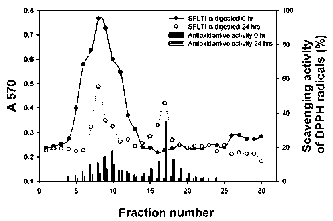
each fraction contained 2 mL of which the absorbance at 280 nm was then determined.
The FTC method was adapted from the method of Osawa and Namiki (1981). Twenty mg/mL of samples dissolved in 4 ml of 99.5% (w/v) ethanol were mixed with linoleic acid (2.51%, v/v) in 99.5% (w/v) ethanol (4.1 mL), 0.05 M phosphate buffer pH 7.0 (8 mL) and deionized water (3.9 mL) and kept in a screw-cap container at 40°C in the dark. Then, to 0.1 mL of this solution was added 9.7 mL of 75% (v/v) ethanol and 0.1 mL of 30% (w/v) ammo-nium thiocyanate. Precisely 3 min after the addition of 0.1 mL of 20 mM ferrous chloride in 3.5% (v/v) hydrochloric acid to the reaction mixture, the absorbance at 500 nm of the resulting red color [Fe (SCN)2+, Fe3+ was formed after linoleic acid peroxide was produced and Fenton reaction occurred.] was measured every 24 h until the day when the absorbance of the control reached the maximum value. The inhibition of linoleic acid peroxidation was calculated as (%) inhibition = 100 - [(absorbance increase of the sam-ple/absorbance increase of the control) x 100]. All tests were run in duplicate and analyses of all samples were run in triplicate and averaged.
Statistical analysis
Means of triplicates were calculated. Student's t test was used for comparison between two treatments. All data (expressed as percent of control value) were means± SE. A difference was considered to be statistically significant whenp < 0.05, p < 0.01 or p < 0.001.
RESULTS and DISCUSSION
Purification of expressed SPLTI-a
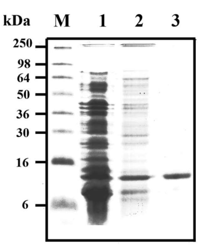
SPLTI-a cDNA clones from sweet potato leaf was isolated. SPLTI-a was subcloned in a pQE-32 expression vector in E. coli and SPLTI-a was produced with a 6x His-tag at the N-terminus. SDS-PAGE analysis of crude extracts from transformed E. coli (M15) showed a high level of a polypeptide with the expected molecular mass (ca. 8 kDa). This polypeptide was found as a soluble pro­tein in the supernatant (Figure 1, lane 2), and was absent in protein extracts obtained from E. coli transformed with
Protection of SPLTI-a against hydroxyl radical-induced calf thymus DNA damage
The hydroxyl radical was generated by Fenton reaction according to the method of Kohno et al. (1991). The 15 μL reaction mixture containing SPLTI-a (1.25, 2.5, 5, and 10 mg/mL), 5 μL of calf thymus DNA (1 mg/mL), 18 mM FeSO4, and 60 mM hydrogen peroxide were incubated at room temperature for 15 min. Then 2μL of 1 mM EDTA was added to stop the reaction. Blank test contained only calf thymus DNA and the control test contained all reac­tion components except SPLTI-a. The treated DNA solu­tions were subjected to agarose electrophoresis and then stained with ethidium bromide and examined under UV light.
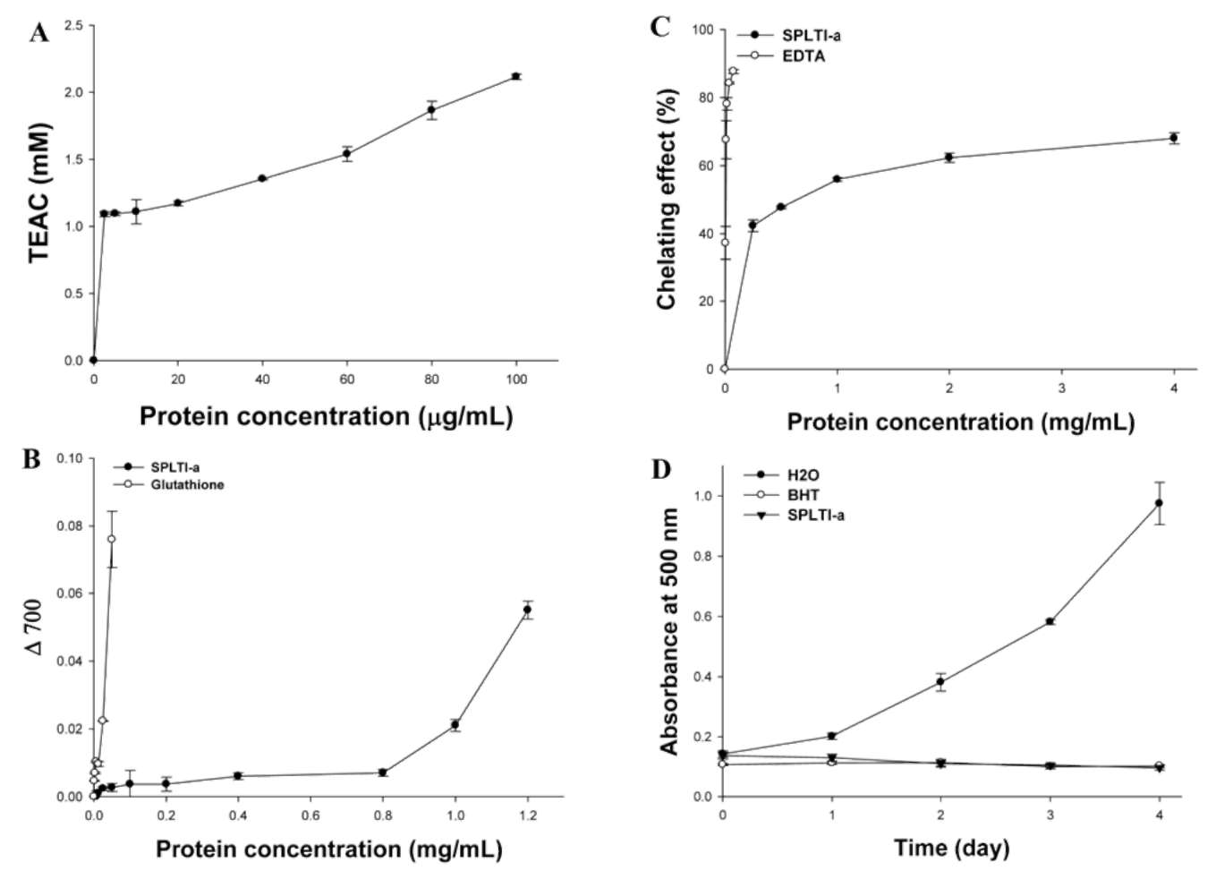
Determination of the antioxidative activity of SPLTI-a tryptic hydrolysates
Six mg of SPLTI-a was dissolved in 1 mL of 0.1 M KCl buffer (pH 8.0). Then 0.1 mL (12 mg) of trypsin was add-ed at 37°C for 0 and 24 h. After hydrolysis, 0.5 mL of 0.5 M Tris-HCl buffer (pH 8.3) was added, and the solution was heated at 100°C for 5 min to stop enzyme reaction. The trypsin was heated before SPLTI-a hydrolysis for the 0 h reaction. Each of the 60 μL SPLTI-a hydrolysates was used for determinations of the DPPH antioxidative activi-ties by spectrophotometry (Mine et al., 2004; Qian et al., 2008).
Figure 1. SDS-PAGE analysis of purified recombinant sweet potato leaf trypsin inhibitor (SPLTI-a). Crude extracts from E. coli (M15) transformed with pQE30 (lane 1) or with pQE30-SPLTI-a (lane 2) were analyzed by 15% (w/v) SDS/PAGE with 10 μg protein applied on each lane, and then the gel was stained with Coomassie blue G-250. Molecular masses of standard pro-teins are indicated at the left of the figure. His-tagged SPLTI-a was purified by Ni2+-chelated affinity chromatography (lane 3). The experiments were done twice and a representative one is shown.
Chromatograms of tryptic hydrolysates of SPLTI-a on a Sephadex G-50 column
The unhydrolyzed SPLTI-a and tryptic SPLTI-a hydro-lysates at 24 h were separated by Sephadex G-50 chroma-tography (1 x 60 cm). The column was eluted with 20 mM Tris-HCl buffer (pH 7.9). The flow rate was 30 mL/h, and
218
Botanical Studies, Vol. 53, 2012
pQE-32 vector (Figure 1, lane 1). The expressed protein was purified from crude extracts by Ni2+-chelate affinity chromatography, which yielded highly purified His-tagged SPLTI-a (Figure 1, lane 3).
1999). This was measured using the total antioxidant status assay kit (Figure 2A). SPLTI-a protein exhibited a dose-dependent total antioxidant activity within the applied con­centrations (0, 2.5, 5, 10, 20, 40, 60, 80, and 100 [ig/mL): the highest at 100 [ig/mL (expressed as 2.12 0.02 mM Trolox equivalent antioxidative value, TEAC). At 2.5 [ig/ mL, SPLTI-a displayed the lowest total antioxidant status (1.09 ± 0.01 mM TEAC).
Measurement of total antioxidant status using ABTS assay
Several methods have been developed to determine the antioxidant potential of natural products. The trolox equivalent antioxidant capacity (TEAC) using ABTS as an oxidant, the ferric reducing antioxidant power (FRAP), and the DPPH free radical scavenging assays are some of the most commonly used. Antioxidants can reduce radicals primarily by two mechanisms: single electron transfer and hydrogen atom transfer. ABTS, FRAP, and DPPH are methods that measure the former (Ozgen et al., 2006).
Measurement of reducing power
In this assay, the yellow color of the test solution changes to various shades of green and blue, depending on the reducing power of each compound. Presence of reduc­ers causes the conversion of the Fe3+/ferricyanide complex used in this method to the ferrous form. By measuring the formation of Perl's Prussian blue at 700 nm, it is possible to determine the Fe2+ concentration (Gülçin et al., 2003). We investigated the Fe3+-Fe2+ transformation in the pres­ence of the samples of SPLTI-a to measure its reducing capacity. The reducing capacity of a compound may serve as a significant indicator of its potential antioxidant activ­ity (Meir et al., 1995). The antioxidant activity of putative
ABTS assay is often used in evaluating total antioxi-dant power of single compounds and complex mixtures of various plants. It is used to estimate the total antioxidant power because the method is quick and simple to perform, and the reaction is reproducible and linearly related to the molar concentration of the antioxidants (Benzie et al.,
Figure 2. Radical scavenging activity of the recombinant SPLTI-a determined by TEAC (A), reducing power (B), Fe2+-chelating abil­ity (C) and inhibition of linoleic acid peroxidation (D). Each absorbance value represents average of triplicates of different samples analyzed. Results represent the means ± SE from at least 3 separate experiments. *p < 0.05, **p < 0.01 and ***p < 0.001 (unpaired t test) compared to SPLTI-a unsupplemented samples.
HUANG — Leaf trypsin inhibitor and its peptides with antioxidant activities
219
antioxidants have been attributed to various mechanisms, among them are prevention of chain initiation, binding of transition metal ion catalysts, decomposition of peroxides, prevention of continued hydrogen abstraction, and radi­cal scavenging (Liu et al., 2006). The reducing power of SPLTI-a is shown in Figure 2B with glutathione served as a positive control. The reducing power activity of SPLTI-a exhibited a dose-dependence (significant at p< 0.05) with­in the applied concentrations (0, 0.1, 0.2, 0.4, 0.8, 1.0, and 1.2 mg/mL).
Figure 3. Protection against hydroxyl radical-induced calf thy-mus DNA damage by recombinant SPLTI-a. Sample lanes 1-4 contained 1.25, 2.5, 5, and 10 mg/mL SPLTI-a, respectively. Blank (B) contained calf thymus DNA only; while the control (C) contained all reaction components except SPLTI-a.
Measure of Fe2+-chelating ability
Direct reaction of a substance is not the only mecha­nism by which the antioxidants may display their activity. Antioxidants act through numerous possible mechanisms. Some antioxidants do not convert free radicals to more stable products but slow the rate of oxidation by several different mechanisms. One of the most important mecha­nisms of action of secondary antioxidants is chelation of pro-oxidant metals. Iron and other transition metals (copper, chromium, cobalt, vanadium, cadmium, arsenic, nickel) promote oxidation by acting as catalysts of free radical reactions. These redox-active transition metals transfer single electrons during changes in oxidation states. Chelation of metals by certain compounds decreases their pro-oxidant effect by reducing their redox potentials and stabilizing the oxidized form of the metal. Chelating com­pounds may also sterically hinder formation of the metal hydroperoxide complex (Blokhina et al., 2003).
density lipoprotein (LDL) peroxidation has been reported to contribute to atherosclerosis development (Steinbrecher, 1987). Therefore, delay or prevention of LDL peroxida-tion is an important function of antioxidants. Figure 2D shows the time-course curve for the antioxidative activity of the SPLTI-a from sweet potato leaf, BHT, and H2O by the FTC method. The BHT was used as a positive control, and H2O as a negative control. The results indicate that SPLTI-a has antioxidative activity. SPLTI-a may act as a significant LDL peroxidation inhibitor (P < 0.05).
Protection against hydroxyl radical-induced calf thymus DNA damage by SPLTI-a
Free radicals could damage macromolecules in cells, such as DNA, protein, and lipids in membranes (Kohno et al., 1991). The oxidative damage of DNA is one of the most important mechanisms in the initiation of cancer. The damage is usually caused by hydroxyl radicals. The activ­ity of these radicals can be reduced by natural antioxidants found in plants including herbs. The Fenton reaction in­volves the reaction between hydrogen peroxide and Fe2+ to form hydroxyl radicals. Scavengers of hydroxyl radicals inhibit this reaction through the reduction of Fe2+. Figure 3 shows that SPLTI-a protected calf thymus DNA against hydroxyl radical-induced damages. The blank contained calf thymus DNA only, and the control contained all com­ponents except SPLTI-a. Compared to the blank and con­trol, it was found that 2.5 mg/mL SPLTI-a could protect against hydroxyl radical induced calf thymus DNA dam­ages during 15-min reactions.
Fe2+ ion is the most powerful pro-oxidant among the various species of metal ions. Ferrozine can quantita­tively form complexes with Fe2+. Therefore, the measure­ment of color reduction allowed the estimation of the metal chelating activity of the coexisting chelator. Metal chelating capacity was significant because it reduced the concentration of the catalyzing transition metal in lipid peroxidation (Halliwell and Gutteridge, 1984). The metal chelating capacity of SPLTI-a and standard antioxidants was determined by assessing their ability to compete with ferrozine for the ferrous ions. The Fe2+-chelating ability of the SPLTI-a is shown in Figure 2C. EDTA was used as a positive control. The Fe2+-chelating ability of SPLTI-a was lower than that of EDTA and this difference was statisti­cally significant (P < 0.05). SPLTI-a at doses of 0.25, 0.5, 1, 2, and 4 mg/mL exhibited 42.36, 44.71, 55.94, 62.31, and 68.02% iron binding capacity, respectively. On the other hand, EDTA at doses of 0.05, 0.1, 0.2, 0.4, and 0.8 mg/mL had 37.24, 67.59, 78.12, 84.25 and 87.68 % chelat-ing activity of iron, respectively.
Determination of the antioxidative activity of tryptic SPLTI-a hydrolysates and their peptide distributions
We used trypsin to hydrolyze SPLTI-a to mimic the hydrolysis course during digestion in human's (or ani­mal's) intestine. Figure 4 shows the antioxidative activity of tryptic SPLTI-a hydrolysates and the antioxidative ac­tivity (scavenging activity of DPPH radicals, percent) of tryptic SPLTI-a hydrolysates collected at different trypsin hydrolysis times. From the results, it was found that the antioxidative activity increased from 18% (0 h) to about 35% (24 h); at the same time, smaller peptides increased
Ferric thiocyanate (FTC) method
Malondialdehyde formed from the breakdown of poly-unsaturated fatty acids was served as a convenient index for determining the extent of lipid perxoidation reaction. The ferric thiocyanate method measured the amount of peroxide produced during the initial stages of oxidation which was the primary product of oxidation. So far low-
220
Botanical Studies, Vol. 53, 2012
the number of phenolic hydroxyl groups. Tyrosine residue could be oxidized to a tyrosyl radical through an electron-transfer process. Nitration may follow from the reaction of a tyrosyl radical with -NO2 (Lin et al., 2003; Pietraforte et al., 2001). Our results further indicate that tyrosine resi­dues (EYIFDR) in sweet potato SPLTI-a contribute to the antiradical activity. The synthetic peptide, EYIFDR, has the highest antioxidant activity (IC50 is 0.73 mM) as good as reduced glutathione (IC50 is 0.07 mM). These results demonstrated that deduced synthetic peptides from tryptic SPLTI-a hydrolysates exhibited antioxidative activity.
Especially, EYIFDR was more potent for scavenging hydroxyl and peroxyl radicals. It is generally accepted that the chemical activity of hydroxyl radical is the strongest among ROS. Hydroxyl radical reacts easily with biomol-ecules, such as amino acids, proteins and DNA. Therefore, the removal of hydroxyl radical is probably one of the most effective defenses of a living body against various diseases (Cacciuttolo et al., 1993). Carbon-centered radi­cals that represent R-, RO- and ROO- could be quenched by EYIFDR. This result agreed with a lipid peroxidation inhibition assay finding that EYIFDR inhibited lipid per-oxidation by scavenging lipid-derived radicals.
Figure 4. Antioxidative activity of tryptic hydrolysates of re-combinant SPLTI-a. The plot shows the antioxidative activity (%) of SPLTI-a hydrolysates at different trypsin hydrolysis time (0 hr and 24 hr). The proteins and the scavenging activity of DPPH radicals (%) were shown. The scavenging effect (%) was calculated according to the equation [1-(Abs 517 nm of sample + Abs 517 nm of control)] x 100%.
with trypsin hydrolytic time. Here, 100% activity was de­fined as the concentration of substrate that causes 100% loss of the DPPH activity. The purifications of potential peptides of antioxidative activity need further investiga­tions. We used synthetic peptides to measure antioxidative activity. Synthetic peptides were designed by mimicking trypsin cutting sites of SPLTI-a gene products from sweet potato (http://expasy.nhri.org.tw/tools/peptidecutter/). New peptides (Table 1) for antioxidative activity, that is, VR, STIEK, ITDGK and EYIFDR were synthesized according to deduction. IC50 values of individual peptides were 5.83, 3.75, 2.65, and 0.73 mM, respectively, when scavenging activity of DPPH radicals (%) was measured. Tyrosine residues with free -OH were reported to have antioxidant activities. Tyrosine had an effect on DPPH radical scav­enging, ABTS radical scavenging, superoxide anion radi­cal scavenging, H2O2 scavenging, total ferric ions reducing power and metal chelating on ferrous ions activities (Gul-cin, 2007). It was reported that the radical scavenging and antioxidant activities of tyrosine were highly controlled by
In conclusion, the results from in vitro experiments, including total antioxidant status assay (Figure 2A), re­ducing power method (Figure 2B), Fe2+-chelating abil­ity (Figure 2C), FTC method (Figure 2D), and hydroxyl radical-induced calf thymus DNA damage (Figure 3), demonstrated that SPLTI-a in sweet potato leaf may have significant antioxidant activities. SPLTI-a may contribute significantly to change the redox states and as a potent an-tioxidant against both hydroxyl and peroxyl radicals when people consume sweet potato. The ex vivo or in vivo anti-oxidant activity of SPLTI-a should be performed in near further.
Acknowledgements. The authors want to thank the finan­cial supports from the National Science Council (NSC100-2313-B-039-004- and NSC 100-2320-B-039-033-), China Medical University (CMU) (CMU99-TC-35, CMU99-S-29, CMU99-S-29, CCM-P99-RD-042, and CMU99-COL-10) and Taiwan Department of Heath Clinical Trial and Research Center of Excellence (DOH100-TD-B-111-004).
Table 1. SPLTI-a peptides with antioxidant activity.

Peptides

Scavenging activity of DPPH radicals (%), IC50 (mM)

GSH (control)

0.07 ± 0.01
VR
5.83 ± 0.57
STIEK
3.75 ± 0.35
ITDGK
2.65± 0.24
EYIFDR
0.73 ± 0.02
LITERATURE CITED
Blokhina, O., E. Virolainen, and K.V. Fagerstedt. 2003. Anti-oxidants, oxidative damage and oxygen deprivation stress. Ann. Bot. 91: 179-194.
Benzie, I.F.F., Y. Wai, and J.J. Strain. 1999. Antioxidant (reduc­ing) efficiency of ascorbate in plasma is not affected by concentration. J. Nutr. Biochem. 10: 146-150.
Cacciuttolo, M.A., L .Trinh, J.A. Lumpkin, and G. Rao. 1993. Hyperoxia induces DNA damage in mammalian cells. Free Radic. Biol. Med. 14: 267-276.
Chang, H.Y., Y.L. Ho, M.J. Sheu, W.H. Pen, S.H. Wu, G.J.

Note: The sequence of SPLTI-a contains pre-pro-sequence. This sequence was retrieved from the NCBI (National Center for Biotechnology Information, http://www.ncbi.nlm.nih.gov) with the accession number AF330700.
HUANG — Leaf trypsin inhibitor and its peptides with antioxidant activities
221
Huang, and Y.S. Chang. 2007. Antioxidants and free radi­cal scavenging activity of Phellinus merrillii extracts. Bot. Stud. 48: 407-417.
Constabel, C.P. 1999. A survey of herbivore-inducible defensive proteins and phytochemicals. In A.A. Agrawal, S. Tuzun, and E. Bent (eds.), Induced Plant Defenses Against Patho­gens and Herbivores-/Biochemistry, Ecology, and Agricul­ture, The American Phytopathological Society, APS Press, USA, pp. 137-166.
Dunaevsky, Y.E., E.B. Pavlukova, G.A. Beliakova, T.A. Tsybina, T.N. Gruban, and M.A. Belozersky. 1998. Proteinase inhibi­tors in buckwheatseeds: comparison of anionic and cationic inhibitors. J. Plant Physiol. 152: 696-702.
Espin, J.C., C. Soler-Rivas, H.J. Wichers, and C. Viguera-Garcia. 2000. Anthocyanin-based natural colorants: a new source of antiradical activity for foodstuff. J. Agric. Food Chem. 48:
1588-1592.
Giil"n, I., M. Oktay, E. K—ci, and O.I. Kufreviolu. 2003. Screening of antioxidant and antimicrobial activities of an­ise (Pimpinella anisum L.) seed extracts. Food Chem. 83:371-38.
Gulcin, I. 2007. Comparison of in vitro antioxidant and antiradi-cal activities of L-tyrosine and L-Dopa. Amino Acids. 32:
431-438.
Halliwell, B. and J.M. Gutteridge. 1984. Oxygen toxicology, oxygen radicals, transition metals and disease. Biochem. J.
219: 1-4.
Hsieh, P.C., G.J. Huang, Y.L. Ho, Y.H. Lin, S.S. Huang, Y.C. Chiang, M.C. Tseng, and Y.S. Chang. 2010. Activities of
antioxidants, a-Glucosidase inhibitors and aldose reductase inhibitors of the aqueous extracts of four Flemingia species
in Taiwan. Bot. Stud. 51: 293-302.
Huang, D.J., C.D. Lin, H.J. Chen, W.C. Hou, and Y.H. Lin. 2004a. Active recombinant thioredoxin h protein with anti-oxidant activities from sweet potato (Ipomoea batatas [L.] Lam 'Tainong 57') storage roots. J. Agric. Food Chem. 52:
4720-4724.
Huang, D.J., H.J. Chen, C.D. Lin, and Y.H. Lin. 2004b. Antioxi-
dant and antiproliferative activities of sweet potato (Ipom-oea batatas [L.] Lam 'Tainong 57') constituents. Bot. Bull. Acad. Sin. 45: 179-186.
Huang, D.J., H.J. Chen, C.D. Lin, and Y.H. Lin. 2005. Antioxi-
dative and antiproliferative activities of water spinach (Ipo-moea aquatica) constituents. Bot. Bull. Acad. Sin. 46: 99­106.
Huang, D.J., H.J. Chen, W.C. Hou, and Y.H. Lin. 2006. Sweet
potato (Ipomoea batatas [L.] Lam 'Tainong 57') storage roots mucilage with antioxidant activities in vitro. Food Chem. 98: 774-781.
Huang, G.J., H.J. Chen, Y.S. Chang, M.J. Sheu, and Y.H. Lin.
2007. Recombinant Sporamin and its synthesized peptides with antioxidant activities in vitro. Bot. Stud. 48: 133-140.
Huang, S.S., G.J. Huang, Y.L. Ho, Y.H. Lin, H.J. Hung, T.N. Chang, M.J. Chan, J.J. Chen, and, Y.S. Chang. 2008. Anti-
oxidant and antiproliferative activities of the four Hydro-
cotyle species from Taiwan. Bot. Stud. 49: 311-322.
Huang, G.J., C.S. Chiu, C.H. Wu, S.S. Huang, W.C. Hou, S.
Amegaya, M.J. Sheu, J.C. Liao, and Y.H. Lin.. 2010. Redox Status of Bowman-Birk Inhibitor from soybean Influence its in vitro antioxidant activities. Bot. Stud. 51: 431-437.
Koncic, M.Z., M. Barbaric, I. Perkovic, and B. Zorc. 2011. An-tiradical, chelating and antioxidant activities of hydroxamic acids and hydroxyureas. Molecules 16: 6232-6242.
Kehrer, J.P. 1993. Free radicals as mediators of tissue injury and disease. Crit. Rev. Toxicol. 23: 21-48.
Kohno, M., M. Yamada, K. Mitsuta, Y. Mizuta, and T. Yo-shikawa. 1991. Spin-trapping studies on the reaction of iron complexes with peroxides and the effects of water-soluble
antioxidants. Bull. Chem. Soc. Jpn. 64: 1447-1453.
Lin, HI., U.M. Kent, H. Zhang, L. Waskell, and P.F. Hollen-berg. 2003. Mutation of tyrosine 190 to alanine eliminates the inactivation of cytochrome P450 2B1 by peroxynitrite. Chem. Res. Toxicol. 16: 129-136.
Liu, Y.H., H.J. Liang, H.C. Cheng, Y.W. Liu, and W.C. Hou.
2006. Comparisons of in vitro antioxidant activities of stor­age proteins in tuber of two Dioscorea species. Bot. Stud. 47: 231-237.
Meir, S., J. Kanner, B. Akiri, and S.P. Hadas. 1995. Determina­tion and involvement of aqueous reducing compounds in oxidative defense systems of various senescing leaves. J. Agric. Food Chem. 43: 1813-1817.
Mine, Y., F. Ma, and S. Lauriau. 2004. Antimicrobial peptides re­leased by enzymatic hydrolysis of hen egg white lysozyme.
J. Agric. Food Chem. 52: 1088-1094. Oliva, M.L.V., M.C.C. Silva, R.C. Sallai, M.V. Brito, and M.U.
Sampaio. 2010. A novel subclassification for Kunitz pro-teinase inhibitors from leguminous seeds. Biochimie 92:1667-1673.
Osawa, T. and M. Namiki. 1981. A novel type of antioxidant isolated from leaf wax of Eucalyptus leaves. Agric. Biol.
Chem. 45: 735-739.
Ozgen, M., R.N. Reese, A.Z. Jr Tulio, J.C. Scheerens, and A.R. Miller. 2006. Modified 2, 2-azino-bis-3-ethylbenzothiazo-line-6-sulfonic acid (abts) method to measure antioxidant capacity of Selected small fruits and comparison to ferric reducing antioxidant power (FRAP) and 2,2'-diphenyl-1-picrylhydrazyl (DPPH) methods. J. Agric. Food Chem. 54:
1151-1157.
Pietraforte, D., A.M. Salzano, G. Scorza, G. Marino, and M. Minetti. 2001. Mechanism of peroxynitrite interaction with ferric hemoglobin and identification of nitrated tyrosine residues. CO2 inhibits heme-catalyzed scavenging and isomerization. Biochemistry 40: 15300-15309.
Qian, Z.J., W.K. Jung, and S.K. Kim. 2008. Free radical scav­enging activity of a novel antioxidative peptide purified from hydrolysate of bullfrog skin, Rana catesbeiana Shaw. Bioresour Technol. 99: 1690-1698.
Repetto, M.G. and S.F. Llesuy. 2002. Antioxidant properties of natural compounds used in popular medicine for gastric ul­cers. Braz J. Med. Biol. Res. 35: 523-534.
222
Botanical Studies, Vol. 53, 2012
Richardson, M. 1991. Seed storage proteins: the enzyme inhibi­tors. Methods Plant Biochem. 5: 259-305.
Ryan, C.A. 1989. Proteinase inhibitor gene families: strategies for ransformation to improve plant defenses against herbi­vores. BioEssays 10: 20-24.
Steinbrecher, U.P. 1987. Oxidation of human low-density lipo-
protein results in derivatization of lysine residues of apo-lipoprotein B by lipid peroxide decomposition products. J.
Biol. Chem. 262: 3603-3608.
Valko, M., C.J. Rhodes, J. Moncol, and M. Izakovic. 2006. Free radical, metals and antioxidants in oxidative stress-induced cancer. Minireview. Chem.-Biol. Interact. 160: 1-40.
甘薯葉中新型胰蛋白酶抑制因子和其合成之胜肽
含有抗氧化的活性
黃冠中1 林穎志2 鄧正賢3 陳顯榮4 廖容君5
黃世勳5 張淑貞5 林耀輝6
1中國醫藥大學中國藥學暨中藥資源系
2仁德醫護管理專科學校視光學科
3亞洲大學保健營養生技學系
4中山大學生命科學系
5中國醫藥大學藥學系
6中央研究院植物暨微生物研究所
在大腸桿菌(M15)中大量表現重組蛋白質SPLTI-a ,然後利用鎳離子螯合之親和性管柱純化。
SPLTI-aSDS-PAGE分析其分子量約爲8 kDa 。本硏究利用不同的抗氧化方法評估(總抗氧化能力、
還原力、亞鐵離子螯合能力、抑制過氧化物形成能力,和保護DNA免於氫氧自由基傷害)。SPLTI-a
在總抗氧化能力分析上在100 ig/mL時可達最高的抗氧化活性(2.12 0.02 mM Trolox equivalent
antioxidative value, TEAC,
表示)。在所有分析項目中'重組之SPLTI-a蛋白質都具有顯著的的抗氧化
活性。利用胰蛋白酶水解SPLTI-a時,小分子的胜肽會隨著水解時間增加。24小時後抗氧化活性(
DPPH之清除能力)可以從18 % (0 h)增加到35% (24 h)。利用電腦模擬胰蛋白酶水解SPLTI-a蛋白質
的結果,四種人工合成具有抗氧化活性胜肽:VR, STIEK, ITDGKEYIFDR ,利用DPPH自由基清除
率測定其ICs。爲5.83, 3.75, 2.65, and 0.73 mM 。結果發現胜肽上具有酪胺酸基者具有很好的抗自由基活
性。本篇文章建議,SPLTI- a可能有助於抗氧化活性。
關鍵詞:抗氧化活性;葉子之胰蛋白酶抑制因子;重組蛋白質;甘藷。