Botanical Studies (2012) 53: 353-362.
MORPHOLOG
Developmental changes in cell wall of bundle sheath fibers close to phloem of Fargesia yunnanensis
Shu-Guang WANG1, Shu-Yan LIN2, Xiao-Lan PU1, Yu-Long DING2 *, and Xian-Chong WAN3
1Key Laboratory for Forest Resources Conservation and Use in the Southwest Mountains of China, Minstry of Education, Southwest Forestry University, Kunming Yunan 650224, P.R. China
2Nanjing Forestry University, Bamboo Research Institute, Nanjing Jiangsu 210037, P.R. China
3Institute of New Forest Technology, Chinese Academy of Forestry, Beijing 10009, P.R. China
(Received August 16, 2011; Accepted May 18, 2012)
ABSTRACT. The developmental changes of fiber wall in the bundle sheathes close to phloem of Farge­sia yunnanensis including wall thickness and number of cell wall layers from 0.5 months of bamboo shoots emerged from the ground to 36 months old were investigated. The study revealed that culms of F. yunnanensis have two types of vascular bundles and the fibers adjacent to the phloem developed earlier than other fibers. There are two obviously developmental stages in fiber wall thickening during the first growth year. Six fiber wall types could be distinguished in the bundle sheath fibers close to phloem of 36 months old culms and lots of fibers with relatively big lumina at the periphery still could be observed. Two main deposition ways could be found in the developmental phase of bundle sheath fibers close to phloem.
Keywords: Development; Fiber wall; Wall layers; Wall thickness.
INTRODUCTION
geneity in the layering structure of fiber cell walls and six main fiber types were identified in mature culms of Den-drocalamus asper. However, Murphy and Alvin (1992) categorized fibers of Phyllostachys viridi-glaucescence into four major types. It can be concluded that the fibers of different bamboo species have different developmental mechanism from these observations and no consistent de­velopment model was found for the fiber development.
The bamboo culm consists of ground parenchyma tis­sues enclosing a lot of vascular bundles that vary in size and structure and richly distribute in different parts of the culm. The main change occurred during culm matura­tion is the thickening of cell walls and lignifications. The fibers are characteristically thick-walled at maturity and the high tensile strength of bamboo tissue is attributed mainly to their multilayered cell wall structure, which is also typical of parenchyma cells (Gritsch et al., 2004). Fi­ber characteristics such as fiber lumen diameter, cell wall thickness, number of cell wall layers and lignifications of cell wall have been shown to vary according to their loca­tion in the culm and within vascular bundles, as well as with state of maturation of the culm (Parameswaran and Liese, 1976; Murphy and Alvm, 1992; Latif, 2001; Bhat, 2003). Itoh (1990) concluded that lignifications increased progressively in both fibers and parenchyma cells and was completed in one growing season. However, few studies have attempted to characterize accurately the layering patterns and the mechanism of fiber wall thickening in fiber sheaths. Murphy and Alvin (1992) pointed out that the degree of layering in fiber cell walls of Phyllostachys viridi-glaucescens varied according to the fiber position within the vascular bundle, the position of vascular bundle in the culm wall and also with state of maturity. Gritsh et al. (2004) reported that there was a great degree of hetero-
This paper mainly revealed the mechanism of fiber wall development during the fiber maturation in the culms of F. yunnanensis and assessed their distribution by map­ping them in order to show the dynamic change and probe the fibers developmental mechanism and the correlation between the increasing of the fiber wall thickness and number of wall layers, according to the number of cell wall layers and wall thickness of bundle sheath fibers close to phloem in the vascular bundles of culms at different de­velopmental stages.
MATERIALS AND METHOEDS
Plant material and samples
The species F. yunnanensis is one alpine bamboo, about ten meters tall and near solid in its bottom culm, distributed mainly in Sichuan and Yunnan province, China. It is one of the most easily available resources to supplement local woods and delicious bamboo shoots. Culm samples of F. yunnanensis were gathered from the bamboo garden of Southwest Forestry University in Yun­nan Province, China.

*Corresponding author: E-mail: ylding@vip.163.com; Tel: +86-25-85427787.
354
Botanical Studies, Vol. 53, 2012
Table 1. The culm age and the corresponding season.

Age

Calendar

Season

Mean temperature

Mean precipitation

Growth state
(month)
(°C)
(mm)

0.5-1*

September-October

Autumn

17.5

120.8

Young shoots sprout and grow fast
1-2
October-November
Autumn
15.0
88.4
Young culms grow continuously
2-3
November-December
Winter
11.3
42.2
Young culms grow slowly and start the dormancy
3-6
December-March
Winter
8.5
11.8
Young culms enter the dormancy
6-9
March-June
Spring
16.2
44.2
Culms grow fast and branches stretch out
9-12*
June-September
Summer
19.5
197.4
New branches and leaves germinate constantly
12*
September-October
Autumn
17.5
120.8
New shoots sprout extensively

*These seasons are the local rainy seasons and others are dry seasons in Yunnan province.
The culm age was determined by monitoring the growth starting from new shoots that emerged from the ground. The shoots were recorded and marked with color paint. Culms (0.5, 1, 2, 3, 5, 7, 10, 12, 24 and 36 months old) were sampled and as one alpine bamboo, the bam­boo shoots usually sprout in autumn and the relationships between the culm age and the corresponding seasons are presented in Table 1.
Table 2. The morphological characteristics of the culms used as samples.

Age (month)

Sample

Internodal
length of 8th (cm)

Internodal diameter of 8th (cm)

Height of culm (m)

0.5

1

12.25

2.85

0.98
2
12.84
2.78
1.02
3
13.27
2.71
1.09
1
1
27.54
3.23
6.78
2
28.01
3.27
6.96
3
27.51
3.09
7.05
2
1
39.87
3.15
8.01
2
38.54
3.23
7.98
3
38.09
3.34
8.12
3
1
38.24
3.24
7.98
2
37.56
3.21
8.05
3
38.69
3.26
8.21
6
1
39.79
3.12
7.94
2
39.45
3.04
8.12
3
40.24
3.25
8.05
9
1
40.02
2.86
7.98
2
41.21
2.94
8.11
3
39.89
3.12
8.23
12
1
41.04
3.09
8.14
2
40.21
2.85
8.10
3
40.26
2.91
8.23
24
1
41.13
2.78
8.42
2
41.27
3.11
8.15
3
40.18
2.89
8.30
36
1
40.24
3.15
8.42
2
41.28
3.20
8.15
3
40.03
2.98
8.30


Three culms of each age class were obtained from the same clump and are likely to be the same genetic indi­vidual. Young shoots and culms with the same diameter and height were chosen in order to decrease the influence of the variation among bamboo culms on the experimental data. Due to the selection of bamboo culms with similar diameter and height, the developmental stage of each in-ternode from the base to middle of the culms is similar and blocks of the internodes areas of 8th (counted from the base, which is usual the longest internode of the culm) were cut. The morphological characteristics of the culms used as samples are presented in Table 2.
Young and soft bamboo shoots of F. yunnanensis were cut into small pieces, soaked in FAA fixative (45% alco­hol, 0.25% acetic acid and 1.85% formaldehyde) and then were dehydrated in a graded series of alcohol (begins at 50%) and technology of paraffin section was used. Trans-vers sections (7 (im) were cut using a rotary microtome and stained with toludine blue O. Toluidine blue O stains lignified cell walls blue-green and unlignified cell walls reddish purple (O'Brein et al., 1964; Lybeer et al., 2006).
From the older samples preserved in a mixture of 50% alcohol, 10% glycerin and 40% water (Lybeer et al., 2006), transverse sections were cut to 17(im thickness using a sliding microtome (Leica) and double stained with 1% al­coholic Safranin O (Sigma S-2255) (in 50% ethanol), dis­tilled water, and 1% aqueous Alcian Blue (Fluka 05500) and dehydrated in a graded series of ethanol. The sections were permanently mounted in Canada balsam.
Image analysis and scoring protocols
Three vascular bundles containing bundle sheathes close to phloem of similar shape and size located close to each other and 2-3 bundles away from the inner culm cav-
WANG et al. ― Developmental changes in cell wall of bundle sheath fibers close to phloem
355
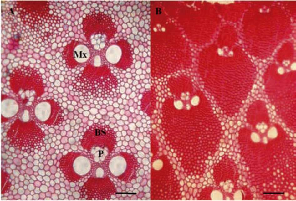
Figure 1. Vascular bundles from the inner and outer culm walls of F. yun-nanensis in 36 months old culms. (A) Open type vascular bundles from the inner culm wall; (B) Semi-open type vascular bundles from the outer culm wall. Mx: metaxylem; P: phloem; Px: protoxylem; BS: bundle sheath. Scale bars = 100 (im.
ity were selected from each age category (Gritsch, 2004). Image of all the fibers in the bundle sheathes as seen in cross-section were captured via a video camera linked to a converted fluorescence microscope (Zeiss Axiovert 200M) and a Lenovo computer. Three images of each bundle sheathes were taken and the cells wall thickness of each fiber cell was measured and marked on the images using the Carl Zeiss Imaging systems, in order to carry out the correlation analysis conveniently.
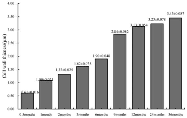
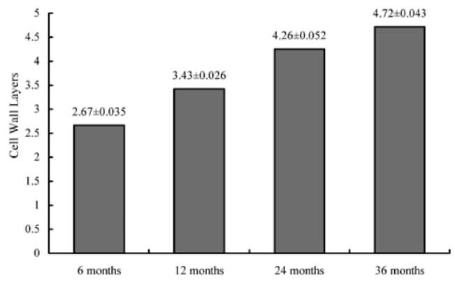
Cell wall layers for each numbered cell were counted as observed under the microscope using the x100 oil-immersion objective. Data were graphically represented
using Microsoft Excel 7.0 and the correlation between the number of wall layers and thickness were verified using SPSS 13.0.
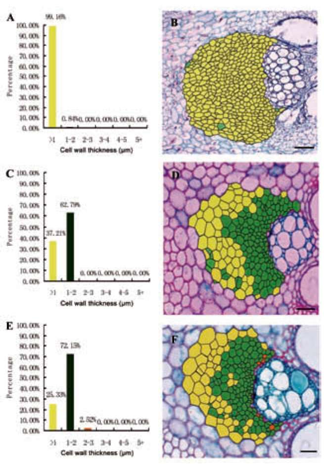
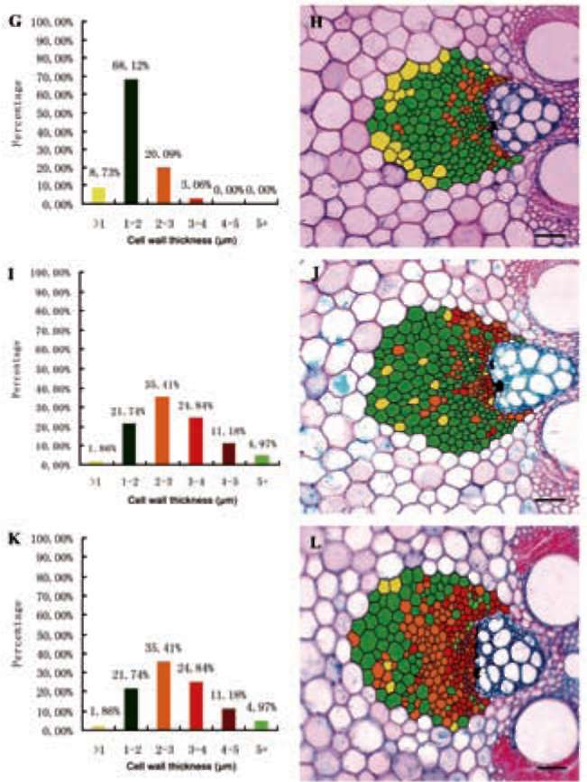
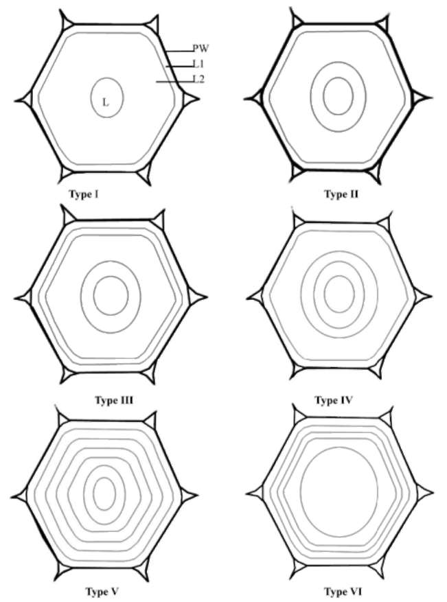
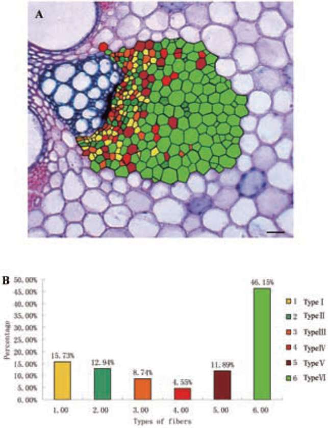
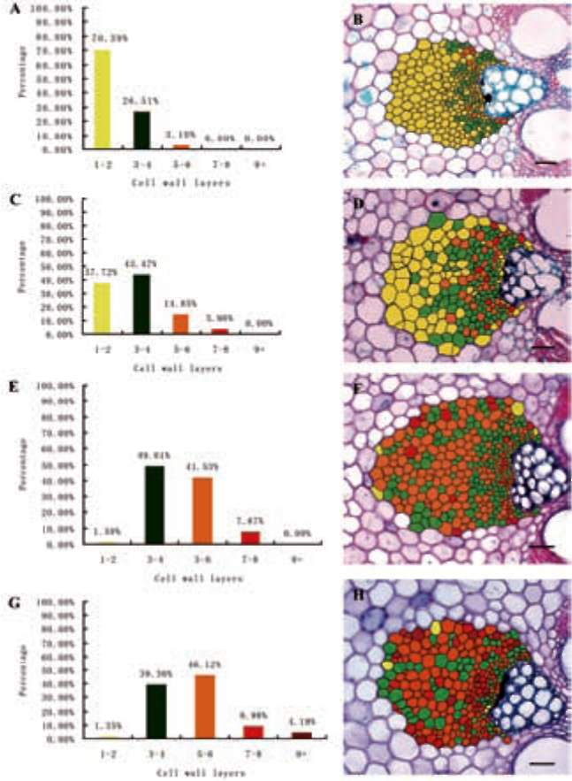
All images for two-dimensional synthesis were taken using x 10 objective and then were processed using Adobe Photoshop 7.0 (Gritsch, 2003). A printed enlargement was used to trace each cell in the bundle sheath onto tracing paper. The traced drawings of the bundle sheathes were then scanned and overlaid onto the original digital photo­graph. Fibers were categorised according to the number of cell wall layers (1-2, 3-4, 5-6, 7-8, 9 or more) and a colour was assigned to each category.
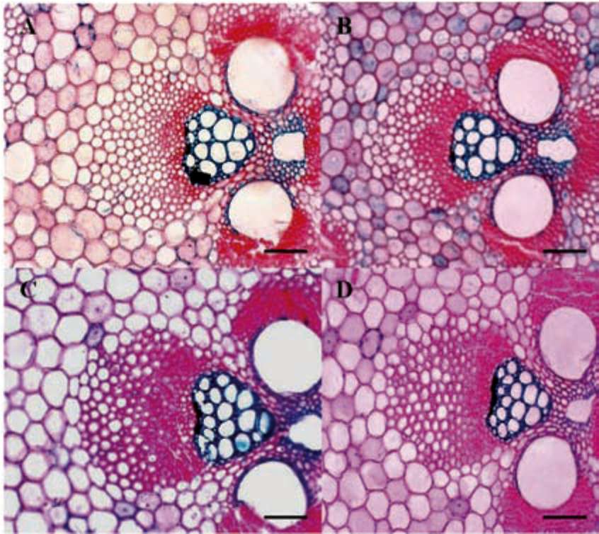
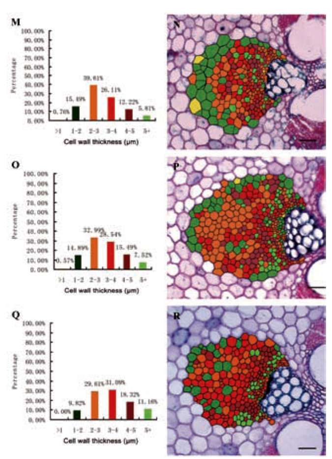
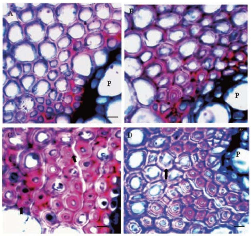
Figure 2. Vascular bundles from the inner culm wall. (A) 6 months old culm (Elongating culm). Fibers with the thickest cell walls are found close to the vascular elements and cells adjacent to phloem remain relatively thin-walled (arrow­head); (B) 12 months old culm (Young culm). The majority of fiber cell walls have begun to thicken; (C) 24 months old culm. Most of fiber cell walls located in the periphery have high potential for further thickening; (D) 36 months old culm. Many fiber cell walls including some immediately adjacent to the phloem still have not reached their maximum thickness. Scale bars = 75 (im.
356
Botanical Studies, Vol. 53, 2012
RESULTS
thick cell wall decreased. At the same time, the percent­ages of fibers with 2-3 (im and 3-4 (im thick cell wall in­creased significantly and an increase of over 4 (im in cell wall thickness occurred between 6 months and 9 months (Figure 3, Figure 4-2K, L).
Cell wall thickness
The bamboo culms of F. yunnanensis usually have two types of vascular bundles-open type and semi-open type (Figure 1).
After 12 months, the fibers had an average cell wall thickness of 3.13±0.054 (im and the fibers wall began to deposit slowly. Only 0.76% of fibers had the wall thick­ness of 1-2 (im and the percentage of fibers with 2-3 (im thick walls had been the highest, which got up to 39.61%. Fibers with <1 (im thick wall had not been found in 36 months old culms, which had an average wall thickness of 3.45±0.087 (im (Figure 3, Figures 4-3 Q, R).
Figure 2 shows representative vascular bundles from culms of the four ages analyzed. Within the bundle sheathes, the typical pattern of wall thickening was ob­served, with fibers close to xylem and phloem elements maturing first. The walls of some fibers immediately adjacent to the phloem developed slowly even did not complete their thickening after two years. Gritsch (2004) reported that these fibers would complete their thickening at the same time as large-diameter fibers at the periphery of the bundle sheath close to phloem.
The main period of secondary wall deposition and thickening in phloem fibers of F. yunnanensis occurred during the first year of growth, mainly including two de­velopmental stages. The fiber cell wall thickness increased significantly from 0.5 months to 1 month and from 6 months to 12 months of age and the mean wall thickness increased slowly after 12 months (Figure 3).
The mean cell wall thickness of phloem fibers was 0.61±0.016 (im in new germinated bamboo shoots (0.5 months old) and most fiber cell wall thickness were less than 1 (m, which got up to 99.16% (Figure 4-1A). After 1 month, the mean cell wall thickness (1.09±0.021 im) increased sharply due to the fact that 62.79% of fibers had the wall thickness of 1-2 (m and the percentage of fibers wall thickness less than 1(im had decreased quickly from 99.16% to 32.71%. On the face of it, the deposition speed of fiber wall thickness in this developmental stage was very quick. When at 2 months old, the developmental speed of fiber wall began to decrease. However, at the age of 6 months, the mean cell wall thickness was 1.900.048 (im and at 9 months old was 2.84±0.082 (im, thus the mean fiber wall thickness increased steeply in this devel­opmental stage and great percentage of fibers with 1-2 (im
Figure 4-1. Data regarding the fiber wall thickness (A, C and E) and two-dimensional maps (B, D and F) of the distribution of fi­bers in bundle sheath close to phloem according to the fiber wall thickness. (A, B) 0.5 months old culm showing a high percent­age of fibers with thin wall thickness (<1 (im). Scale bars = 100 (m; (C, D) 1 month culm showing a substantial increase in the percentage of fibers with wall thickness of 1-2 (im close to phlo­em and fibers with wall thickness less than 1(im still accounted for considerable proportion. Scale bars = 60 (im; (E, F) 2 months old cum showing the highest percentage (72.15%) of fibers with wall thickness of 1-2 (m. The percentage of fibers with wall thickness less than 1 (im further decreased and some fibers with wall thickness of 2-3 (m occurred close to the phloem. Scale bars = 40 (im.
Figure 3. Change in average cell wall thickness of bundle sheath fibers from 0.5 months to 36 months. The fiber cell wall thicken­ing tendency from 0.5months to 1 month and from 6 months to 12 months of age increased significantly.
WANG et al. ― Developmental changes in cell wall of bundle sheath fibers close to phloem
357
seen clearly and were counted (Figure 5). The layering pattern among individual fibers varied in terms of number of layers present and thickness of individual layers. At maturity (36 months old culm), several different cell wall layering patterns could be observed. In order to estab­lish the degree of variation in cell wall patterning among fibers, a classification system of the most commonly ob­served patterns was devised based on light microscopy observations in bundle sheath close to phloem of mature culms (Figure 6).
The distribution of the six types of fibers (Type I to VI) and their proportion in mature culms were presented in Figure 7. Fiber patterns from I to VI were mainly close to
Figure 4-2. Data regarding the fiber wall thickness (G, I and K) and two-dimensional maps (H, J and L) of the distribution of fibers in bundle sheathes close to phloem according to the fiber wall thickness; (G, H) 3 months old culm showing the per­centage of fibers with wall thickness less than 1 (im decreased greatly and only located in the periphery of the bundle sheath and fibers with wall thickness of 3-4 (im occurred close to the phloem; (I, J) 6 months old culm showing the percentage of fibers with wall thickness of 3-4 (im and 4-5 (im increased; (K, L) 9 months old culm showing the percentage of fibers with wall thickness of 1-2 (im decreased steeply and fibers with wall thickness>2 (im increased greatly. Fibers with wall thickness>4 (im occurred adjacent to phloem element. Scale bars = 60 (im.
Two-dimensional maps regarding fiber wall thickness
Two-dimensional maps of representative bundle sheath fibers close to phloem for each age, showing the distribu­tion pattern of fibers according to their cell wall thickness, are presented in Figure 4 (B, D, F, H, J, L, N, P and R). In the young, elongating culm the colors used to illustrate those fibers with the thickest cell wall are located close to the phloem elements. The middle and outer parts of the bundle sheathes are composed of cells with thinner cell wall. There is a strong tendency that the distribution is more heterogeneous with age.
Figure 4-3. Data regarding the fiber wall thickness (M, O and Q) and two-dimensional maps (N, P and R) of the distribution of fibers in bundle sheathes close to phloem according to the fiber wall thickness. (M, N) 12 months old culm showing the high­est percentage of fibers with wall thickness of 2-3 (im and the percentage of fibers with wall thickness less than 1(im continued to decrease; (O, P) 24 months old culm showing the proportion of fibers with wall thickness of 1-2 (im and 2-3 (im continuing to decrease and fibers with wall thickness>3 (im continuing to increase. The fibers with wall thickness<1 (m still could be find; (Q, R) 36 months old culm showing the percentage of fi­bers with wall thickness<3 (im decreasing. The fibers with wall thickness<1 (im could not be find any more and most of fibers with thicker wall were close to the phloem and the minority lo­cated at the periphery. Scale bars = 60 (m.
Cell wall layering structure
Cell wall layers for each numbered cell under the mi­croscope using the x100 oil-immersion ojective could be
358
Botanical Studies, Vol. 53, 2012
phloem and fully developed fibers (i.e. with small lumina) immediately adjacent to the phloem had highly multi-layered walls with regularly spaced layers of type V. In mature culms of F. yunnanensis, a lot of fibers still had not completed their cell wall thickening and had relatively big lumina, thus had high potential to deposit and these fibers were classified to type VI. Type V fibers were usually de­veloped from type VI.
which were mainly located at the periphery and close to the phloem.
Two-dimensional maps regarding fiber wall layers
Two-dimensional maps of representative bundle sheathes close to phloem for each age, showing the dis­tribution pattern of fibers according to their number of wall layers, are presented in Figure 8 (B, D, F, H). In the 6 months old culm the colors used to illustrate the fibers with the highest layers were located close to the phloem elements (Figure 8B). The middle and outer parts of the bundle sheath were composed of cells with only one or two layers (yellow color) accounting for 70.39%. In the 12 months old culm the distribution was more heterogeneous. Nevertheless, there is a strong tendency for fibers with the highest number of layers to be near the phloem. In the 24 months old culm the fibers with 5-6 layers illustrated by the orange color increased significantly. In the 36 months old culm most fibers with nine or more layers were located close to the phloem and the periphery of the bundle sheath. Meanwhile, small quantities of thin wall fibers with 1-2 layers still could be observed.
Cell wall layering and development
The average cell wall layers after 6 months were 2.67±0.035 layers in phloem fibers, accounting for 70.39%, among which the most fibers had 2 layers (Figure 9). However, the percentage of fibers with wall layering>3 layers was small, mainly close to the phloem and no fibers with 7-8 layers occurred in this period. After 1 year, the percentage of fibers with 1-2 layers decreased drastically from 70.39% to 37.72%. At the same time, the fibers with 3-6 layers increased significantly and fibers with 7-8 lay­ers began to form. The main change in 24 months old culm were the significant decrease of fibers with 1-2 layers and the significant increase of fibers with 5-6 layers. The fibers with wall layering >9 layers were not found. Small quantities of fibers with 1-2 layers still can be observed, immediately adjacent to the phloem and in the periphery of the bundle sheathes, in three years old culm. The per­centage of fibers with 3-4 layers began to decrease and the fibers with wall layering >9 layers could be observed,
Cell wall thickness and number of cell wall layers
According to the distribution pattern within the mature bundle sheath fibers close to phloem (Figure 4 R and 8 H)
Figure 5. Layering patterns in the bundle sheathes close to phloem. (A) 6 months old culm. Fibers close to the phloem (P). The majority of fibers have two to three cell wall layers; (B) 1 2 months old culm showing fibers close to phloem; (C) In 24 months old culm, fiber close to the phloem showing the cell with layering pattern type I (arrowhead); (D) In 36 months old culm, large-lumina fibers (type VI, arrowhead) still could be found that indicated great potential to wall thickening for mature culm. Scale bars = 15 (m.
WANG et al. ― Developmental changes in cell wall of bundle sheath fibers close to phloem
359
Table 3. The correlation between cell wall thickness and layers at different age classes.
Fiber wall thickness
Age
6 months 12 months 24 months 36 months
Fiber wall layers
6 months
0.762**
12 months
0.554*
24 months
0.339
36 months
0.295
Figure 6. The fiber layering patterns in the bundle sheathes close to phloem of 36 months old culm of F. yunnanensis. The por­tion between two black lines was one secondary wall layer. The thickness of the broad layers is variable, whereas the thin layers appear to have a more constant thickness. Fiber pattern of type V was usually developed from type VI. PW: primary cell wall; L1, L2: secondary cell wall layers; L: lumen.
both the fibers with thicker wall and more layers mainly distributed around the phloem, but the location of their distribution is not the same, which indicated that there was no significant correlation between cell wall thickness and number of cell wall layers in 36 months old culm.
Figure 7. The two dimensional map of 6 different types of fi­bers (type I to VI) in mature culms and their proportion in fiber bundles. Lots of fibers in the periphery still have high potential to deposit. Scale bars = 30 pm.
The correlation between cell wall thickness and number of cell wall layers was significant in 6 and 12 months old culms and decreased with age (Table 3). In 6 months old culm the number of fiber wall layers was generally low and the fiber wall thickness was interrelated with the number of fiber layers. In 12 months old culm the correlation was still significant but decreased that implicated the main morphological change of fibers in this period was the number increment of fiber layers and the secondary cell wall layer thickening, which had a significant impact on the correlation between cell wall thickness and number of cell wall layers. In 24 and 36 months old culms the correlation were not significant any more that indicted the wall thickness was not associated with the number of wall layers. It can be observed that some fibers close to phloem had thick cell wall but with few cell wall layers. However, the fibers distributed in the periphery of bundle sheathes
had thin wall but with more layers. It implicated that the fiber wall thickness may be associated with the location of a fiber within the bundle.
DISCUSSION
The present studies on the development of the bundle sheath fibers close to phloem has shown that changes in cell wall layering across the bundle sheathes are simi­lar but somewhat more pronounced than in the xylem sheathes (Murphy and Alvin, 1992). The development in xylem and phloem fibers is similar and the sequence of development for phloem fibers can be considered as a reasonable representation for bamboo fibers in general (Gritsch et al., 2004).
360
Botanical Studies, Vol. 53, 2012
Fiber cell wall thickness
The deposition of bundle sheath fibers close to phloem is mainly during the first year including two developmen­tal stages. The first developmental stage is just the period of the bamboo shoots sprouting out from 2 weeks to 1month. Bamboo shoots grow fast in this developmental phase, so are the fiber cell walls, and this phase is usually the end of the local rainy season in Yunnan province (from September to October). The second developmental stage is the phase of the bamboo shoots sprouting out from 6 months to 9 months (from March to June). It is the phase from the spring season to the beginning of rainy season in the second year and the phase of fiber cell walls in bundle sheathes close to phloem thickening obviously and new branches and leaves of young bamboos stretching out rap­idly at the same time. Gan and Ding (2006) reported that the deposition of fibers wall is analogous with the growth rule of tree rings and the main fiber wall deposition stage in Phyllostachy edulis is from March to June.
In subsequent several growing seasons, fibers wall will thicken constantly with a slow pace. In 36 months old bamboo many fibers remaining relatively thin walled, usu­ally of larger diameter, still can be observed. They are usu­ally located at the periphery of the bundle sheathes close to parenchyma cells but some are close to the phloem elements and it indicates that fibers of F. yunnanensis take longer time to mature than traditional opinions. Gan and Ding (2008) have pointed that the fiber cell in culms of P. edulis is one special long-lived cell in which the protoplast could survive for a long time. It indicated that the fibers wall of P. edulis needs long time to finish their fully depo­sition as alike as F. yunnanensis if the death of fiber cells in bamboo culms was considered as maturation.
Figure 8. Data regarding the layering of fiber cell walls (A, C, E and G) and two-dimensional maps (B, D, F and H) of the distri­bution of fibers in bundle sheathes close to phloem according to the number of cell wall layers. (A, B) 6 months old culm show­ing a high percentage of fibers with a low number of layers. The majority of fibers with more layers were close to the phloem; (C, D) 12 months old culm showing a substantial increase in the number of layers. The fibers with 7-8 layers began to form; (E, F) 24 months old culm showing the significant decrease of fibers with 1-2 layers and the remarkable increase of fibers with 5-6 layers; (G, H) 36 months old culm showing the occurence of fibers with nine or more layers and the constant increase of the numbers of fibers with 5-6 layers and 7-8 layers. Scale bars = 60 pm.
Bhat (2003) reported that fiber thickening in the inner culm wall bundles of Bambusa bambos and Dendrocala-mus strictus starts in the vicinity of the phloem and xylem tissues and proceeds toward the outer parts of bundle. The present observations in F. yunnanensis confirm the same general pattern. However, some fibers immediately adja­cent to the phloem matured slowly and even completed their thickening at the time as the large diameter fibers in the periphery of the bundle. Gritsch et al. (2004) reported similar observations in Dendrocalamus asper and pointed out that such a developmental pattern might be linked to a parenchymatous origin of the cells and there is a certain degree of intergradations between fibers and parenchyma cell types and result in a development pattern with aspects similar to true parenchyma cells.
Layering structure of phloem fiber cell wall
There was a great degree of heterogeneity in the layer­ing structure of fiber cell walls according to the distribu­tion of fiber cell wall layers in F. yunnanensis and six main types of fiber walls were identified including one type with large lumina located at the periphery of the bundle sheath, which is similar with the fiber types in Dendro-calamus asper depicted by Gritsch et al. (2004). Murphy
Figure 9. Change in number of cell wall layers of bundle sheath fibers for each growth period, showing the great increase from 6 months to 24 months and low increase from 24 months to 36 months.
WANG et al. ― Developmental changes in cell wall of bundle sheath fibers close to phloem
361
and Alvin (1992) reported that the fibers with large lumina have intermediate characteristics between parenchyma and fibers and at the same time they pointed out that there is much variation in wall structure of Phyllostachys viridi-glaucescens and categorized fibers into four major types according to their layering structure. Kawase et al. (1986) reported that in the bamboo Sasa kurilensis the fiber cell walls after 4 years are composed of 7-8 layers and Gritsch et al. reported the the average number of wall layers was two in the young culm and increased to five in the fully elongated older culms. In the present study, the fibers in the young culms of Fargesia yunnanensis are composed of one or two layers and the average number of wall lay­ers increased to 4.72±0.043 in mature culms (36 months old). This average masks the real increase of the fiber wall layers with more than 4.72±0.043 layers in mature culms and a lot of fibers had nine or more layers, which is similar with that of Dendrocalamus asper (Gritsch et al., 2004).
The fiber wall thickening of Fargesia yunnanensis is not totally dependent on the layer number increase and sometimes, they reply on the widening between layers. Ac­cording to the actual observation of the bundle sheathes in mature culms, some fibers close to the phloem with simi­lar diameter and wall thickness have huge disparity in the wall layer numbers. Some cell walls have 6 or more layers and other cell walls, however, have only 2-3 layers. At the same time, we have found that some fibers with thin wall may have more layers than that of fibers with thick wall. It can be concluded that there are two ways in the fiber wall thickening: (1) the widening between layers; (2) the in­creasing number of fiber wall layers. It is difficult to verify that which the main way is or both are the main ways in the fiber wall thickening, which need to be further studied in future.
Two-dimensional maps regarding fiber wall layers
Cell wall thickness and number of wall layers
Fibers with the thickest wall often appeared firstly in the location near the phloem in the development of fiber wall. However, fibers with the highest number of wall lay­ers were located both adjacent to the phloem and in the periphery of the bundle sheathes in mature culms. Most of fibers in the periphery of the bundle sheathes have rela­tively big lumina in mature culms (>3 years), which indi­cated that the main way of the fibers' wall thickening in the periphery is the increasing number of fiber wall layers in the early developmental stage of the culms. However, both two main ways of the fibers' wall thickening can be observed in the region adjacent to the phloem. The fibers distributed in the periphery of the bundle sheath close to phloem will develop into type V, which is similar with the depiction about the fibers of type VI in Dendrocalamus asper (Gritsch et al., 2004) and type IV in Phyllostachys viridi-glaucescens depicted by Murphy and Alvin (1992).
Parameswaran and Liese (1976) have ever described two kinds of fibers, namely thick-walled 'polylamellated' fibers with up to 18 'lamellae' and non-lamellated thin-walled fibers in several bamboo species. However Murphy and Alvin (1992), Gritsch et al. (2004) pointed out that the number of layers within any one wall does not depend entirely on its thickness, since very thick walls can pos­ses anything from one or two layers up to more than nine. According to the two-dimension maps of cell wall thick­ness, it can be demonstrated that the thickness the fiber wall thickness is related not only to the location within the bundle sheathes but also to the diameter of the fibers. Usu­ally, the lumen diameter increased with the diameter of the fibers and it needs to more time to complete their thicken­ing and the cell wall thickness will be higher.
Liese and Weiner (1996) pointed out that the develop­ment of bamboo fibers occurs in two main phases. The main one happens during the first 2 years and leads to the development of fibers with lignified cell walls. A second phase follows for several years after during which ad­ditional lamellae are formed. Conversely, Gritsch et al. (2004) reported that it seems unlikely that significant wall deposition in later years (i.e. beyond 36 months) would have been possible and then was not possible to verify the second phase of fiber wall development due to the small lumina in bundle sheath fibers close to phloem of 36 months old culms. In the present study it was not verified the second phase of fiber development due to the age range of the test materials (0-36 months) of Fargesia yunnanensis. However, some fibers on the periphery of the bundle sheathes close to phloem in 36 months old culms of F. yunnanensis still have large lumina diameter and they maybe occur the main second phase that need to be verified. Although there are two significant devel­opmental phase in the first growth year, both of them are due to the increase of the fiber thickness not the number of wall layers.
Usually, type I and type II developed from relatively small fiber cells and their formation time is earlier than other types. No direct relation could be found between the number of layers and the types of fibers. The fibers of type VI always have many layers although they have big lumina, which maybe develop through the main way of the widening between layers in the subsequent developmental stage that need further confirmation in future.
CONCLUSION
Six different fiber wall types could be distinguished in the bundle sheath fibers close to phloem of 36 months old culms. The correlation between the number of wall layers and thickness was not obvious in mature culms. Usually, thinner-walled fibers could have a large number of layers and thicker-walled fibers could also have less number of layers. The fibers close to the phloem began to develop earlier than other fibers. The 36 months old culms still have lots of fibers remaining relative thin-walled and sub­stantial further thickening occurs in subsequent growing years.
362
Botanical Studies, Vol. 53, 2012
The development of fibers has two main phases in the first growth year. The first main developmental phase is the period from 0.5 months for bamboo shoots germination to 1 month. The second developmental phase is the period that the bamboo shoots sprouting out from 6 months to 9 months. There are two main ways in the fiber wall thick-ening, i.e. the widening between layers and the increasing number of fiber wall layers.
(Schultes F.) Backer ex Heyne. PhD thesis, Imperial Col­lege, University of London, UK.
Gritsch, C.S., G. Kleist, and R.J. Murphy. 2004. Developmental
changes in cell wall structure of phloem fibres of the bam­boo Dendrocalamus asper. Ann. Bot. 94: 497-505.
Itoh, T. 1990. Lignification of bamboo (Phyllostachys heterocyla Mitf.) during its growth. Holzforschung 44: 191-200.
Kawase, K., K. Sato, H. Imagawa, and U. Ujiie. 1986. Studies on utilisation of Sasa-bamboos as forest resources. 4. Pulp­ing of young culms and histological change of cell structure of the culms in growing process. Res. Bull. Coll. Exp. For­ests, Hokkaido University 43: 73-96.
Latif, M.A. 2001. Anatomical features of Bambusa vulgaris and Gigantochloa scortechinii from four harvesting sites in Pen­insular Malaysia. J. Trop. For. Products 7: 10-28.
Liese, W. and G. Weiner. 1996. Ageing of bamboo culms. A re­view. Wood Sci. Technol. 30: 77-89.
Lybeer, B., G. Koch, J.V. Acker, and P. Goetghebeur. 2006. Lig-
nification and cell wall thickening in nodes of Phyllostachys viridiglaucescens and Phyllostachys nigra. Ann. Bot. 97:529-539.
Murphy, R.J. and K.Alvin. 1992. Variation in fibre wall struc­ture in bamboo. IAWA Bull. 13: 403-410.
O'Brein, T.P., N. Fedr, and M.E. McCully. 1964. Polychromatic staining of plant cell walls by toluidine blue O. Protoplasma 59: 367-373.
Parameswaran, N. and W. Liese. 1976. On the fine structure of bamboo fibres. Wood Sci. Technol. 10: 231-246.
Acknowledgements. We are thankful to the related anon­ymous reviewer for their valuable comments on the earlier version of this paper. The project was fully funded by the National Natural Science Fund of China (31100453) and the national "twelfth Five-Year" scientific and technologi­cal support plan subject (2012BAD23B05). We are grate­ful to all of them.
LITERATURE CITED
Bhat, K.V. 2003. Anatomical changes during culm maturation in Bambusa bambos (L.) Voss and Dendrocalamus strictus Nees. J. Bamboo Rattan 2: 153-166.
Gan, X.H. and Y.L. Ding. 2006. Ultrastructural study of the fiber developmental process in the culms of Phyllostachys edulis. Chinese Bull. Bot. 21(2): 180-188.
Gan, X.H. and Y.L. Ding. 2008. A special long-lived cell: the culm fiber cell of Phyllostachys edulis. Acta Bot. Yunn. 30(2): 151-158.
Gritsch, C.S. 2003. Aspects of cell wall formation during culm development in the Bamboo Dendrocalamus asper
雲南箭竹韌皮部纖維鞘纖維細胞壁發育變化
王曙光1 林樹燕2 普曉蘭1 丁雨龍2 萬賢崇3
1雲南昆明西南林業大學西南山地森林資源保育與利用省部共建教育部重點實驗室
2江蘇南京林業大學竹類研究所
3北京中國林業科學研究院新技術研究所
對雲南箭竹從剛萌發0.5個月竹筍至36個月竹稈韌皮部纖維鞘纖維細胞壁包括細胞壁厚度和層數
發育之變化進行研究。發現雲南箭竹稈中存在兩種維管束類型,且臨近維管束鞘纖維細胞發育早於其他
纖維。在竹子生長的第一年纖維細胞壁增厚可分為兩個顯著地發育階段。在三年生竹稈韌皮部纖維鞘中
可見6種纖維細胞壁類型並且纖維鞘外圍許多纖維細胞仍然存在較大的細胞腔。在韌皮部纖維鞘纖維細
胞壁發育過程中有兩種主要的沉積途徑。
關鍵詞:纖維細胞壁;細胞壁厚度;細胞壁層;發育。