Botanical Studies (2012) 53: 459-465
BIOCHEMISTRY
Toxicity attenuation of atractyloside in traditional chinese medicinal herbs after hydrothermal processing
Chien-Hua CHU1, Chi-Ming CHIU4, Anren HU3, Hui-Chung WU4, Shu-Ping YE4, Kuo-Chieh HO1'2 *, and Liang-Yu CHEN4 *
1Institute of Plant Biology, National Taiwan University, Taipei, 106, Taiwan
2Department of Food Science, National Quemoy University, Kinmen, 892, Taiwan
3Institute of Medical Biotechnology, Tzu-Chi University, Hualien, 970 Taiwan
4Department of Biotechnology, Ming-Chuan University, Gui-Shan, 333, Taiwan
(Received February 14, 2012; Accepted June 21, 2012)
ABSTRACT.
Atractyloside (ATR) is a diterpenoid glycoside and occurs naturally in Asteraceae plants, many
of which are used in foods and ethnomedicines. The toxicity of ATR has caused fatal renal proximal tubule necrosis and/or centrilobular hepatic necrosis in man and farm animals. A GC/MS screening method was op­timized to detect the presence of ATR in Atractylodes species which are important herbs used in traditional Chinese medicine. The stability and hydrothermal detoxification of ATR in practical processing were also evaluated quantitatively by chromatographic analysis. In addition, the effects of hydrothermal detoxification of ATR from different processing methods on cell viability were performed. The results suggest that degradation of ATR is a way to increase pharmaceutical safety for the Chinese medicinal industries.
Keywords:
Atractylodes lancea; Atractylodes macrocephala; Detoxification; Hydrothermal processing; Solid
phase extraction; Xanthium strumarium.
INTRODUCTION
of two biologically active parts, an aglycone with a perhy-droplenanthrene structure, and a glycoside moiety made up of glucose, isovalerate and sulfate. The 4-carboxy-atractyloside (CATR) is more toxic than the ATR, and is found in fresh but not in dried plants because it is deoxy-lated to ATR during ageing or desiccation (Obatomi and Bach, 1998; Steenkamp et al., 2004). The interest in these compounds was stimulated by the high toxicity of both glucosides, which has been responsible for many deadly poisonings (Obatomi and Bach, 1998; Laurens et al., 2001; Popat et al., 2001; Steenkamp et al., 2004; Daniele et al.,2005; Steenkamp et al., 2006).
The use of medicinal plants has increased significantly in many developed countries. There is a predominant view that herbal medicines are harmless and free of side effects because they have a natural source. However, numerous cases of hepatic injury and even death were associated with the use of herbal medicines (Larrey and Faure, 2011; Stickel and Schuppan, 2007; Stickel et al., 2005). The safety of several commercially available herbs has recently come into question due to reports of adverse reactions with prescription drugs. The safe and effective use of medicinal herbs has therefore been identified as a top research prior­ity (Burns et al., 2010; Cheng et al., 2010).
ATR occurs naturally in many Asteraceae plants and many of these plants are important herbal medicines (Atractylis species, Wedelia species, and Xanthuim spe­cies, as shown in Figure 2) (Obatomi and Bach, 1998; Han et al., 2009; Cao et al., 2010; Meng et al., 2010). Among them, dried rhizomes of A. macrocephala and A. lancea are important crude drugs used in herbal medicine. A. macrocephala is commonly used in traditional Chinese formulas to treat phlegm retention, asthma, cough with excessive sputum production, and to stop sweating. The pharmacological human clinical studies have shown A. macrocephala to exert a diuretic effect only in patients with edema and not in normal subjects. It has also been shown to be liver protective and to be a mild hypoglyce-mic agent (Shan and Tian, 2003). A. lancea is mainly used for the treatment of indigestion and stomach disorders
Traditional Chinese medicines (TCMs) are popularly used in Chinese society and countries in eastern Asia. The safety of TCMs is a major concern in Taiwan. Because of the complexity of the components in TCMs, it is often difficult to establish quality control standards for TMCs (Chau and Wu, 2006; Hsiao, 2007).
The structures of Atractyloside (ATR) salt is illustrated in Figure 1. ATR is a diterpenoid glycoside that consists

*Corresponding authors: Tel: +886-233662508, E-mail: kch@ntu.edu.tw (Kuo-Chieh HO); Tel: +886-33507001 ext. 3773; E-mail: loknath@mcu.edu.tw (Liang-Yu CHEN).
460
Botanical Studies, Vol. 53, 2012
MATRIALS AND METHODS
Chemicals, reagents and apparatuses
The potassium salt of atractyloside (>99% by TLC), two internal standards (5a-androstane-3p,17p-diol and cholesterol) and the derivative reagent, trimethylsilyl imi-dazole (TMSI), were purchased from Sigma Chemical-Aldrich (ST. Louis, MO, USA). Ethyl acetate, methanol and pyridine (Silylation grade) were obtained from Merck (Darmstadt, Germany). All chemicals were analytic or ultrapure grade and used without purification. The wa­ter used was the ultrapure deionised water prepared by Milli-Q filtration. The standard solutions of ATR were 100 and 1000 |ig/ml, and stored in the dark at -20°C. Hyper-Sep C18 SPE cartridge (200 mg bed weight /3 ml volume) were purchased from Thermo Scientific Corp. (USA). A 12-port vacuum manifold was used for all extractions.
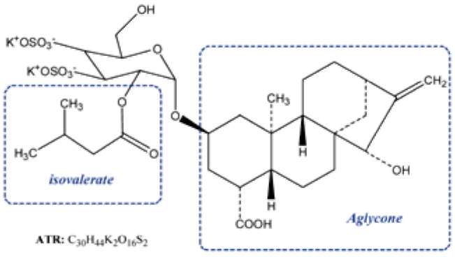
Figure 1.
The chemical structures of atractyloside (R= H) and
corboxy-atractyloside (R= COOH).
(Duan et al., 2008; Wang et al., 2008).
The toxicity and biochemistry of ATRs have been re­viewed (Obatomi and Bach, 1998; Daniele et al., 2005). ATR specifically binds to adenine nucleotide translocation in the inner mitochondrial membrane and competitively inhibits ADP and ATP transport. The complete mechanism of toxicity of ATR is not fully understood. However, crude herbal medicines are brewed (infused and extracted with hot water) before drinking. The toxicity and chemical profiles of ATRs are highly dependent on the variations of both species and processing (Obatomi and Bach, 1998; Steenkamp et al., 2004; Daniele et al., 2005).
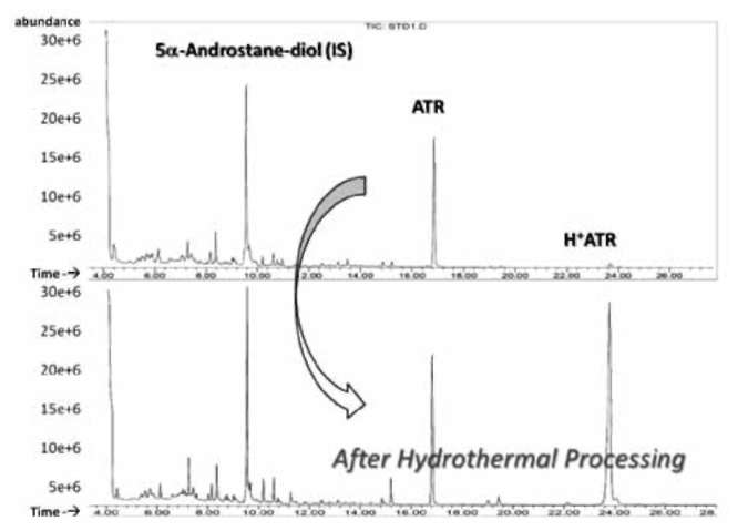
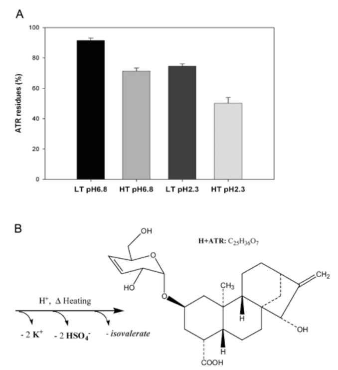
Stability of ATR in processing
Five 200 ml aliquots of 100 [ig/ml ATR were prepared to evaluate the kinetic stability of ATR by heating and acidification. One aliquot was untreated and stored at am­bient temperature (about 22°C) as the negative control. The other samples were adjusted to pH 6.8 and pH 2.3 in 100mM phosphate buffered saline (PBS), and the solutions were incubated at 98°C and 65°C, respectively. Three 2 ml samples were collected after heating for 120 minutes. Then, the samples were extracted using SPE and deriva-tized for GC/MS analysis.
Gas chromatography-mass spectrometry (GC/MS) is widely used in forensic, doping, and toxic screening laboratories because it has the advantage of high sample throughput, rapid detection, and economic feasibility (Lau-rens et al., 2001; Hsiao, 2007; Chen et al., 2011). The aim of this work was to develop a rapid method for determina­tion of ATR in TCMs by GC/MS. Also, the quantitative data acquired will help us to assess the safety and risk of medicinal herbs in pharmaceutical industries and ethnop-harmacology (Chau and Wu, 2006; Hsiao, 2007; Han et al., 2009; Jiang et al., 2010).
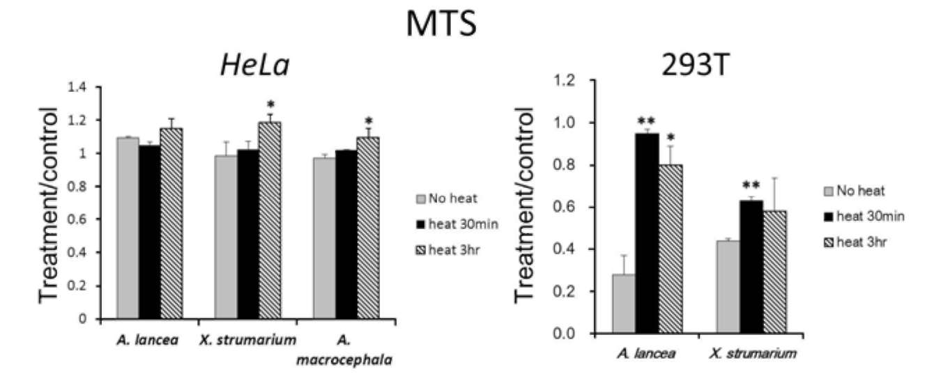
Sample preparation and aqueous infusion
The rhizomes of the A. macrocephala and A. lancea and the fruits of Xanthium strumarium were harvested in the greenhouse of Institute of Plant Biology in NTU when the active principles are believed to reach the highest. After cleaning and drying, the specimens of herbs were pulver­ized to a powder before the infusion process (hot water extraction). Five grams of pulverized powder and 40 ml of water were added into a 250 ml round bottomed flask and heated to boiling for 15 min. Then, the hot solution was
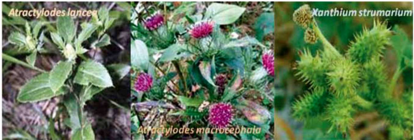
Figure 2.
The Asteraceae plants used in TCM.
CHU et al. ― Detoxification of atractyloside
461
sonicated for 30 seconds and filtered through two layers of gauze, and adjusted to 50 ml by adding water.
plates respectively. The medium was replaced with com­plete medium containing 1/5 diluted extracts of Cocklebur (Xanthium strumarium) seeds, dried rhizomes of the A. macrocephala or A. lancea according to the procedures of sample preparation. The incubation for cell proliferation assay was performed for 48 hours.
Solid phase extraction (SPE)
Three 2.0 ml aliquots of the solution were removed into 10 ml polypropylene centrifuge tubes and acidified with hydrochloric acid (2 ml, 2.0 N). After vortex mixing, the samples were centrifuged at 2500 rpm for 5 minutes. SPE cartridges were conditioned by washing consecutively with 2ml of methanol and 2ml of deionised water. The pre­conditioned cartridges were prevented from drying before applying the supernatants, spiked and standard solutions. The cartridges were washed with 3 ml deionised water before elution. After drying for 2 minutes, two consecutive elutions (2 ml each) were carried out with a mixture of methanol (80:20, v/v) containing 2% ammonium hydroxide, and maintained under vacuum for at least 30 min. The extracts were then evaporated to dryness at 40°C under a stream of nitrogen.
Cell proliferation assay
HeLa cells (1x10⁴ cells/well) and 293T cells (2x10⁴ cells/well) in 96-well plates were incubated with different filtrated extracts for 48 hours. Cell number/ viability were examined by the MTS assay kit (Promega, USA). 20 jjj of Cell Titer 96® AQueous One solution reagent was added in each well at 37°C for 1.5 hours. The reaction products were measured at 450nm by a 96-well plate ELISA reader (GE Healthcare, USA). Cells were inoculated in 96-well plates and then treated with various hot-water extracting conditions (heat 30 minutes, 3 hours, and no heat treat­ments) of medicinal herbs (Atractylodes lancea, Xanthium strumarium seeds, Atractylodes macrocephala). The rela­tive viable cell amount determined by MTS formazan production was converted into relative cell number using the cell titration curve in each plate. The Y axis represents the ratio of relative cell number versus the cell number without medicinal treatment. The collected data were ex­pressed as the mean S.E.M. of at least three wells in two independent experiments.
Derivatization
The dry residues were redispensed in 0.5 ml of metha-nol, and transferred to derivatizing vials. 50 μl aliquots of internal standards solution were spiked in each vial for der-vatization and quantitative analysis. The solvent evaporated to dryness again with nitrogen before 200 μl of pyridine and TMSI was added. The vials were well mixed by vortex. The derivatization was carried out at 100°C for 2 hours.
RESULTS AND DISCUSSION
GC-MS analysis
Analyses were performed with a HP-6890 gas chro-matograph and a HP-5973 mass-selective detector (MSD) from Agilent Technologies (Palo Alto, CA, USA) equipped with a capillary gas chromatography column DB-1MS (30 mx0.25 mm I.D., 0.25 [im film thickness) (J&W Scien­tific, USA). The carrier gas was helium at a flow rate of 1 ml/min and the injection volume was 2 μ Injection was made in splitless mode (0.5 min delay) and the tempera­ture of the inlet was 250°C. The oven temperature was programmed as follow: initial temperature at 210°C for 3 min and increased at 10°C/min up to 270°C, then at 30°C /min until 310°C and held for 15 min. The analysis was performed in the EI mode and the ionization voltage was fixed at 70 eV The scan acquisition (m/z 50-800) of MSD was performed by the HP chemistation software. The quantitative data was calculated by the internal standard method based on integrated area of peaks in selected ion monitoring (SIM) mode.
The natural materials are usually formulated and pro­cessed before their use as medicines in ethnopharmacol-ogy. However, the evaluation of the stability of toxic compounds in processing is very difficult to analyze be­cause of the complicated reactions in the material (Franje et al., 2010; Toh et al., 2010). Pure ATR and the phosphate buffers were used to avoid interferences and optimize ex­perimental conditions.
Cell culture and treatment
The human cervical cancer and kidney cell lines - HeLa and 293T were cultured in DMEM medium supplemented with 10% fetal bovine serum (Hyclone/ Logan, Ut) and penicillin/ streptomycin at 37°C, 5% CO2 incubation. Cells were seeded in 96-well plates and maintained to attach overnight in an incubator. For cell proliferation assay, two cells were seeded at 1x10⁴ and 2x10⁴ cells/well in 96-well
Figure 3.
Chromatograms of the TMS derivatives of standards.
The lower chromatogram shows the acidified and hydrothermal-ly treated ATR (pH 2.3, 98°C) in contrast to the upper one (pH 6.8, 65°C).
462
Botanical Studies, Vol. 53, 2012
Hydrolysis of ATR and mass spectral interpreta­
tions
cates the hydrolysis of isovalerate by acidification before TMS derivatization.
The GC chromatograms obtained from the TMS de­rivatives of ATR standards are shown in Figure 3. The complicity of chromatograms also demonstrates that the degradation of ATR could generate multiple products dur­ing hydrothermal processing. A new peak was observed in the chromatogram of acidified and 120 minute hydrother-mally treated ATR and named as the H+ATR.
Degradation of ATR
The hydrothermal processing is a very convenient pro­tocol to be applied to TCM's. According to the literature reviews (Obatomi and Bach, 1998; Daniele et al., 2005), many endogenesis toxins are unstable under high tempera­ture, oxidative, or light irradiated (Buscemi et al., 2003) conditions. Therefore, the appropriate processing condi­tions might be able to attenuate the toxicity of crude herbs. The degradation of ATR by hydrothermal treatments is shown in Figure 4. At high and moderate temperatures, degradation of ATR was observed and increased with incu­bation time. Acidification also enhances the efficiency of degradation. The utility of SPE in sample preparation pro­cedure could avoid the esterification errors derived from the traditional method with liquid-liquid extractions. Ap­proximate 50% of ATR could be removed by hydrothermal processing at 98°C and pH 2.3 for 2 hours. Detoxication results from decomposition, hydrolysis and saponification of toxic analytes, which react in an aqueous solution of a natural mixture. Thus could explain the reason that reports of intoxications are rare from brewing and drinking the herbs in TCMs, owing to the effects of the hydrothermal processing.
Based on our previous study (Chen et al., 2011), the mass spectrum of ATR (retention time at 16.836 min) is characterized by ion fragments at m/z 316, 647, and 663. The difference of the fragments at m/z 647 and 663 were due to the oxygen atom (m/z 16). The ion fragments at m/ z 316 was observed as the TMS derivative of sulfate group (-OS-(O-TMS)3. Therefore, the molecular structure of ATR derivatives should contain one of sulfate group at least.
The mass spectrum of H+ATR (retention time at 23.747 min) is characterized by ion fragments at m/z 289 and 447. The standard spectrum was first reported by Laurens et al. while detecting the acidified ATR in a patient poi­soned with Callipepis laureola (Laurens et al., 2001). The ion fragment at m/z 447 was the aglycone moiety, disrupt­ed on diterpene side of glycosidic oxygen. The other ion fragment at m/z 289 is formed from the glucose moiety due to loss of sulfates. The structural analysis of fragment at m/z 289 with two trimethylsilyl groups (-O-TMS) indi-
We found that the hydrolysis of isovalerate and sulfate groups is increased by acidification and hydrothermal pro­cessing. Therefore, a possible degradation mechanism was revealed to the sodium or potassium salt of ATR in natural medical herbs. The sulphuric acid esters in acidified ART may also be hydrolyzed by heating. A possible mechanism was shown in Figure 4B.
Determination of ATR and method validation
Seven-point calibration curves were prepared by spik­ing corresponding amounts of target compounds in de-ionized water with two internal standards. The internal standard cholesterol was used for quantitative analysis of the samples. The calibration curves had a good linear cor­relation (R2>0.99). The mass spectrometer was operated in SIM mode for quantitative measurements. The limit of detection (LOD) for the standard was about 7 [g/ml and determined experimentally. The LOD was taken as the concentration of the standard which produced a signal-to-noise (S/N) ratio of 3. The recovery of 100 [g-ATR/ml spiked in samples was 87-103%.
The total ion chromatograms (TIC) were obtained from the TMS derivatives of extracts of A. lancea and A. mac-rocephala. The ATR level in each of raw botanic materials were evaluated and reviewed from the previous literatures (Chen et al., 2011; Daniele et al., 2005; Obatomi and Bach, 1998; Steenkamp et al., 2004). The high levels of ATR in two TCM herbs were determined. However, there are ge-netic, histological, environmental, and time of harvest fac-tors which influence the ATR content of botanic samples. Our results indicate that because of toxic materials in
Figure 4.
(A) The ATR in solution were degraded by acidifi-
cation and heating for 120 minutes. The LT and HT are rep­resented as the high temperature (98°C) and low temperature l(65C), respectively. (B) The possible structure of H+ATR and mechanism were depicted after the acidification and hydrother-mal processing.
CHU et al. ― Detoxification of atractyloside
463
Figure 5.
The effect of hydrothermal detoxification of ATR from three different extracts on cell viability in HeLa and 293T cells. The
results are showed as the meansS.D. *, p <0.05; **, p<0.01, control versus treated ones.
TCMs, they should be harvested and stored for extended times before use, since the level of toxic compounds de­creases with ageing.
pharmaceutical safety. The toxic effect of Atractylis spe­cies arises from ATR. This study validates the degradation of ATR as a possible mechanism for reducing its toxicity. Hydrothermal processing could also facilitate the detoxi­fication of the raw materials used in the Chinese medicine industry.
The effect of hydrothermal processing extracts
on cell proliferation
ATR has been demonstrated to cause damage to kid­ney and liver. The detoxifying processing of Atractylodes medicine via hot water boiling is usually observed in the preparation of traditional Chinese medicine. To under­stand whether the procedure will eliminate the toxicity of ATR, we compared the cell viability under the incubation of untreated and heat-processing plant extracts. Interest­ingly, the results showed that the hydrothermal process caused different influences in these two cell lines. With Hela cells, there were no significant changes observed in the cell viability, only slightly increased when Hela cells were cultured in medicinal extracts pre-treated with 3 hours hot water (Figure 5A). With human kidney cell line, the untreated Atractylodes lancea or Xanthium strumarium crude extracts caused a negative effect on the 293T cell growth and this negative effect was removed when the hydrothermal process was applied (Figure 5B). The differ­ent influences of hydrothermal process in the two cell lines correspond to the fact that ATR has been demonstrated to cause damage to kidney, thus the toxicity effect of ATR and the consequence of hydrothermal process is more sig­nificant in the 293T cell. The cell viability results provide evidence that hydrothermal detoxification of ATR should be a useful procedure to decrease the negative effect of ATR in herb medicines of Atractylodes plants.
Acknowledgements.
The authors thank the financial
support from the National Science Council, Taiwan, under contract no. NSC 100-2113-M-130-001-MY2. We would also thank Dr. Michael Conrad for his suggestions and critical reading on the manuscript. A special word of ap­preciation is given to Professor Yi-Ming Chen in the In­stitute of Plant Biology of National Taiwan University for his advising.
LITERATURE CITED
Burns, J.J., L. Zhao, E.W. Taylor, and K. Spelman. 2010. The influence of traditional herbal formulas on cytokine activity. Toxicology 278: 140-159.
Buscemi, S., S. Rosselli, M. Bruno, N. Vivona, and F. Piozzi. 2003. Photoinduced functionalization of diterpenes: trans­formation of the C-20 methyl of atractyligenin into a carbomethoxymethyl or carbamoylmethyl group. J. Photo-
chem. Photobiol. A: Chemistry 155: 145-149.
Cao, H., X. Chen, H. Sun, T. Sakurai, J. Zhou, W. Sun, H. Lv, and X. Wang. 2010. Pharmacokinetics-based elucidation on disparity in clinical effectiveness between varieties of Zhi Zhu Wan, a Traditional Chinese Medical formula. J. Eth-nopharmacol. 128: 606-610.
Chau, C.F. and S.H. Wu. 2006. The development of regulations of Chinese herbal medicines for both medicinal and food uses. Trends Food Sci. Technol. 17: 313-323.
Chen, L.-Y., C.-X. Chen, and C.-K. Lee. 2011. Quantitative anal­ysis and mass-spectra explanation for diterpenoid glycoside
The analysis of atractyloside in natural materials has al­ways been problematic. The GC-MS method coupled with SPE and TMS derivatization allows for the detection of ATR in traditional Chinese medicinal herbs. The method is suited as a general screening and a quantitative evaluation for hazard analysis and critical control point (HACCP) in
464
Botanical Studies, Vol. 53, 2012
atractyloside (ATR) in traditional Chinese herbs. MC Trans. Biotechnol. 3: e2.
Cheng, H.-M., C.-C. Li, C.Y.-C. Chen, H.-Y. Lo, W.-Y. Cheng, C.-H. Lee, S.-Z. Yang, S.-L. Wu, C.-Y. Hsiang, and T.-Y.
Ho. 2010. Application of bioactivity database of Chinese herbal medicine on the therapeutic prediction, drug devel­opment, and safety evaluation. J. Ethnopharmacol. 132: 429-437.
Daniele, C., S. Dahamna, O. Firuzi, N. Sekfali, L. Saso, and G. Mazzanti. 2005. Atractylis gummifera L. poisoning: an ethnopharmacological review. J. Ethnopharmacol. 97: 175­181.
Duan, J.-A., L. Wang, S. Qian, S. Su, and Y. Tang. 2008. A new
cytotoxic prenylated dihydrobenzofuran derivative and oth­er chemical constituents from the rhizomes of Atractylodes lancea DC. Archives Pharm. Res. 31: 965-969.
Franje, C.A., S.-K. Chang, C.-L. Shyu, J.L. Davis, Y.-W. Lee,
R.-J. Lee, C.-C. Chang, and C.-C. Chou. 2010. Differential
heat stability of amphenicols characterized by structural degradation, mass spectrometry and antimicrobial activity. J. Pharm. Biomed. Anal. 53: 869-877.
Han, T., Q.-Y. Zhang, H. Zhang, J. Wen, Y. Wang, B.-K. Huang,
K. Rahman, H.-C. Zheng, and L.-P. Qin. 2009. Authentica­tion and quantitative analysis on the chemical profile of Xanthium fruit (Cang-Er-Zi) by high-performance liquid chromatography-diode-array detection tandem mass spec-trometry method. Anal. Chim. Acta 634: 272-278.
Hsiao, J.I.H. 2007. Patent protection for Chinese herbal medi­cine product invention in Taiwan. J. World Intellectual Property 10: 1-21.
Jiang, Y., B. David, P. Tu, and Y. Barbin. 2010. Recent analytical approaches in quality control of traditional Chinese medi­cines~A review. Anal. Chim. Acta 657: 9-18.
Larrey, D. and S. Faure. 2011. Herbal medicine hepatotoxicity: A new step with development of specific biomarkers. J. He-patol. 54: 599-601.
Laurens, J.B., L.C. Bekker, V. Steenkamp, and M.J. Stewart. 2001. Gas chromatographic-mass spectrometric confirma-
tion of atractyloside in a patient poisoned with Callilepis laureola. J. Chromatogr. B: Biomed. Sci. Appl. 765: 127­133.
Meng, H., G. Li, R. Dai, Y. Ma, K. Zhang, C. Zhang, X. Li, and J. Wang. 2010. Chemical constituents of Atractylodes chinen-sis (DC.) Koidz. Biochem. Syst. Ecol. 38: 1220-1223.
Obatomi, D.K. and P.H. Bach. 1998. Biochemistry and toxicol­ogy of the diterpenoid glycoside atractyloside. Food Chem. Toxicol. 36: 335-346.
Popat, A., N.H. Shear, I. Malkiewicz, M.J. Stewart, V. Steen-kamp, S. Thomson, and M.G. Neuman. 2001. The toxicity of Callilepis laureola, a South African traditional herbal medicine. Clinical Biochem. 34: 229-236.
Shan, J.J. and G.Y. Tian. 2003. Studies on physico-chemical properties and hypoglycemic activity of complex polysac-charide AMP-B from Atractylodes macrocephala Koidz.
Yao Xue Xue Bao 38: 438-441.
Steenkamp, P.A., N.M. Harding, F.R.v. Heerden, and B.-E.v. Wyk. 2004. Determination of atractyloside in Callilepis laureola using solid-phase extraction and liquid chromatog-raphy-atmospheric pressure ionisation mass spectrometry. J. Chromatogr. A 1058: 153-162.
Steenkamp, P.A., N.M. Harding, F.R. van Heerden, and B.E. van Wyk. 2006. Identification of atractyloside by LC-ESI-MS in alleged herbal poisonings. Forensic Sci. Inter. 163: 81-92.
Stickel, F. and D. Schuppan. 2007. Herbal medicine in the treat­ment of liver diseases. Digest. Liver Dis. 39: 293-304.
Stickel, F., E. Patsenker, and D. Schuppan. 2005. Herbal hepato-toxicity. J. Hepatol. 43: 901-910.
Toh, D.-F., L.-S. New, H.-L. Koh, and E.C.-Y. Chan. 2010. Ul­tra-high performance liquid chromatography/time-of-flight mass spectrometry (UHPLC/TOFMS) for time-dependent profiling of raw and steamed Panax notoginseng. J. Pharm. Biomed. Anal. 52: 43-50.
Wang, H.-X., C.-M. Liu, Q. Liu, and K. Gao. 2008. Three types
of sesquiterpenes from rhizomes of Atractylodes lancea.
Phytochemistry 69: 2088-2094.
CHU et al. ― Detoxification of atractyloside
465
熱液處理對傳統中草藥之蒼朮苷減毒作用
朱建華1 邱啟銘4 胡安仁3 吳慧中4 葉舒平4 何國傑1,2 陳良宇4
1國立台灣大學植物科學研究所
2國立金門大學食品科學系
3慈濟大學醫學生物技術研究所
4銘傳大學生物科技學系
蒼朮苷(ATR)是存在於許多菊科植物中的二萜醣苷(diterpenoid glycoside)天然物而這類植物常使
用於中藥材或一般食物中。ATR是致命性劇毒性物質,會造成人類及畜養動物之腎小管或是肝細胞肝
小葉中心壞死。本研究運用氣相層析質譜(GC/MS)法分析常見藥用菊科植物(蒼朮、白朮及蒼耳)的
ATR含量,藉以評估熱液處理後ATR分子的穩定性及含量變化,並以細胞實驗驗證熱液處理對細胞毒
性與活性的影響。質譜分析及定量結果顯示酸化及熱液處理可以破壞ATR的化學穩定性,進一歩有效
地降解其細胞毒性。本研究針對現代中草藥在使用安全上提出合理的解釋與應用熱液處理的方式。
關鍵詞:
蒼朮;白朮;蒼耳;減毒;熱液處理;固相萃取。