Botanical Studies (2012) 53: 479-488.
MORPHOLOGY
Topography and nanosculpture of petals' surfaces of short-lived flowers of the wild species Cistus creticus, Cistus salviifolius, Eruca sativa and Sinapis arvensis
Apostolos ARGIROPOULOS and Sophia RHIZOPOULOU*
National and Kapodistrian University of Athens, Department of Biology, Section of Botany, Panepistimiopolis, Athens, 15784, Greece
(received January 19, 2011; Accepted April 10, 2012)
ABSTRACT. The adaxial and the abaxial petal surface of short-lived flowers of the successively blossom­ing species Sinapis arvensis, Eruca sativa, Cistus creticus and Cistus salviifolius were examined using light microscopy, scanning electron microscopy and atomic force microscopy. The topography of petals revealed a submicron relief that is expected to influence the visual appearance and the wettability of floral tissues. Adaxial, papillate epidermal cells of petals and mesophylls consistent of loosely arranged cells and large in­tercellular spaces produce conditions of coordinated light trapping areas, affecting the light use efficiency and the likelihood of changing optical properties of the tissues. Visualization of the petals' epidermises using an atomic force microscope revealed a microrelief that increases the cell surface area of the epidermal cells and this may be a well adapted mechanism to a short floral span. Distinct striations on the petal surfaces of Sima-pis arvensis and Eruca sativa may strengthen the delicate tissues and influence the adhesive contacts, during a three-day floral span. Smooth petal surfaces of ephemeral flowers of Cistus creticus and Cistus salviifolius may show strong reflections. High resolution imaging shows that roughening of the adaxial surface of petals is higher than that of the abaxial surface, in all the above mentioned species. Traits of micromorphology of the epidermal surface of short-lived petals may be particularly important for the performance of flowers of wild species grown under ambient conditions.
Keywords: Adaxial; Abaxial; Flower; Folding; Microsculpture; Petal; Repellent; Surface; Wild species.
INTRODUCTION
Epidermal cells and cuticular surface of petals are re­lated to the capture of incident light and serve as interfaces between the tissues and their environment (Pfundel et al., 2006; Bhushan, 2009; Koch and Barthlott, 2009). The mul­tifunctional cuticle has attracted the attention of several plant disciplines (e.g. taxonomy, morphology, physiology, biochemistry and evolution), due to a wide range of traits reasonably constant for each species (Olowokudejo, 1993; Barthlott et al., 1998; Mill and Stark Schilling, 2009).
It has been argued that the relief, frequently observed on the surfaces of petals minimizes water loss across the epidermis, it protects the tissues (against physical, chemi­cal and biological attack) and influences their optical properties (Gorton and Vogelmann, 1996; Whitney and Glover, 2007; Zhang et al., 2008; Whitney et al., 2009). Also, it reduces the absorbance of ultraviolet radiation that reaches the cells and it forms favourable sculptures for in-
sect pollinators to walk on petals (Kevan and Lane, 1985; Petanidou and Lambron, 2005; Kerstiens, 2006; Jacobs et al., 2007). The cuticular boundary layer combines many aspects attributed to smart materials (Benitez et al., 2004; Derdej and Koch, 2007) and the way it has evolved seems to be well suited to playing many different roles at a time (Kerstiens, 1996). Hence, major processes contributing to the subtleties of floral life span are related to hydrophobic properties of the petal surfaces (Wagner et al., 2005; Feng et al., 2008; Whitney et al., 2011). The cuticle -consisting of cutin, polysaccharide micro fibrils and waxes- responds to both intrinsic and extrinsic factors in the course of tis­sues' development (Martens, 1936; Lolle and Pruitt, 1999) and our best understanding comes from cultivated plant species grown under controlled conditions (Li-Beisson et al., 2009).
The aim of this study was to identify structural and functional features of short-lived flower petals of wild spe­cies, blossoming in the field, early in the spring. Epidermal cells of petals can influence a diverse set of properties and the life span of floral tissues (Nieto Feliner and Aedo, 1995; Davies and Turner, 2004). We studied the abaxial and the adaxial petal surface of four successively flower-

*Corresponding author: E-mail: srhizop@biol.uoa.gr; Tel: 0030210-7274513; Fax: 0030210-7274702.
480
Botanical Studies, Vol. 53, 2012
ing species, consistent floristic elements of the Mediter­ranean landscape. Light microscope, scanning electron microscope (SEM) and atomic force microscope (AFM) were used to study the topography of the adaxial and the abaxial petal surface. Scientific work has demonstrated the suitability of SEM and AFM, for observations of structural traits in leaves (Mechaber et al., 1996; Koch et al., 2004; Solga et al., 2007; Agrawal et al., 2009) and petals (Kay et al., 1981; Gale and Owens, 1983; Kaplan, 2008; Whitney et al., 2009; Argiropoulos and Rhizopoulou, 2012). High-resolution imaging using AFM reveals hierarchical micropapillae and striated nanosculptures that increase the size of surface area of petal epidermises, and influence optical and adhesive properties of the delicate tissues (Miller et al., 2011; Rands et al., 2011; Argiropoulos and Rhizopoulou, 2012; Chimona et al., 2012). To the best of our knowledge, structural and functional properties of the petals' surfaces of the examined species (cited alphabetically) Cistus creticus, Cistus salviifolius, Eruca sativa, and Sinapis arvensis, using high resolution imaging at the nanometer scale, which may greatly expand our understanding about the microsculpture of the delicate tissues, have not been hitherto reported.
MATERIALS AND METHODS
Plant material
The study was carried out at the Campus of the Uni­versity of Athens in Greece (3857' N, 2348' E, altitude 250 m). Expanded, turgid flowers were harvested from four plant species that grow in an open field and are pre­sented here according to the succession of their flowering period: A) Sinapis arvensis L., Cruciferae (Figure 1A). B) Eruca sativa (Miller) Thell., Cruciferae (Figure 1B). C) Cistus creticus L. (C. incanus subsp. creticus), Cistaceae (Figure 1C). D) Cistus salviifolius L., Cistaceae (Figure 1D). Flowering was observed on a regular basis, every day during the blossoming period, of the above mentioned spe­cies. Flowers of S. arvensis and E. sativa exhibit a three-day life span, while those of C. creticus and C. salviifolius are ephemeral, by exhibiting one-day floral span. Sampling was made at the end of March 2009 and 2010. The above mentioned species begin to bloom in the end of February, when some appearance of spring is seen; their flowering period coincides with a monthly precipitation that varies from 70 mm (February) to 45 mm (March), while the av­erage monthly temperature varies between 10C and 15C, respectively.
Microscopy
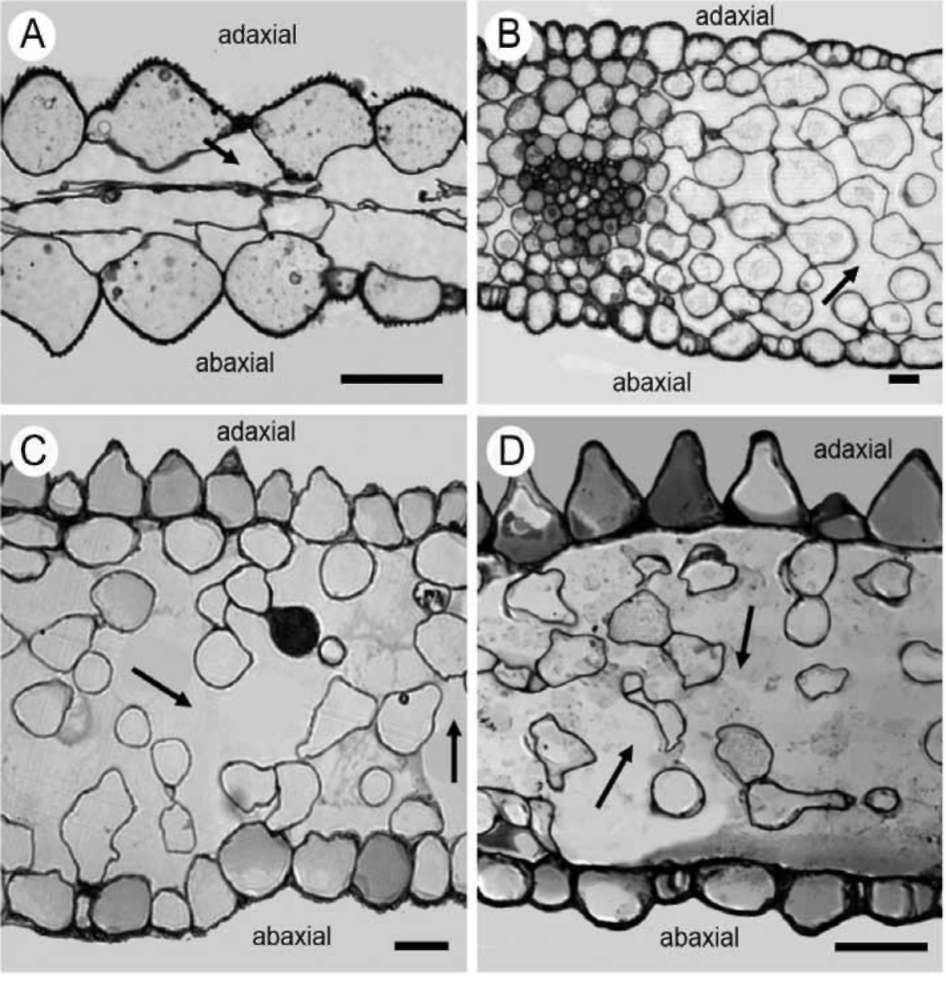
The study was carried out in developed petal regions (Figure 1). Samples from the petal blade were carefully cut in square pieces (4 mm2) and fixed in 3% glutaraldehyde in Na-phosphate buffer at pH 7, at room temperature, for 2 h. Plant material was washed three times by immersion in buffer for 30 min each time; then, it was post fixed in 1% OsO4 in the same buffer at 4°C and dehydrated in acetone
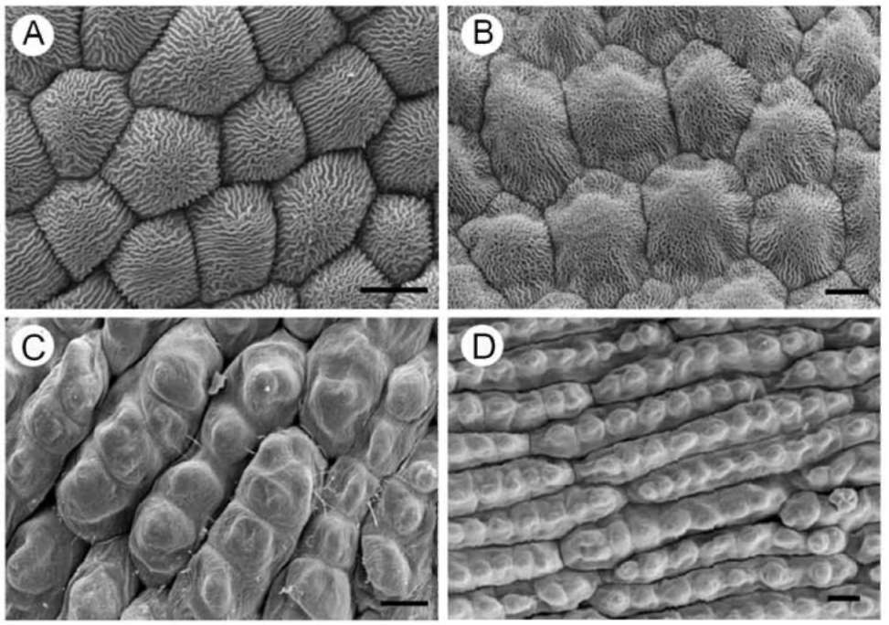
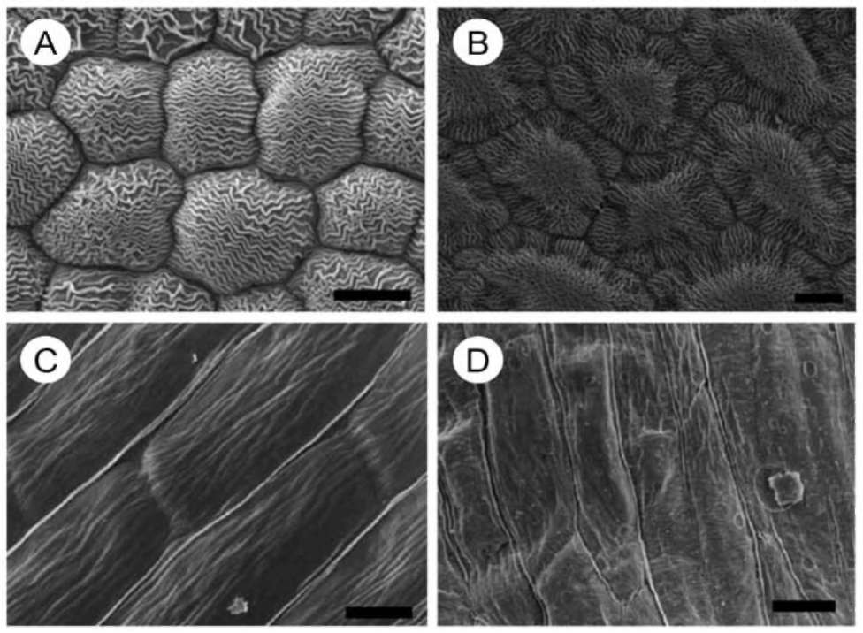
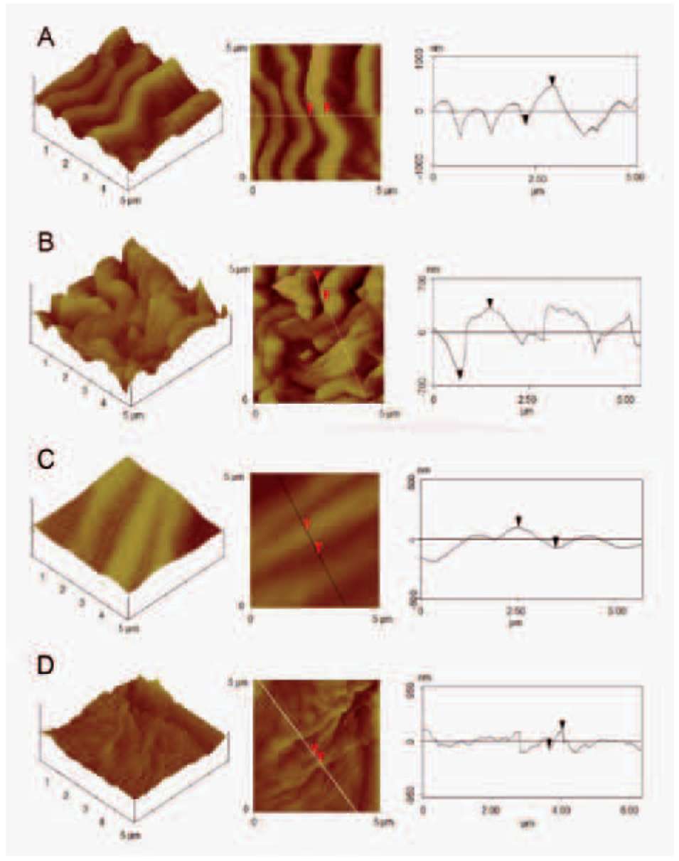
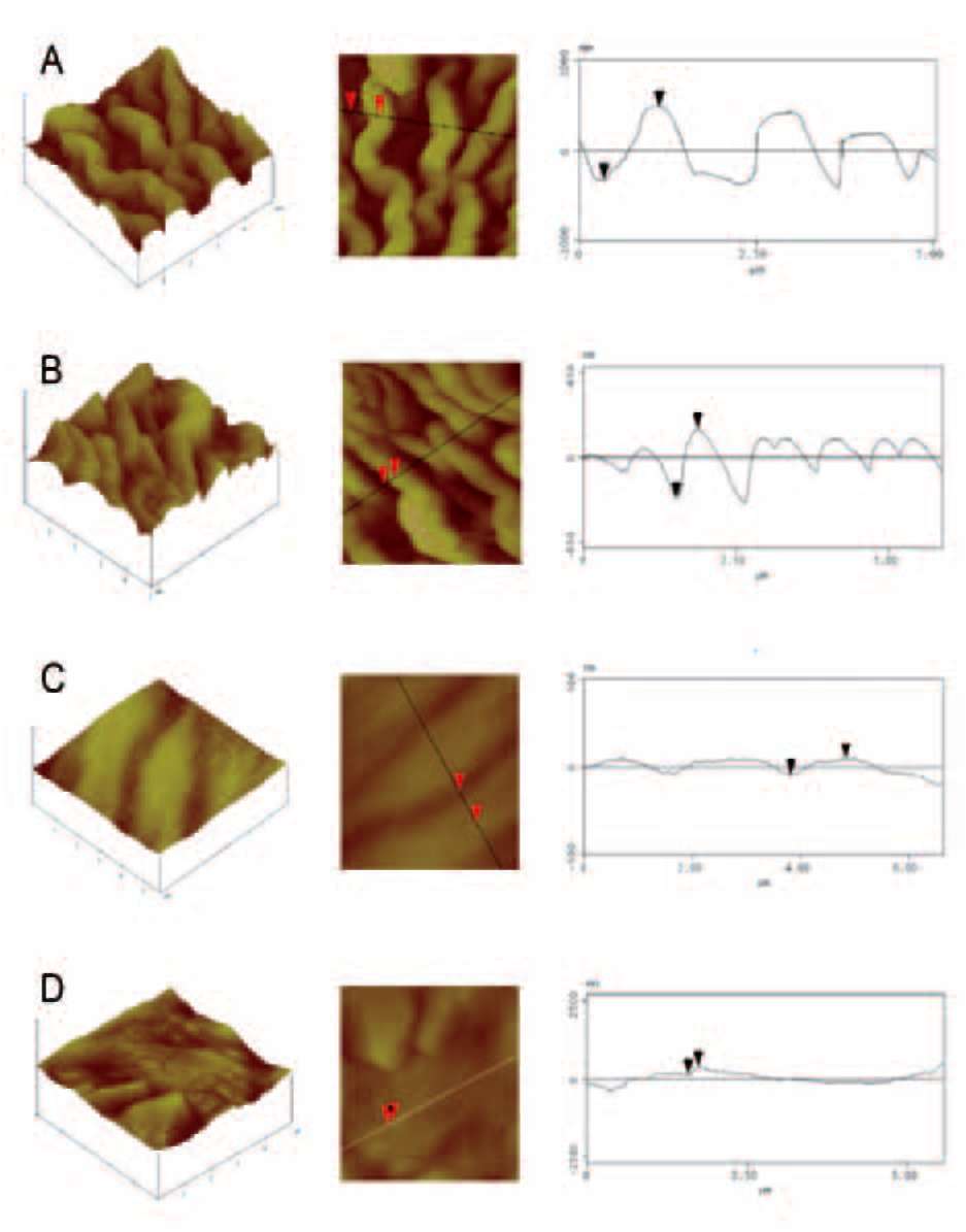
solutions. Dehydrated tissues were embedded in SPURR (Serva) resin. Semi-thin sections of resin-embedded tissue (LKB Ultratome III microtome) were stained in Toluidine Blue '0', in 1% borax solution, photographed and digi­tally recorded using a Zeiss Axioplan light microscope (Carl Zeiss Inc., Thornwood, N.Y.) equipped with a digi­tal camera (Zeiss AxioCam MRc5). Dehydrated samples were dried at the critical point in a Bal-tec CPD-030 dryer, mounted with double adhesive tape on stubs, sputter coated with 20 nm gold in a Bal-tec SCP-050. The adaxial and abaxial epidermises of petals were viewed using the scanning electron microscope JEOL JSM 840 (JEOL Ltd, Tokyo, Japan). Also, the adaxial and the abaxial petal areas (25 (im2) were imaged by using a tap mapping atomic force microscope (Multimode SPM; Veeco, Santa Barbara, CA, USA). Several parameters were analysed and processed, using the software package Nanoscope III (Veeco, USA), in order to detect detailed information for the surfaces of petals. The quantitative measurements in­clude surface roughness (Ra) of the tissues, horizontal and vertical distances that represented the height of a step be­tween nanofolds, and the length between the markers that represented the surface distance. The surface area ratio (Sr), representing the density of microfolding, was the per­centage of the three-dimensional surface, compared to the projected flat surface area, on the threshold plane. Angles (。) between a straight line connecting the cursors and the horizontal surface were measured on both petal surfaces
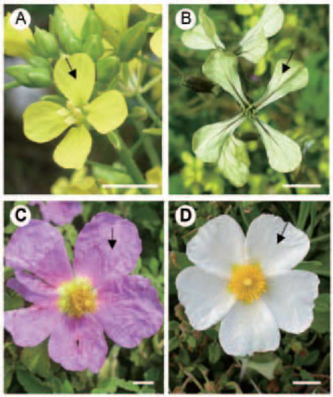
Figure 1. Flowers of S. arvensis (A), E. sativa (B), C. creticus (C) and C. salviifolius (D); sampling regions of adaxial, fully expanded petal tissues are indicated by arrows. Scale bars: 1 cm.
ARGIROPOULOS and RHIZOPOULOU ― Micromorphology of short-lived petals
481
and varied among the species and between adaxial and abaxial epidermis, i.e. being 47° and 46° respectively for S. arvensis, 56° and 51° respectively for E. sativa, 10° and 5° respectively for C. creticus and 48° and 40° respectively for C. salviifolius. Traits, obtained from nine different samples, are given in the representative micrographs (Fig­ures 5, 6) and Table 2. Mean values in Tables 1, 2 are fol­lowed by standard errors ± S.E.).
Statistical analysis
One-way ANOVA was used to analyse differences in traits of petals among species. Then, the data were analy­sed by Duncan's new multiple range test and the signifi­cant difference was defined at P<0.05. Statistical analysis has been realized with the SPSS statistical program.
RESULTS
Flower petals of S. arvensis (Figure 1A) possess a narrow mesophyll (Table 1) with a loosely arranged pa­renchyma between two epidermises (Figure 2A); the thickness of each of the adaxial and the abaxial epidermis is the same order of magnitude with that of the mesophyll.
Petals of E. sativa (Figure 1B), C. creticus (Figure 1C) and C. salviifolius (Figure 1D) possess a large mesophyll, with loosely arranged cells and wide intercellular spaces (Figures 2B, 2C, 2D, respectively); the mesophyll thick­ness is three (C. salviifolius), four (C. creticus) and five (E. sativa) folds thicker, than that of either the adaxial or the
Table 1. Mean values of thickness of the mesophyll, the adax­ial and the abaxial epidermis of petals of flowers of four suc­cessively blossoming species. Each value is the mean of nine measurements S.E. Means followed by the same letters are not statistically different at P=0.05.


Petals

Species


Thickness


Mesophyll
(μm)

Adaxial epidermis (μm)

Abaxial epidermis (μm)

S. arvensis

15.50 0.45a

19.260.28a

15.75 0.51a
E. sativa
152.50 6.50b
25.50 0.45a
19.40 0.30a
C. creticus
113.50 9.50c
21.50 0.35a
20.50 0.55a
C. salviifolius
64.00 4.00d
21.20 0.20a
11.25 0.18e

Figure 2. Transverse sections through the expanded region of petals of S. arvensis (A), E. sativa (B), C. creticus (C) and C. salviifolius (D); intercellular spaces are indicated by arrows. Scale bars: 20 (im.
482
Botanical Studies, Vol. 53, 2012
Figure 3. Scanning electron mi­crographs of adaxial, epidermal cells of petals of S. arvensis (A), E. sativa (B), C. creticus (C) and C. salviifolius (D). Scale bars: 10 μm.
abaxial epidermis of each of the above mentioned species (Table 1). The adaxial epidermis of the petals is composed of conical-papillate (Figures 2A, 2C, 2D) and lenticular (Figure 2B) cells, while the abaxial epidermis is composed of papillate (Figure 2A), rectangular (Figure 2B) and len­ticular cells (Figures 2C, 2D). Adaxial and abaxial papil­late cells of S. arvensis bear a row of papillate projections in the inner face (Figure 2A). Adaxial papillate cells of C. creticus and C. salviifolius exhibit relatively flat, lenticular projections on the inner face, facing the mesophyll (Fig­ures 2C, 2D).
Adaxial (Figure 3A) and abaxial (Figure 4A) epidermal
cells of S. arvensis are covered by wavy striations. Also, polygonal, convexly shaped cells on the adaxial (Figure 3B) and the abaxial (Figure 4B) epidermises of petals of E. sativa are covered by densely packed, wavy striations. Cell wall tortuosities were detected on the basal area of the adaxial (Figure 3B) and the abaxial (Figure 4B) epidermal cells of petals of E. sativa.
The adaxial surfaces of petals of C. creticus and C. salviifolius show certain peculiarities in the shape of epi­dermal cells. Thus, adaxial epidermal cells of petals of C. creticus are elongated and four micro-papillae are arranged in a single row per cell (Figure 3C). Adaxial epidermal
Figure 4. Scanning electron mi­crographs of abaxial, epidermal cells of petals of S. arvensis (A), E. sativa (B), C. creticus (C) and C. salviifolius (D). Scale bars: 10 μm.
ARGIROpouLOS and RHIzpuLu ― Micromorphology of short-lived petals
483
cells of petals of C. salviifolius are extremely elongated and seven to nine micro-papillae are arranged in a single row per cell (Figure 3D). Abaxial epidermal cells of C. creticus and C. salviifolius possess a flat and smooth sur­face (Figures 4C, 4D).
Petal surfaces of the plant species mentioned above pos­sess a different nanosculpture, as indicated by projections in the shape of peaks and cavities which vary in height, density and arrangement (Figures 5, 6; Table 2). The adaxial (Figure 5A) and the abaxial (Figure 6A) surfaces of flower petals S. arvenis exhibit the highest roughness (Table 2), among the examined species. The adaxial and the abaxial surfaces of E. sativa (Figures 5B, 6B) possess a smaller roughness (Table 2), when compared to rough­ness of S. arvensis. Also, the density of forms on epider­mal cells with striated surfaces (represented by values of surface area ratio) differs between the petals' surfaces of the above mentioned species (Table 2).
Micromorphology of the adaxial epidermal cells of ephemeral petals of C. creticus and C. salviifolius (Fig-
ures 3C, 3D) differs from that of the abaxial epidermal cells (Figures 4C, 4D); in C. creticus, the abaxial vertical distance is ten-fold smaller than the abaxial horizontal dis­tance (Table 2). Vertical and horizontal distances on both the adaxial and the abaxial surfaces of the white petals of C. salviifolius (Figures 5D, 6D) indicate a quite parallel arrangement of folds (Table 2).
DISCUSSION
The most prominent feature of the microsculpturing of petal's surface is the epidermal cell shape (Barthlott, 1981). Papillate, epidermal cells of the adaxial surface may absorb light over a greater part of petal surface (Pfundel et al., 2006; Glover, 2007). A papillae shape is expected to reduce reflectance and increase the proportion of incident light that enters the epidermal cells, enhancing light absorption by floral pigments and produce maximum brilliance (Noda et al., 1994; Lee, 2007). In the loosely arranged mesophyll of petals of the examined species
Figure 5. Atomic force micrographs of adaxial petal surface of S. arvensis (A), E. sativa (B), C. creticus (C) and C. salviifolius (D): three dimensional profile (left), integrated line of mea­sured points on plane profile (middle) and profile view of the line section (right).
484
Botanical Studies, Vol. 53, 2012
(Figures 2B, 2C, 2D) numerous light reflecting interfaces may increase light scattering within the petal (Kay et al., 1981; Weston and Ryke, 1999; Rhizopoulou et al., 2006). Convexly shaped adaxial and abaxial epidermal cells re-fract light into focused areas and in combination with cell interiors that exhibit higher refractive indices than air, may act as condensing lenses, sustaining light intensities within petals via intercellular reflectance (Vogelmann, 1993; Lee, 2007).
The papillate inner face of the epidermal cell of S. ar­vensis will act as a light-trap both for light reflected from the mesophyll and for light transmitted from below, via the narrow mesophyll (Figure 2A). Papillate epidermal cells of petals of S. arvensis and C. creticus absorb light over the greater part of their exposed surface, acting as a light-trap for incident light and, in conjunction with the reflective mesophyll, light is guided through the pigments contained in the epidermal cells and returns to the exterior by a combination of external reflection and refraction (Kay et al., 1981).
It appears that papillate epidermal cells compensate for an increase in the cell surface area, by increasing the area of anticlinal undulations, as in S. arvensis and E. sativa, modifying the outline of the cells and contributing to the capture of light and wettability of petals (Barthlott, 1981; Gale and Owens, 1983; Whitney et al., 2011).
The longitudinally elongated, epidermal multiple-papillate cells of petals of C. creticus and C. salviifolius (Figures 3C, 3D) seem to be a characteristic of Cistaceae, which may aid the rapid petal expansion of flowers of Cis­tus species (Barthlott, 1981; Kay et al., 1981; Guzman and Vargas, 2005). Papillate cells of the adaxial epidermises of C. creticus and C. salviifolius will absorb incident light (Figures 2C, 2D, 3C, 3D), while flat cells of the abaxial epidermises of Cistus species (Figures 2C, 2D, 4C, 4D) may reflect light waves at different angles. Also, surface microsculpture of epidermal cells (Figures 5C, 5D, 6C, 6D) affects the firmness of adhesive contact with droplets of water; thus, smooth surfaces of epidermal cells of C. creticus and C. salviifolius decline water retention on the
Figure 6. Atomic force micrographs of abaxial petal surface of S. arvensis (A), E. sativa (B), C. creticus (C) and C. salviifolius (D): three dimensional pro­file (left), integrated line of measured points on plane profile (middle) and profile view of the line section (right).
ARGIROPOULOS and RHIZOPOULOU ― Micromorphology of short-lived petals
485
Table 2. Estimates of roughness on adaxial and abaxial petal surfaces of four successively blossoming species using atomic force microscopy; also, horizontal, vertical and surface distances representing dimensions between nanofolds, and surface area ratio rep­resenting density of nanofolds, are given. Mean values (nine samples S.E.) followed by the same letters are not statistically differ­ent at P=0.05.

Adaxial petal surface
Species

Roughness (Ra) (nm)

Vertical distance (nm)

Horizontal distance
(nm)

Surface distance (nm)

Surface area ratio (Sr)

S. arvensis

2322a

7054g

6543g

10825k

2.440.04n
E. sativa
1633b
9443h
7912g
15729l
2.160.06n
C. creticus
524c
1713b
9556h
9802h
1.020.02o
C. salviifolius
923 d
3281i
3813i
5424j
1.580.03p


Abaxial petal surface


S. arvensis

2163a

8065g

7813g

13013l

2.060.05n
E. sativa
1273e
5274j
3531i
6993m
1.430.06p
C. creticus
292f
912d
10187k
10306k
1.010.02o
C. salviifolius
726c,d
1562b
1583b
3435i
1.470.04p

delicate petals, during their short life-span (Argiropoulos and Rhizopoulou, 2012).
Striations densely arrayed towards the apex of the adax-ial and the abaxial surfaces of S. arvensis (Figures 3A, 4A) and E. sativa (Figures 3B, 4B) support the delicate tissues with water-repellent properties (Wagner et al., 2003; Koch and Barthlott, 2009) and some extra strength (Gale and Owens, 1983) during their three-day life span. Similar wavy striations exist on the petal surfaces of Asphodelus ramosus (Rhizopoulou et al., 2008), which blossoms dur­ing the same period in Eastern Mediterranean. In some cases, striated patterns of petals (Figure 3B) may exhibit an illusive, vibratile movement, by possessing a figural intensity, which may help flower discrimination and attrac­tion of pollinators (Dafni et al., 1997). This may be advan­tageous for E. sativa, because the striated, lenticular petal surfaces show weak reflectance (Kay et al., 1981).
Imaging of the relief using atomic force microscopy revealed detailed surface patterns, which may have a great influence on their attributes as interfaces (Glover, 2007; Zhang et al., 2008; Bhushan, 2009). It appears that mi-crofolding, i.e. patterns of ridges and striations, increases cell surface area of the short lived petals and this may be particularly important for their performance in the field. A sculpturally increased surface area of light absorbing papillae cells of adaxial epidermises increases the energy exchange of petals with the surrounding environment and supports the warming of flowers at low ambient tempera­tures, in the early spring (Barthlott, 1981; McKee and Richards, 1998; Rands and Whitney, 2008).
The adaxial and the abaxial relief of C. creticus pos­sess a horizontal distance that is 8-10 folds higher than the vertical distance. In C. creticus, vertical distances between folds are comparable to the sub-wavelength regime, i.e. being approximately 170 nm, and thus effective in reflect­ing radiation of shorter rather than longer wavelengths; if the distance between folds (grating period) is smaller than
the visible wavelength spectrum, then refraction, scattering and polarisation of light of wavelengths greater than the grating period may occur (Groning, 2005). Traits of pet­als of C. creticus viewed with AFM appear to be capable of producing some structural colour effects (Glover and Whitney, 2010; Feng et al., 2010; Lee et al., 2011).
Adaxial and abaxial wrinkled petal surfaces of C. creti-cus are covered by a smooth relief (Figures 5C, 6C), which coincides with small roughness, low vertical distances and elevated horizontal distances (Table 2) that enhance the cell surface area of adaxial and abaxial petal epidermises, of the short lived, fuchsia flowers. Hence, the nanostruc-ture of petals of C. creticus increases the reflectability of the ephemeral, wrinkled, fuchsia tissues (Figure 1C). It seems likely that ephemeral and delicate petals of C. cre-ticus blowing in the wind are well adapted to the ambient conditions.
The surface area ratios, which represent the density of forms on the abaxial epidermis, as in C. salviifolius and E. sativa, are not statistically different (mean values: 1.47 and 1.43 respectively); however, the abaxial surface of E. sativa exhibits a significantly higher roughness than that of C. salviifolius (mean values: 126 nm and 73 nm respec­tively). Features of petal surfaces at the nanoscale level indicate adaptation to the environment mostly combined with non wetted tissues. Microsculpturing increases in size the area of the epidermal cells, which aids optical proper­ties and assists the water status of petals (Herminghaus, 2000; Groning, 2005; Argiropoulos and Rhizopoulou, 2012; Chimona et al., 2012). Conical-papillate cells have a significant impact on how water is retained on the petal surface (Whitney et al., 2011; Argiropoulos and Rhizopou-lou, 2012). Adaxial petal surfaces with papillate cells and wavy striations, as in S. arvensis, might have developed to profit from sunshine and to not be harmed when exposed to unfavourable environmental conditions, during the early spring flowering period. Abaxial, flat epidermal surfaces of
486
Botanical Studies, Vol. 53, 2012
petals of the examined species seem to be less susceptible to wet conditions. Connected to the reduced wettability appears to be a minimal ability of adhesion of pathogens and dust particles, which may be washed off by rain drop­lets. Also, surface microsculpture may be a tactile cue for insects to approach sites of rewards, while at the same time the delicate, ephemeral petals should remain unaf­fected (Bargel et al., 2006; Kutschera, 2008; Harder, 2009; Stelzer et al., 2010).
The topography of the adaxial and the abaxial petal surfaces of the examined species reveal traits linked to an astonishing performance of ephemeral corollas and func­tionality of boundary layers, which significantly influence the physical properties of petal tissues. In this context, the use of AFM improves the accuracy of "vision" related with biological structures and has greatly increased our knowledge about the function of floral tissues (Bhushan and Nosonovsky, 2010). Nanoridges, playing an impor­tant role in optical properties and adhesive contacts, differ among the examined species and between their adaxial and abaxial petal surfaces; roughness (Ra) and density of folds (Sr) of adaxial petal surfaces of the examined spe­cies were statistically different at P<0.001, while Ra and Sr of the abaxial surfaces at P<0.05. Previous studies have described flower petals possessing striations on both the adaxial and the abaxial epidermises and exhibiting a three-day life span, as in Allium species, Ornithogalum umbel-latum and Trifolium repents (Kay et al., 1981; Petanidou et al., 1995). While, petals lacking striations expand on ephemeral and short-lived corollas, as in Erodium cicu-tarium, Hypericum perforatum and Silene alba (Kay et al., 1981; Petanidou et al., 1995). However, striations of petal surfaces and floral life span have never been hitherto cor­related. It is noteworthy that texture of petals of S. arvensis and E. sativa with a greater life span exhibit higher surface roughness (Ra) and density of nanofolds (Sr), while the opposite holds true for ephemeral petals of C. creticus and C. salviifolius; Ra and Sr of petal surfaces of the examined species are positively correlated with flowers' life span (r2=0.73 and r2=0.58, respectively).
Adaxial surface distances of the examined petals -which are exposed to the ambient environmental conditions and seen by potential pollinators as they approach flowers國,dif­fer from the abaxial surface distances. In addition, smooth surfaces composed of folding larger than the wavelength of the incident light, as in S. arvensis, reflect the light ra­diation, while, surfaces composed by gratings smaller than the wavelength spectrum, as in C. creticus, are more ef­fective in reflecting radiation of shorter rather than longer wavelengths (Table 2). Different features of petal surfaces at the nanoscale level may be species specific and related with their lifespan and adaptations to climatic conditions, mostly combined with light absorption and wettability of tissues grown in the field. It is worth mentioning that submicron patterns of petals' surfaces, mostly from roses, have already been transferred to biomimetic materials, throughout a rapidly growing and enormously promising field of research (Fratzl, 2007; Feng et al., 2010; Koch et
al., 2009; Stratakis et al., 2009; Qian et al., 2011; Zhang et al., 2012). Further investigation will be required to test these hypotheses in wild species.
Acknowledgements. The authors thank Dr. A. Siakouli and Dr. E. Spanakis (IESL-FORTH, Heraklion, Crete) for help with microscopic techniques. This work was sup-ported by the grant PENED 03174 to S.R., co-financed by European Social Fund and GSRT.
LITERATURE CITED
Agrawal, AA., M. Fishbein, R. Jetter, J.P. Salminen, J.B. Gold­stein, A.E. Freitag, and J.P. Sparks. 2009. Phylogenetic ecology of leaf surface traits in the milkweeds (Asclepias spp.): chemistry, ecophysiology, and insect behavior. New Phytol. 183: 848-867.
Argiropoulos, A. and S. Rhizopoulou. 2012. A study on the mi­cromorphology of petals of the invasive weed Oxalis pes-caprae. Weed Biol. Manag. 12: 47-52.
Bargel, H., K. Koch, Z. Cerman, and C. Neinhuis. 2006. Struc­ture-function relationships of the plant cuticle and cuticular waxes - a smart material? Funct. Plant Biol. 33: 893-910.
Barthlott, W. 1981. Epidermal and seed surface characters of plants: systematic applicability and some evolutionary as­pects. Nord. J. Bot. 1: 345-355.
Barthlott, W., C. Neinhuis, D. Cutler, F. Ditsch, I. Meusel, I. Theisen, and H. Wilhelmi. 1998. Classification and termi­nology of plant epicuticular waxes. Bot. J. Linn. Soc. 126:237-260.
Benitez, J.J., A.J. Matas, and A. Heredia. 2004. Molecular char­acterization of the plant biopolyester cutin by AFM and spectroscopic techniques. J. Struct. Biol. 147: 179-184.
Bhushan, B. 2009. Biomimetics: lessons from nature-an over­view. Phil. Trans. R. Soc. A 367: 1445-1486.
Bhushan, B. and M. Nosonovsky. 2010. The rose petal effect and the modes of superhydrophobicity. Phil. Trans. R. Soc. A 368: 4713-4728.
Chimona, C., A. Stamellou, A. Argiropoulos, and S. Rhizopou-lou. 2012. Study of variegated and white flower petals of Capparis spinosa expanded at dusk in arid landscapes. J. Arid Land 4: 171-179.
Dafni, A., M. Lehrer, and P.G. Kevan. 1997. Spatial flower pa­rameters and insect spatial vision. Biol. Rev. 72: 230-282.
Davies, K.L. and M.P. Turner. 2004. Morphology of floral papil­lae in Maxillaria Ruiz & Pav. (Orchidaceae). Ann. Bot. 93:75-86.
Derdej, A. and K. Koch. 2007. Molecular structure of isolated plant cuticles visualised by atomic force microscopy (AFM). Microsc. Microanal. 13: 204-205.
Feng, L., Y. Zhang, J. Xi, Y. Zhu, N. Wang, F. Xia, and L. Jiang. 2008. Petal effect: a superhydrophobic state with high adhe­sive force. Langmuir 24: 4114-4119.
Feng, L., Y. Zhang, M. Li, Y. Zheng, W. Shen, and L. Jiang.
ARGIROPOULOS and RHIZOPOULOU ― Micromorphology of short-lived petals
487
2010. The structural solor of red rose petals and their dupli­cates. Langmuir 26: 14885-14888.
Fratzl, P. 2007. Biomimetic materials research: what can we really learn from nature's structural materials? J. R. Soc.
Interface 4: 637-642.
Gale, R.M.O. and S.J. Owens. 1983. Cell distribution and sur­face morphology in petals, androecia and styles of Com-melinaceae. Bot. J. Linn. Soc. 87: 247-262.
Glover, B.J. 2007. Understanding flowers and flowering, an inte­grated approach. Oxford: Oxford University Press.
Glover, B.J. and H.M. Whitney. 2010. Structural color and iri­descence in plants: the poorly studied relations of pigment colour. Ann. Bot. 105: 505-511.
Gorton, H.L. and T.C. Vogelmann. 1996. Effects of epidermal cell shape and pigmentation on optical properties of Anthir-rhinum petals at visible and ultraviolet wavelengths. Plant Physiol. 112: 879-888.
Groning, P. 2005. Nanotechnology: an approach to mimic natu­ral architectures and concepts. Adv. Eng. Mater. 7: 279-291
Guzman, B. and P. Vargas. 2005. Systematics, character evolu­tion, and biogeography of Cistus L. (Cistaceae) based on ITS, trnL-trnF, and matK sequences. Mol. Phylogenet.
Evol. 37: 644-660.
Harder, L.D. 2009. The legacy of Darwin's studies of floral ad­aptation and plant mating. S. Afr. J. Bot. 75: 387-388.
Herminghaus, S. 2000. Roughness-induced non-wetting. Euro-
phys. Lett. 52: 165-170.
Jacobs, J.F., G.J.M. Koper, and W.N.J. Ursem. 2007. UV protec­tive coatings: a botanical approach. Prog. Org. Coat. 58:166-171.
Kaplan, M. 2008. Raindrops on roses. Nature News doi: 10.1038/news.2008.778.
Kay, Q.O.N., H.S. Daoud, and C.H. Stirton 1981. Pigment dis­tribution, light reflection and cell structure in petals. Biol. J. Linn. Soc. 83: 57-89.
Kerstiens, G. 1996. Cuticular water permeability and its physi­ological significance. J. Exp. Bot. 47: 1813-1832.
Kerstiens, G. 2006. Water transport in plant cuticles: an update. J. Exp. Bot. 57: 2493-2499.
Kevan, P.G. and M.A. Lane. 1985. Flower petal microtexture is
a tactile cue for bees. Proc. Natl. Acad. Sci. USA 82: 4750­4752.
Koch, K. and W. Barthlott. 2009. Superhydrophobic and supe-rhydrophilic plant surfaces: an inspiration for biomimetic materials. Phil. Trans. R. Soc. A 367: 1487-1509.
Koch, K., C. Neinhuis, H.-J. Ensikat, and W. Barthlott. 2004.
Self assembly of epicuticular waxes on living plant surfaces imaged by atomic force microscopy (AFM). J. Exp. Bot.
55: 711-718.
Koch, K., B. Bhushan, and W. Barthlott. 2009. Multifunctional
surface structures of plants: An inspiration for biomimetics.
Prog. Mater. Sci. 54: 137-178.
Kutschera, U. 2008. The growing of the outer epidermal wall:
design and physiological role of a composite structure. Ann.
Bot. 101: 615-621.
Lee, D. 2007. Nature's palette, the science of plant color. Chi­cago: The University of Chicago Press.
Lee, S.-M, J. tipping, A. Bielawny, and M. Knez. 2011. Struc­ture-based color of natural petals discriminated by polymer replication. Appl. Mater. Interfaces 3: 30-34.
Li-Beisson, Y., M. Pollard, V. Sauveplane, F. Pinot, J. Ohlrogge, and F. Beisson. 2009. Nanoridges that characterize the sur­face morphology of flowers require the synthesis of cutin polyester. Proc. Natl. Acad. Sci USA 106: 22008-22013.
Lolle, S.J. and R.E. Pruitt. 1999. Epidermal cell interactions: a case for local talk. Trends Plant Sci. 4: 14-20.
Martens, P. 1936. Reserches sur la cuticule. IV. Le relief cuticu-laire et la differenciation epidermique des organs floraux. Cellule 43: 289-320.
McKee, J. and A.J. Richards. 1998. Effect of flower structure and flower colour on intrafloral warming and pollen germi­nation and pollen-tube growth in winter flowering Crocus L. (Iridaceae). Bot. J. Linn. Soc. 128: 369-384.
Mechaber, W.L., D.B. Marshall, R.A. Mechaber, R.T. Jobe, and F.S. Chew. 1996. Mapping leaf surface landscapes. Proc. Natl. Acad. Sci USA 93: 4600-4603.
Mill, R.R. and D.M. Stark Schilling. 2009. Cuticle micromop-
hology of Saxegotheae (Podocarpaceae). Bot. J. Linn. Soc.
159: 58-67.
Miller, R., S.J. Owens, and B. Rorslett B. 2011. Plants and colour: flowers and pollination. Opt. Laser Technol. 43:
282-294.
Nieto Feliner, G. and C. Aedo. 1995. A cladistic analysis of Geranium subgen. Erodioidea (Picard) Yeo (Geraniaceae). Bot. J. Linn. Soc. 119: 195-212.
Noda, K.I., B.J. Glover, P. Linstead, and C. Martin 1994. Flower colour intensity depends on specialized cell shape con-trolled by a Myb-related transcription factor. Nature 369:661-664.
Olowokudejo, J.D. 1993. Comparative epidermal morphology of West African species of Jatropha L. (Euphorbiaceae). Bot. J. Linn. Soc. 111: 139-154.
Petanidou, T., W.N. Ellis, N.S. Margaris, and D. Vokou. 1995.
Constraints on flowering phenology in a phryganic (East Mediterranean shrub) community. Am. J. Bot. 82: 607-620.
Petanidou, T. and E. Lambron. 2005. A land for flowers and bees: studying pollination ecology in Mediterranean com­munities. Plant Biosyst. 139: 279-294.
Pfundel, E.E., G. Agati, and Z.G. Zerovic. 2006. Optical proper­ties of plant surfaces. In M. Riederer and C. Muller (eds.), Biology of the Plant Cuticle. Blackwell, Oxford, pp. 216­247.
Qian, J., F. Chen, X. Zhao, and Z. Chen. 2011. China rose petal as biotemplate to produce two-dimensional ceria nano-
sheets. J. Nanoparticle Res. 12: 7149-7158.
Rands, S.A. and H.M. Whitney. 2008. Floral temperature and optimal foraging: Is heat a feasible floral reward for pollina-
488
Botanical Studies, Vol. 53, 2012
tors? PLoS ONE 3: 1-7.
Rands, S.A., B.J. Glover, and H.M. Whitney. 2011. Floral epi­dermal structure and flower orientation: getting to grips with awkward flowers. Arthropod-Plant Interact. 5: 279­285.
Rhizopoulou, S., E. Ioannidi, N. Alexandredes, and A. Argiropo-ulos. 2006. A study on functional and structural traits of the nocturnal flowers of Capparis spinosa L. J. Arid Environ. 66: 635-647.
Rhizopoulou, S., A. Argiropoulos, E. Spanakis, D. Gikas, N. Alexandredes, D. Koukos, and D. Anglos. 2008. Visual perception of colourful petals reminds us of classical frag­ments. Nature Precedings hdl:10101/npre.2008.1523.1
Stelzer, R.J., R. Stanewsky, and L. Chittka. 2010. Circadian for­aging rhythms of bumblebees monitored by radio-frequency identification. J. Biol. Rhythm. 25: 257-267.
Solga, A., Z. Cermanm, B.F. Strifflerm, M. Spaeth, and W. Bar-thlott. 2007. The dream of staying clean: Lotus and biomi-metic surfaces. Bioinspir. Biomim. 2: S126-S134.
Stratakis, E., V. Zorba, M. Barberoglou, E. Spanakis, S. Rhizo-poulou, P. Tzanetakis, S. Anastasiadis, and C. Fotakis. 2009. Laser structuring of water-repellent biomimetic surfaces. SPIE Newsroom 10.1117/2.1200901.1441
Vogelmann, T.C. 1993. Plant tissue optics. Ann. Rev. Plant Phys-iol. Mol. Biol. 44: 231-251.
Wagner, P., R. Furstner, W. Barthlott, and C. Neinhuis 2003.
Quantitative assessment to the structural basis of water re-pellency in natural and technical surfaces. J. Exp. Bot. 54:
1295-1303.
Weston, E.L. and K.A. Pyke. 1999. Developmental ultrastructure of cells and plastids in the petals of wallflower (Erysimum cheiri). Ann. Bot. 84: 763-769.
Whitney, H.M. and B.J. Glover 2007. Morphology and develop­ment of floral features recognised by pollinators. Arthropod-Plant Inte. 1: 147-158.
Whitney, H.M., M. Kolle, P. Andrew, L. Chittka, U. Steiner, and B.J. Glover. 2009. Floral iridescence, produced by diffrac-tive optics, acts as a cue for animal pollinators. Science 323: 130-133.
Whitney, H.M., R. Poetes, U. Steiner, L. Chittka, and B.J. Glov­er. 2011. Determining the contribution of epidermal cell shape to petal wettability using isogenic Anthirrhinum lines. PLoS ONE 6: e17576.
Zhang, Y., T. Hayashi, M. Inoue, Y. Oyama, M. Hosokawa, and S. Yazawa. 2008. Flower color diversity and its optical mechanism. Acta Hortic. 766: 469-476.
Zhang, Y., Y. Chen, L. Shi, J. Li, and Z. Guo. 2012. Recent
progress of double-structural and functional materials with
special wettability. J. Mater. Chem. 22: 799-815.
野生種Cistus creticus, Cistus salviifolius, Eruca sativaSinapis arvensis之短命花之花瓣表面的立體構造及聚合物微泡
Apostolos ARGIROPOULOS and Sophia RHIZOPOULOU
National and Kapodistrian University of Athens, Department of Biology, Section of Botany,
Panepistimiopolis, Athens, 15784, Greece
連續開花之野生種Sinapis arvensis, Eruca sativa, Cistus creticus禾口 Cistus salviifolius的短命花之花瓣
的向軸面及離軸面分別以光學顯微鏡,電子顯微鏡和原子力顯微鏡檢視。花瓣之立體構造顯示一種次微
米之緩解結構,由此可預期其可視外形和花之組織的可溼性。花瓣和葉肉之向軸,乳突的表皮細胞。含
鬆散地安排之細胞及大的細胞間隙;由此產生了共軛捕捉光的區域之環境,因此影像日光使用效率以及
改變組織之光學性質的可能性。使用原子力顯微鏡檢視花瓣之表皮顯示出一種次微米緩解結構;此結構
增加表皮細胞之細胞表面面積,此特徵很可能是短命花之演化適應機制。Sinapis arvensisEruca sativa
花瓣表面之不同分層帶可能加強組織之巧妙性和影響黏密的接觸,這些都發生在短命花之三天開發期。
Cistus creticusCistus salviifolius之短暫的花之光滑光瓣表面可能顯示強之反射作用高解析圖像顯示
在上述所有花種花瓣向軸面之粗糙化現象都比離軸面高。知命花瓣表面之微細構造之特性可能對生長在
自然環境下之野生種的花的表現特別重要。
關鍵詞:向軸的;離軸的;花;摺;聚合物微泡;花瓣;反撥的;表面;野生種。