Botanical Studies (2012) 53: 535-539.
SYSTEMATICS
Pileomyces formosanus gen. et sp. nov. (Halosphaeri-aceae, Ascomycota) from a rocky shore of Taiwan
Ka-Lai PANG* and Jen-Sheng JHENG
Institute of Marine Biology, National Taiwan Ocean University and Center of Excellence for Marine Bioenvironment and Biotechnology, 2 Pei-Ning Road, Keelung 20224, Taiiwan
(Received May 10, 2011; Accepted June 29, 2012)
ABSTRACT. In Taiwan, knowledge of marine fungi is fragmentary with recent studies documenting 69 spe­cies from mangrove habitats, of which 34 taxa were new records for the country. Marine fungi on trapped substrates from rocky shores of Taiwan are comparatively less known. In a fieldtrip to Yingkeshih (New Tai­pei City, northeastern Taiwan), a new fungus belonging to the Halosphaeriaceae was discovered. This fungus is characterized by dark-coloured ascomata, broadly clavate asci with an apical thickening, pore and retraction of plasmalemma, and ellipsoidal ascospores with a sheet-like appendage covering one end, which eventually detaches from the ascospore. This distinctive ascospore appendage morphology is unique in the Halospha­eriaceae and Pileomyces formosanus gen. et sp. nov. is introduced to accommodate this fungus. Pileom^yces resembles Aniptodera and Phaeonectriella in the morphology of the ascus, and Tirisporella (T beccariana) in gross morphology of the ascospore appendage, however, the appendage is attached to the ascospores at the apex in Tirisporella.
Keywords: Aquatic fungi; Ascomycota; Microascales; Taxonomy.
INTRODUCTION
Obligate marine fungi grow and sporulate exclusively in a marine or marine-related habitat (Kohlmeyer and Kohlmeyer, 1979). Habitats of marine fungi include rocky shores, sandy beaches, mangrove forests, and marine fungi occur primarily on woody tissues, but leaves, fruits, reeds, sea grasses, algae and seaweeds are also substrates (Vrij-moed, 2000). Marine fungi can also be isolated from ani­mal exoskeletons, keratinaceous substrates, sediments, sea foam and seawater (Vrijmoed, 2000). Marine fungi play a major role in the decomposition of complex organic mat­ter in the marine ecosystem by producing wood-modifying enzymes (cellulases, laccases and Mn-dependent peroxi­dases) that degrade lignocellulosic materials (Bucher et al., 2004; Luo et al., 2005). All contribute to the production of particulate and dissolved organic matter for the food web of the oceans (Jones and Pang, 2010).
Diversity of marine mangrove fungi is comparatively well-known in Taiwan (Pang et al., 2010; 2011b), howev­er, information on their diversity on rocky shores is scarce. Hsieh et al. (2002) documented marine fungi from rocky shores of Taiwan, many of which were typical temperate water species, including Halosphaeria appendiculata and Remispora spp. (Koch and Petersen, 1996). During a field-
trip to a rocky shore in northeastern Taiwan, a fungus with characteristics of the Halosphaeriaceae was discovered. Ascospore appendage of this taxon was distinctive and significantly different from other genera in the family, and this therefore warrants the establishment of a new genus.
MATERIALS AND METHODS
Drift and trapped bamboo/wood was collected at Ying­keshih, New Taipei City, Taiwan, on 6 September 2010. Samples were transported to the laboratory and incubated on a tray lined with moist tissue paper for up to 1 month. Fruiting bodies were observed using an Olympus SZ61 stereomicroscope (Tokyo, Japan), sectioned with a razor blade, and centrum material mounted on a slide in sterile seawater. Morphology of the asci and ascospores was observed using an Olympus BX51 microscope (Tokyo, Japan). Photographs were taken with an Olympus DP20 Microscope Camera (Tokyo, Japan).
Small pieces of decaying bamboo with ascomata of the new fungus were fixed by immersion in FAA (50% ethanol, 5% glacial acetic acid and 5% paraformaldehyde) overnight at 4°C. Fixed samples were washed three times with 50% ethanol. Samples were then dehydrated in a graduated ethanol and 1 -butanol series, infiltrated gradu­ally and embedded in paraffin. Paraffin sections (8-10 fim) were cut on a FRM-200P rotary microtome (Japan), floated over a water-bath at 42°C to relax sections, and mounted on slides. Dried sections were deparaffinised and

*Corresponding author: E-mail: klpang@ntou.edu.tw; Tel: +886-02-24622192#5319; Fax: +886-02-24633152.
536
Botanical Studies, Vol. 53, 2012
rehydrated through a graded series of ethanol. Sections were stained with 1% safranin O in 50% ethanol (10 sec) and 0.5% orange G in 95% ethanol (30 sec). After wash­ing and dehydration, each stained section was permanently mounted with a cover slip and Permount (Fisher, Fair Lawn, USA). Specimens were observed under the micro­scope and photographed.
Taxonomy
Pileomyces K.L. Pang & Jheng, gen. nov. (Mycobank
MB519625)
Ascomata fusca, pyriforma, immersa, erumpentia vel exposita, coriacea, ostiolata, laevia. Ostiola brevicollia. Periphyses absentes. Peridium fuscum, unistratum, con-stans ex cellulis elongatis stratorum plurium. Asci unitu-nicati, late clavati, tunica sub apice incrassati, apice poro penetrati, coma brevi, leptodermi, octospori, persistentes vel semi-persistentes, ex pulvino cellularum pseudoparen-chymatarum ad basim ascomati orientes. Catenophyses praesentes, irregulares. Ascosporae ellipsoideae cum apice rotundus, uniseptatae, hyalinae, laeves, leptodermae. Ap­pendices praesentes, ad unum polum.
Typus generis: Pileomyces formosanus K.L. Pang & Jheng
Ascomata dark-coloured, pyriform with globose to subglobose venter, immersed, erumpent or exposed, coria­ceous, ostiolate, smooth. Ostioles short-necked. Periphyses absent. Peridium dark-coloured, one-layered, composed of a few layers of elongated cells. Asci unitunicate, broadly clavate, apically thickened with retraction of plasmalem-ma, with an apical pore, short pedunculate, thin-walled, eight-spored, persistent or semi-persistent, developing from inner wall of ascomal base. Catenophyses present, irregular in shape. Ascospores ellipsoidal with rounded apices, one-septate, hyaline, smooth, thin-walled. Append­ages initially covering one end of ascospores, gradually detached from the ascospore wall to form an ellipsoidal sheet.
Etymology: From the Latin 'pileus', in reference to the shape of the ascospore appendage.
Anamorph: Unknown.
Pileomyces formosanus K.L. Pang & Jheng, sp. nov. (My-
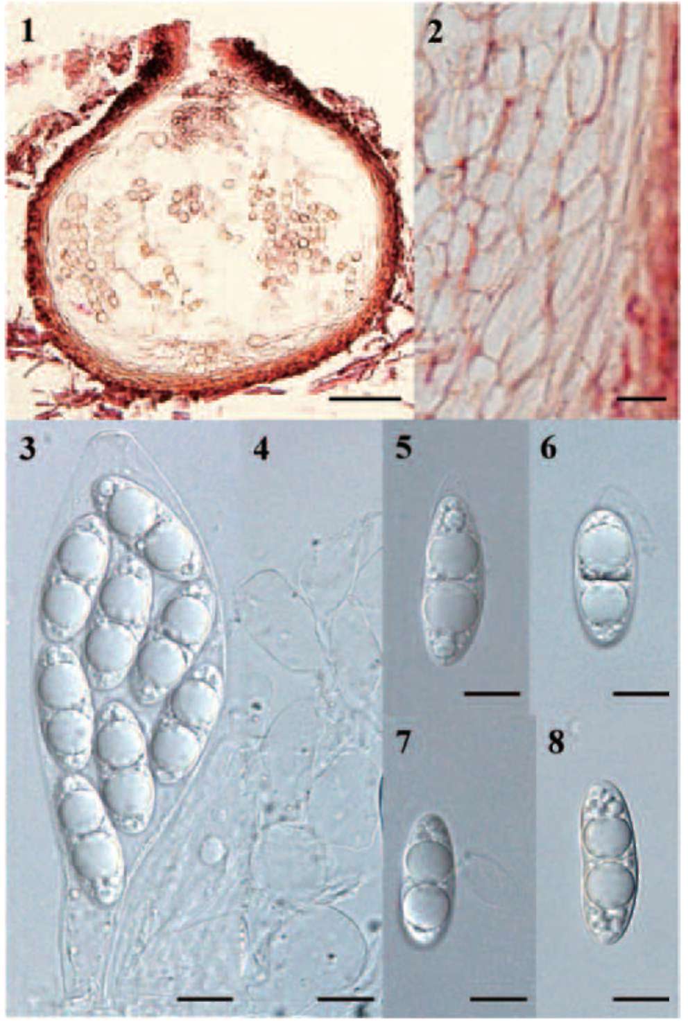
cobank MB563712).                                             Figures 1-8
Ascomata 221-(231)-241 x 182-(234)-285 jam (n=2), fusca, pyriforma, immersa, erumpentia vel exposita, co-riacea, ostiolata, laevia. Ostiola brevicollia. Periphyses absentes. Peridium 15-(19)-24 jam (n=4), fusca, unistra-tum, constans ex cellulis elongatis stratorum plurium. Asci 98-(111)-128 x 27-(31)-37 jam (n=15), umtumcati, late clavati, tunica sub apice incrassati, apice poro pen-etrati, coma brevi, leptodermi, octospori, persistentes vel semi-persistentes, ex pulvino cellularum pseudoparen-chymatarum ad basim ascomati orientes. Catenophyses
praesentes, irregulares. Ascosporae 26-(30)-36 x 9-(10)-12 jam (n=50), ellipsoideae cum apice rotundus, unisepta-tae, hyalinae, laeves, leptodermae. Appendices praesentes, ad unum polum.
Ascomata 221-(231)-241 x 182-(234)-285 jam (n=2), dark-coloured, pyriform with globose to subglobose ven­ter, immersed, erumpent or exposed, coriaceous, ostiolate, smooth. Ostioles short-necked. Periphyses absent. Pe-ridium 15-(19)-24 jam (n=4), dark-coloured, one-layered, composed of a few layers of elongated cells. Asci 98-(111)-128 x 27-(31)-37 jam (n=15), unitunicate, broadly clavate, apically thickened with retraction of plasmalem-ma, with an apical pore, short pedunculate, thin-walled, eight-spored, persistent or semi-persistent, developing from inner wall of ascomal base. Catenophyses present, irregular in shape. Ascospores 26-(30)-36 x 9-(10)-12 j m (n=50), ellipsoidal with rounded apices, one-septate, hya­line, smooth, thin-walled. Appendages initially covering one end of ascospores, gradually detach from ascospore to form an ellipsoidal sheet.
Etymology: In reference to Taiwan where the holotype was collected.
Anamorph: Unknown.
Holotype: TAIWAN: Yingkeshih. On a piece of uniden­tified trapped bamboo culm, 6 September 2010, J.S. Jheng & K.L. Pang, BBH 30192 (BIOTEC Bangkok Herbarium), dried bamboo.
Sequence: JX003862 (internal transcribed spacers (ITS) region of the nuclear rDNA, NCBI).
Known geographical distribution: Yingkeshih, Taiwan.
Substrata'. Trapped bamboo on rocky shore.
RESULTS AND DISCUSSION
Jones et al. (2009) recorded 530 marine fungi with most species described in the period 1980-1989 and 1990-1999 (291 species), but in recent years, this dramatically de-creased. Jones (2011) recently estimated the total number of marine fungi may be in excess of 10,000 and suggested the exploration of wider geographical locations for marine fungi. Pang et al. (2011a) recently recorded six lignicolous marine fungi from an environmentally extreme location, Longyearbyen, inside the Arctic circle and four of these were new to science (Pang et al., 2008; 2009; 2011a).
Taiwan has a long coastline, making it an ideal place for the study of marine fungi. The western Taiwan coast is dominated by mangrove habitats while the eastern and northeastern coasts consist of rocky shores. Pang et al. (2011b) documented 69 species of marine fungi from mangrove habitats of Taiwan while Hsieh et al. (2002) recorded 26 taxa on driftwood from Taiwan rocky shores, making an inventory of 95 taxa. However, many new taxa have been discovered in Taiwan for the last few years and await formal description (K.L. Pang, unpublished results).
The new fungus is a typical member of the Halospha-eriaceae, with its perithecioid ascomata, pseudoparen-
PANG and JHENG ― A new marine fungus from Taiwan
537
chymatous centrum, broadly clavate asci, presence of catenophyses, and ellipsoidal/fusiform, mostly hyaline ascospores with appendages (Spatafora et al., 1998). Ascus of P. formosanus shows great resemblance with that of An-iptodera and Phaeonectriella. In Aniptodera, ascospores are generally thick-walled, with/without unfurling as-
cospore appendages (Shearer and Miller, 1977; Hyde et al., 1999), while ascospores of Phaeonectriella are dark-coloured, with/without unfurling ascospore appendages (Eaton and Jones, 1971; Hyde et al., 1999). The distinctive character of P. formosanus is the sheet-like appendage on one end of the ascospores. However, mechanism on the
Figures 1-8. Pileomyces formosanus gen. et sp. nov. (holotype). 1, Pyriform ascoma with a subglobose venter; 2, One-layered peridi­um of elongate cells; 3, Clavate ascus with an apical thickening, a pore and retraction of plasmalemma; 4, Catenophyses; 5, Ascospore with sheet-like appendage on one end; 6, Appendage starting to detach from ascospore when mounted in seawater; 7, Appendage entirely detached from ascospore forming an ellipsoidal sheet; 8, Ascospore without an appendage. Scale bars: 1= 50 μm, 2=5 μm, 3-8=10 μm.
538
Botanical Studies, Vol. 53, 2012
formation of this ascospore appendage is unknown. The ascospore appendage was not visible inside the ascus, but was evident covering one end of the ascospores (Figure 5) when they were mounted in seawater. The appendage is not attached to the ascospore by a structure, and de­taches from it to form an ellipsoidal sheet. This appendage does not appear to be gelatinous in nature and does not dissolve when mounted in seawater. An ultrastructural study of ascospores of P. formosanus is required to detail formation of the appendage. Corollospora maritima and Chaetosphaeria chaetosa also have sheet-like ascospore appendages resulting from fragmentation of a sheath, but are still attached to the spore at the equatorial position (Jones and Moss, 1978; Jones et al., 1983). In Tirisporella beccariana, the sheath-like apical appendage at one end of the ascospores is secreted through mesosporium (Jones et al., 1996). Gross morphology of ascospore appendage of P. formosanus and T. beccariana is extremely similar, however, the appendage is attached to the ascospores at the apex in the latter.
Many genera in the Halosphaeriaceae have ascospores with unipolar appendages including Moana Kohlm. & Volkm. -Kohlm. , Oceanitis Kohlm. (O. cincinnatula (Shearer & J.L. Crane) J. Dupont & E.B.G. Jones, O. unicaudata (E.B.G. Jones & Le Camp.-Als.) J. Dupont & E.B.G. Jones), Okeanomyces K.L. Pang & E.B.G. Jones, Ophiodeira Kohlm. & Volkm.-Kohlm. and Tirispora E.B.G. Jones & Vrijmoed. However in these genera (with the exception of Okeanomyces), the appendages are hamate, which gradually unfurl to form long thread-like, sticky appendages and therefore differ from that found in Pileomyces. The ascospore appendage of Pileomyces also resembles that of Okeanomyces. Okeanomyces (O. cucul-latus) is characterized by brownish-black to brownish-red ascomata, deliquescing asci and cylindric-fusiform as-cospores with a single deciduous, cap-like or globose ge­latinous appendage (Kohlmeyer, 1964). Pileomyces differs from Okeanomyces in its persistent/semi-persistent asci with an apical thickening, a pore and retraction of plasma-lemma and sheet-like appendage which detaches from the ascospores to form an ellipsoidal sheet.
Acknowledgements. We thank Prof. Gareth Jones for a pre-submission review and valuable comments. K.L. Pang would like to thank National Science Council of Taiwan for a research grant (NSC-99-2611-M-019-012), and Na-tional Taiwan Ocean University for providing financial support.
LITERATURE CITED
Bucher, V.V.C., K.D. Hyde, S.B. Pointing, and C.A. Reddy. 2004. Production of wood decay enzymes, mass loss and lignin solubilisation in wood by marine ascomycetes and their anamorphs. Fungal Divers 15: 1-14.
Eaton, R.A. and E.B.G. Jones. 1971. New fungi on timber from water-cooling towers. Nova. Hedw. 19: 779-788.
Hsieh, S.Y., G.F. Yuan, and H.S. Chang. 2002. Higher Marine Fungi from Taiwan. Food Industry Research & Develop­ment Institute, Hsinchu, Taiwan, R.O.C.
Hyde, K.D., W.H. Ho, and C.K.M. Tsui. 1999. The genera Anip­todera, Halosarpheia, Nais and Phaeonectriella from fresh­water habitats. Mycoscience 40: 165-183.
Jones, E.B.G. 2011. Fifty years of marine mycology. Fungal.
Divers. 50: 73-112.
Jones, E.B.G. and K.L. Pang. 2010. Guest editorial: 11th Inter­national Marine and Freshwater Mycology Symposium, Taichung, Taiwan R.O.C., November 2009. Bot. Mar. 53:475-478.
Jones, E.B.G. and S.T. Moss. 1978. Ascospore appendages of marine ascomycetes: an evaluation of appendages as taxo-nomic criteria. Mar. Biol. 49: 11-26.
Jones, E.B.G., J. Sakayaroj, S. Suetrong, S. Somrithipol, and K.L. Pang. 2009. Classification of marine Ascomycota, ana-morphic taxa and Basidiomycota. Fungal Divers. 35: 1-187.
Jones, E.B.G., K.D. Hyde, S.J. Read, S.T. Moss, and S.A. Alias.
1996. Tirisporella gen. nov., an ascomycete from the man­grove palm Nypafruticans. Can. J. Bot. 74: 1487-1495.
Jones, E.B.G., S.T. Moss, and V. Cuomo. 1983. Spore append­age development in the lignicolous marine pyrenomycetes Chaetosphaeria chaetosa and Halosphaeria trullifera.Trans. Br. Mycol. Soc. 80: 193-200.
Koch, J. and K.R.L. Petersen. 1996. A check list of higher ma­rine fungi on wood from Danish coasts. Mycotaxon 15:
397-414.
Kohlmeyer, J. 1964. A new marine Ascomycete from wood. My-cologia 56: 770-774.
Kohlmeyer, J. and E. Kohlmeyer. 1979. Marine Mycology: the Higher Fungi. Academic Press, New York.
Luo, W., L.L.P. Vrijmoed, and E.B.G. Jones. 2005. Screening of marine fungi for lignocellulose-degrading enzyme activi­ties. Bot. Mar. 48: 379-386.
Pang, K.L., M.W.L. Chiang, and L.L.P. Vrijmoed. 2008. Havispora longymrbyenmsis gen. et sp. nov.: an Arctic ma­rine fungus from Svalbard, Norway. Mycologia 100: 291­295.
Pang, K.M.W.L. Chiang, and L.P. Vrijmoed. 2009. Re-mispora spitsbergenensis sp. nov., a marine lignicolous ascomycete from Svalbard, Norway. Mycologia 101: 531­534.
Pang, K.L., S.A. Alias, M.W.L. Chiang, L.L.P. Vrijmoed, and
E.B.G. Jones. 2010. Sedecimiella taiwammsis gen. et sp. nov., a marine mangrove fungus with 16 spores in an ascus. Bot. Mar. 53: 493-498.
Pang, K.L., R.K.K. Chow, C.W. Chan, and L.L.P. Vrijmoed.
2011a. Diversity and physiology of marine lignicolous fungi in Arctic waters: a preliminary account. Polar Res. 30: 1-5.
Pang, K.L., J.S. Jheng, and E.B.G. Jones. 2011b. Marine man­grove fungi in Taiwan. National Taiwan Ocean University Press, Keelung, Taiwan, R.O.C.
Shearer, C.A. and M. Miller. 1977. Fungi of the Chesapeake Bay
PANG and JHENG ― A new marine fungus from Taiwan
539
and its tributaries V. Aniptodera chesapeakensis gen. et sp.
nov. Mycologia 69: 887-898.
Spatafora, J.W., B. Volkmann-Kohlmeyer, and J. Kohlmeyer. 1998. Independent terrestrial origins of the Halosphaeriales (marine Ascomycota). Am. J. Bot. 85: 1569-1580.
Vrijmoed, L.L.P. 2000. Isolation and culture of higher filamen­tous fungi. In K.D. Hyde and S.B. Pointing (eds.), Marine mycology-A practical approach. Fungal Diversity Press, Hong Kong, pp. 1-20.
臺灣岩岸新種海洋海殼菌科子囊真菌Pileomyces formosanus
gen. et sp. nov.
彭家禮 鄭仁勝
國立臺灣海洋大學海洋生物研究所
臺灣對於海洋真菌研究是不足的,最近的研究紀錄了紅樹林環境中69種海洋真菌,其中34
是新紀錄種。相對於紅樹林的研究,關於臺灣海洋岩岸海洋真菌多樣性之文獻是匱乏的。在新北市
鶯歌石的一次野外採集調查中,發現了一種屬於海殼菌科 (Halosphaeriaceae) 的新種海洋真菌,其特
徵為具黑色孢子器(ascomata),孢子囊 (asci)為棒狀,子囊頂端較厚且具開口 ,內細胞質膜呈收縮狀
(retraction of plasmalemma),孢子(ascospores)為橢圓开乡,其一端具有片狀附著器 (sheet-like appendage),
且會脫落。由於此新種的附著器型態在海殼菌科中是相當獨特的,因此建立一個新屬種 Pileomyces
formosanus
Pileomyces屬之孢子囊形態和 AniptoderaPhaeonectriella 兩屬相似,而孢子之附著器形
態跟Tirisporella (T beccariana)類似,皆存在於孢子一端。
關鍵詞:小囊菌目;海殼菌科;海洋真菌;漂流木;多樣性;分類。