|
|||||
Botanical Studies (2010) 51: 45-51.
|
BIOCHEMISTRY
|
|
|||
|
|||||
|
|||||
Cytotoxic constituents of Hydrangea angustipetala on human gastric carcinoma cells
Ching-Ruey HSIEH1, Pi-Ju YU2, Lih-Geeng CHEN3, Shu-Miaw CHAW4, Chun-Chao CHANG1, and Ching-Chiung WANG2'*
1Division of Gastroenterology, Department of Internal Medicine, Taipei Medical University Hospital, Taipei, Taiwan
2School of Pharmacy, College of Pharmacy, Taipei Medical University, 250 Wu-Hsing Street, Taipei, Taiwan 3 Graduate Institute of Biomedical and Biopharmaceutical Sciences, College of Life SSciences, National Chiayi University, Chiayi, Taiwan 4Research Center for Biodiversity, Academia SSinica, Taipei, Taiwan (Received April 23, 2009; Accepted June 25, 2009)
ABSTRACT. Hydrangea angustipetala Hayata (Saxifragaceae) is an anti-malaria folk medicine and grows in mountains of northern Taiwan. The leaves of H. angustipetala were extracted with 70% acetone and column chromatography was combined with cytotoxic bioassay-guided fractionation to isolate the cytotoxic compounds. The Diaion HP-20 column 40% MeOH eluted fraction (D40M) of H. angustipetala inhibited the growth of P-388Di cells in vitro and prolonged the survival days of P-388Di-bearing CDF1 mice. Furthermore, (+)-febrifugine (1) and trans-3 -p-coumaroylquinic acid (2) were isolated from the D40M, and both compounds induced apoptosis in AGS and SNU-1 cells. However, the cytotoxicity of 1 was stronger than 2 and IC50 values of 1 were 0.17 and 0.05 jig/ml in AGS and SNU-1 cells after treatment for 48 h, respectively. The two compounds both induced decreases in PARP and pro-caspase 3 of AGS and SNU-1 cells, and pre-treatment of cells with a caspase-3 -specific inhibitor reduced cytotoxicity by 1 and 2 in AGS and SNU-1 cells. We suggested that the activation of caspase-3 may provide a mechanistic explanation for their (1 and 2) cytotoxic effects in AGS and SNU-1 cells. (+)-Febrifugine was a good lead compound as development an anticancer drug.
Keywords: Apoptosis; (+)-febrifugine; Hydrangea angustipetala; P-388D1-bearing CDF1 mice; trans-3-p-coumaroylquinic acid; Saxifragaceae.
|
|||||
|
|||||
INTRODUCTION
Taiwan is located in the subtropics and there are many endemic plants in the mountains of this island. The unique biodiversity may potentially hold new drugs waiting to be discovered. Malignant tumors are a major cause of death in humans. In clinical studies, many tumors have expressed drug resistance, and serious side-effects can be caused by chemotherapeutic drugs. Hydrangea angustipetala Hayata (Saxifragaceae) grows in mountains of northern Taiwan, and its roots were used as an anti-malaria folk medicine. However, the anticancer effects of its leaves have not been explored before. Therefore, we selected it as the primary material in this study and explored the principal constituents of its antitumor effects by chromatography following in vivo and in vitro models (Pezzuto, 1997).
The principal constituents of H. angustipetala extract were isolated by cytotoxicity-guided chromatography.
|
Second, the potential in vivo antitumor effects of the active fraction determined in the in vitro study were then evaluated by using P-388D1 bearing mice in an in vivo study. The principal compounds were isolated from the active fraction, and the mechanism of the cytotoxicity was analyzed by biochemistry.
Chemicals can induce cell death through two ways: necrosis and apoptosis. Apoptosis is an energy-dependent form of programmed cell death that differs from necrosis (Koh et al., 2005). Necrosis is an acute form of cell death and causes cell lysis that is typically followed by an inflammatory response, which then can produce side effects. Therefore, new types of antitumor drugs should induce apoptosis, not necrosis, in tumor cells. The cleavage of poly(ADP-ribose) polymerase (PARP) by activated caspase 3 provides one of the most recognizable characteristics in early apoptosis (Nunez et al., 1998; Nicotera et al., 2000). In the present study, PARP and caspase 3 expression by tumor cells treated with natural products were measured by Western blot analysis, and these results provided evidence of activation of cell death mechanisms.
|
||||
*Corresponding author: E-mail: crystal@tmu.edu.tw; Tel: 886-2-27361661 ext. 6161; Fax: 886-2-27329368.
|
|||||
|
|||||
|
|||
46
|
Botanical Studies, Vol. 51, 2010
|
||
|
|||
MATERIALS AND METHODS
General
D i m e t h y l s u l f o x i d e ( D M S O ) , M T T
(3-[4,5-dimethylthiazol-2-yl]-2,5-diphenyl- tetrazolium bromide), trypan blue, adriamycin, nutrient mixture F-12 HAM, Kaighn's (F12 K) and other chemicals were purchased from Sigma (St. Louis, MO, USA). The caspase-3-specific inhibitor (Ac-Asp-Glu-Val-Asp-aldehyde, H-2496) was purchased from BACHEM Company. Dulbecco's modified Eagle medium (DMEM), RPMI medium 1 640, fetal bovine serum (FBS), antibiotics, and glutamine were purchased from Gibco (Grand Island, NY, USA). Western blotting was performed using antibodies specific to human poly(ADP-ribose)polymerase (PARP, sc-71 50), pro-caspase 3 (sc-7148), a-actin (sc-8432), anti-rabbit IgG-AP (sc-2007), and anti-mouse IgG-AP (sc-2008) which were purchased from Santa Cruz Biotechnology (Santa Cruz, CA, USA). Reversed-phase high-performance liquid chromatography (HPLC) was performed on a LiChrospher RP-18e column (4.0 x 250 mm, 5 (im) (Merck, Darmstadt, Germany) using 0.05% trifluoroacetic acid: CH3CN (80: 20) as the mobile phase. The flow-rate was 1.0 ml/min with UV absorbance detection at 280 nm. The column temperature was maintained at 40°C. Column chromatography was carried out on Diaion HP-20 (Mitsubishi Chemical, Japan) and LiChroprep RP-18 (40〜63 fim, Merck). All other reagents and chemicals were of analytical grade.
Plant material
The fresh leaves of Hydrangea angustipetala Hayata (Saxifragaceae) was collected from Yangmingshan National Park in Taipei, Taiwan, in May 2004 and dried at below 40°C to yield 950 g of dried leaves. A voucher specimen (HA001) was deposited in the Graduate Institute of Pharmacognosy Science, Taipei Medical University.
Extraction and isolation
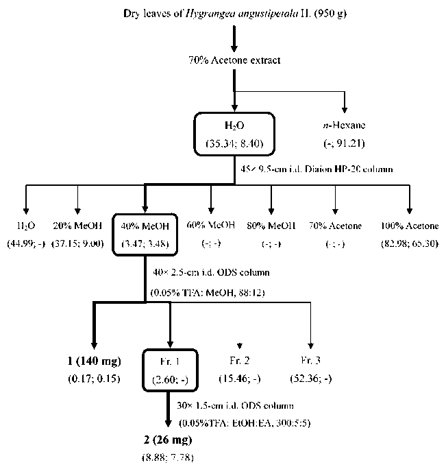
Dried leaves (950 g) of H. angustipetala were homogenized in 70% aqueous acetone (19 l) and filtered. The filtrate was concentrated under a vacuum using a rotary evaporator to remove the acetone. The aqueous solution was partitioned with an equal amount of n-hexane. The aqueous layer was chromatographed over a 45 x 9.5-cm i.d. Diaion HP-20 column (Mitsubishi, Japan) with a stepwise series of H2O ― 20% MeOH ―40% MeOH ― 60% MeOH ― 80% MeOH ― 70% acetone ― 100% acetone. The 40% MeOH eluate (3.895 g) was rechromatographed over a LiChroprep
RP-18 column eluted with 0.05% TFA: MeOH (88:12)
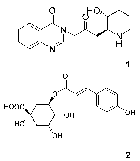
to yield (+)-febrifugine (1, 139.5 mg) and trans-3-p-coumaroylquinic acid (2, 25.6 mg) (Figure 1). Tests of the two compounds showed them to be greater than 95% pure by normal and reversed-phase HPLC chromatography. Retention times for the reversed-phase HPLC were 27.6 and 28.4 min for 1 and 2, respectively. The structure was
|
unambiguously identified by comparing these data with the
literature (Kobayashi et al., 1999; Lu et al., 2000; Norbaek et al., 2002).
Cell cultures
The human gastric carcinomas (AGS and SNU-1), and the mouse lymphoid macrophage (P-388D1) cell lines were obtained from American Type Cell Culture (ATCC, Rockville, MD, USA) and were maintained in F-12K, RPMI 1640, and DMEM (Gibco), respectively,
supplemented with 10% FBS, 100.0 mg/l streptomycin, and 100 IU/ml penicillin (Gibco). All cell cultures were incubated at 37°C in a humidified atmosphere of 5% CO2.
Cytotoxicity assays
A stock solution of test samples (2 x 10⁴ fig/ml) was prepared by dissolving the test samples in dimethyl sulfoxide (DMSO), and then storing the mixture at 4°C until use. Serial dilutions of the stock solution were prepared in the culture medium in 96-well microtiter plates. Test samples at the appropriate concentrations were added to cell cultures for 12, 24, and 48 h without renewing the medium. The number of surviving cells was then counted using the tetrazolium (MTT) assay (Bruggisser et al., 2002). The cytotoxicity index (CI%) was calculated based on the following equation: CI% =[1-(T/C)] x 100%; where T and C represent the mean optical density of the treated and vehicle control groups, respectively. In accordance with the CI% of the concentration-response curve, the concentration of the test compound producing 50% cell growth inhibition (IC50 value) was estimated. Doxorubicin was used as a positive control.
In vivo antitumor assays
P-388D1 cells (1 x 10⁶ cells/mouse) were transplanted
intraperitoneally (i.p.) into 5-week-old CDF1 male mice
(DBA male x BALB/c female) on day 0. The D40M was dissolved in 10% DMSO. The test samples or 1% DMSO
were administered i.p. once a day, on days 0 to 8. DMSO (1%) was used as solvent control. Idarubicin (Zavedos®) was dissolved in distilled water and administered i.p. once a day, on days 0 to 4. The antitumor effect was defined as the percent increase in life span (%ILS) calculated according to the following equation: %IL S = [(T/C)-1] x 100%; where T and C represent the mean survival times (day) of the treated and vehicle control groups, respectively. The body weight of each CDF1 mouse was determined every day using an animal scale. Data are presented as the mean 士 standard deviation (SD). Student's t-test was used to compare survival times (days) between the test and blank groups (Yang et al., 2003).
Western blot analysis
AGS and SNU-1 cells (5 x 10⁵ cells/well) exposed to
test samples for 12 or 48 h were collected into tubes, and then washed with PBS. Protein samples were prepared
|
||
|
|||
|
||||||||||||||||||||||||||||||||||||||||||||||||||||||||||||||||||||||||||||||||||||
HSIEH et al. ― Cytotoxic constituents of Hydrangea angustipetala
|
47
|
|||||||||||||||||||||||||||||||||||||||||||||||||||||||||||||||||||||||||||||||||||
|
||||||||||||||||||||||||||||||||||||||||||||||||||||||||||||||||||||||||||||||||||||
and resolved by denaturing SDS-PAGE, using standard methods (Yang et al., 2003). The proteins were transferred onto an Immobilon-P PVDF 0. 45 -f m membrane
(IPVH00010, Millipore, USA), and Western blotting
was performed using antibodies specific to human PARP, pro-caspase, and a-actin. A goat anti-mouse antibody
conjugated to alkaline phosphatase and BCIP/NBT (BCIP/
NBT, Gibco) were used to visualize the protein bands.
RESULTS AND DISCUSSION
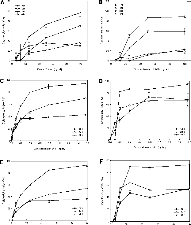
The leaves and flowers of H. angustipetala were extracted with 70% acetone after homogenization and the cytotoxic effects were measured by an MTT assay in AGS cells. The leaves of 70% acetone H. angustipetala extract (70A) exhibited the stronger cytotoxicity against AGS cells than the flowers (Table 1). The 70% acetone extract of its leaves (70A) was more cytotoxic than 50% EtOH extract in AGS cells. Furthermore, the 70A inhibited the growth of AGS and SNU-1 cells in dose- and time-dependent manners (Figure 1A, B, Table 2).
The chemical prescreening tests for H. angustipetala indicated that the flowers were richer in polyphenols than the leaves (Table 1) and suggested that the 70A contained phenol groups. Therefore, the 70% aqueous extract of H. angustipetala leaves was portioned with n-hexane to give n-hexane and aqueous (H2O) layers. Based on the cytotoxicity assay guide for the three fractionations, the H2O layer was more cytotoxic than the n-hexane one (Figure 2). The H2O layer was chromatographed on a
|
Diaion HP-20 column to give seven eluted fractions, and their cytotoxic effects in AGS and SNU-1 cells are shown in Figure 2. Of the seven fractions, fraction III (the 40% MeOH-eluted fraction, D40M) displayed strongest cytotoxic effect. Therefore, the antitumor effects of D40M were evaluated using P-388D1 cells in in vitro and in vivo models. As shown in Table 3, D40M strongly inhibited the growth of P-388D1 cells and the IC50 value was 0.72 fg/ml. Moreover, D40M at 10 mg/kg body weight significantly prolonged the life span of P-388D1 tumor-bearing CDF1 mice by 21.4% compared to the 1% DMSO solvent-treated mice. According to the above results, we suggested that
|
|||||||||||||||||||||||||||||||||||||||||||||||||||||||||||||||||||||||||||||||||||
|
||||||||||||||||||||||||||||||||||||||||||||||||||||||||||||||||||||||||||||||||||||
Table 1. Chemical and bioactivity prescreening tests of Hydrangea angustipetala leaves and flowers.
|
||||||||||||||||||||||||||||||||||||||||||||||||||||||||||||||||||||||||||||||||||||
|
||||||||||||||||||||||||||||||||||||||||||||||||||||||||||||||||||||||||||||||||||||
Figure 1. Cytotoxic effects of tested samples used to treat AGS and SNU-1 cells in dose- and time-dependent manners. The 70% actone extract (70A)-treated AGS (A) and SNU-1 cells (B); (+)-febrifugine (1)-treated AGS (C) and SNU-1 cells (D); and trans-3-p-coumaroylquinic acid (2)-treated AGS (E) SNU-1
cells (F).
|
||||||||||||||||||||||||||||||||||||||||||||||||||||||||||||||||||||||||||||||||||||
a+, the tested solution turned yellow. b+, the test solution turned blackish-green. cIC50 values (ig/ml) of extracts in AGS cells for 48 h. Data were used from three separate experiments.
|
||||||||||||||||||||||||||||||||||||||||||||||||||||||||||||||||||||||||||||||||||||
|
||||||||||||||||||||||||||||||||||||||||||||||||||||||||||||||||||||||||||||||||||||
Table 2. IC50 values of the 70% acetone extract (70A), (+)-febrifugine (1) and trans-3-p-coumaroylquinic acid (2) in gastric carcinoma cell lines (AGS, SNU-1).
|
||||||||||||||||||||||||||||||||||||||||||||||||||||||||||||||||||||||||||||||||||||
|
||||||||||||||||||||||||||||||||||||||||||||||||||||||||||||||||||||||||||||||||||||
|
||||||||||||||||||||||||||||||||||||||||||||||||||||||||||||||||||||||||||||||||||||
|
||||||||||||||||||||||||||||||||||||||||||||||||||||||||||||||||||||||||||||||||||||
aDOX was doxorubicin as a positive control. -, Not detected. Data were used from three separate experiments.
|
||||||||||||||||||||||||||||||||||||||||||||||||||||||||||||||||||||||||||||||||||||
|
||||||||||||||||||||||||||||||||||||||||||||||||||||||||||||||||||||||||||||||||||||
|
||||||||||||||||||||||||||||||||
48
|
Botanical Studies, Vol. 51, 2010
|
|||||||||||||||||||||||||||||||
|
||||||||||||||||||||||||||||||||
the D40M had potential antitumor effects. Therefore, D40M was loaded to the ODS column and eluted with 0.05% TFA:MeOH (88:12), and (+)-febrifugine (1) and trans-3-p-coumaroylquinic acid (2) were obtained (Figure 3). Both compounds showed strong cytotoxic effects and greater sensitivities in SNU-1 than AGS cells (Table 2). Moreover, they also inhibited the growth of AGS and SNU-1 cells in dose- and time- dependent manners (Figure 1C ~F), and 1 exhibited greater cytotoxicity than 2.
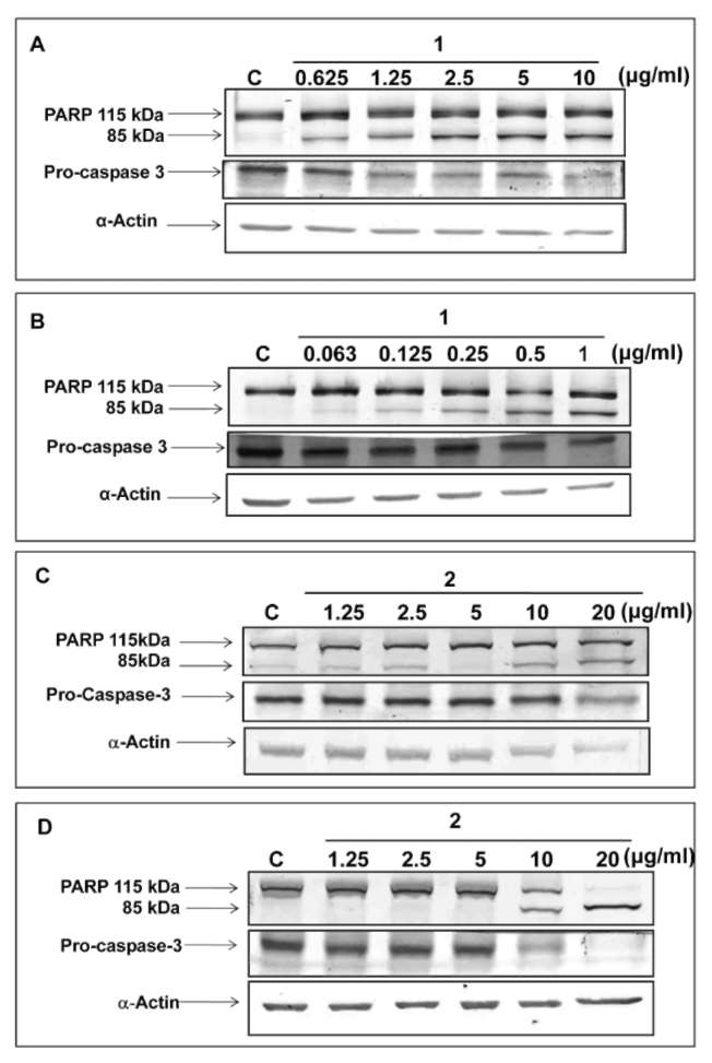
T he highly proteolytic cleavage of PARP and procaspase 3 is a hallmark of apoptosis of tumor cells (Lazebnik et al., 1994; Nunez et al., 1998). In Figure 4, degradation of PARP during treatment with the compounds was investigated by Western blotting in AGS and SNU-1
|
|
|||||||||||||||||||||||||||||||
Table 3. IC50 values of the active fractions in cultured P-388D1 cells and the percentage increase in the lifespan (%ILS) of extract-treated P-388D1-bearing CDF1 mice.
|
||||||||||||||||||||||||||||||||
Figure 3. Chemical structures of (+)-febrifugine (1) and trans-3-p-coumaroylquinic acid (2) isolated from Hydrangea angustipetala.
|
||||||||||||||||||||||||||||||||
|
||||||||||||||||||||||||||||||||
|
||||||||||||||||||||||||||||||||
-, Not detected.
The dosages of D40M and idarubicin were 10 and 0.1 mg/kg, respectively in vivo assay.
Tested groups: There were 6 mice in each D40M and 1%
DMSO group. *Student's t-test,p < 0.05. Positive control groups: There were 3 mice in each idarubicin
and distillation water group. *Student's t-test,p <- 0.05.
|
||||||||||||||||||||||||||||||||
|
||||||||||||||||||||||||||||||||
Figure 4. Western blot analysis of PARP, pro-caspase 3, and a-actin proteins in (+)-febrifugine (1)-treated AGS (A) and SNU-1 (B) cells for 12 h; trans-3-p-coumaroylquinic acid
(2)-treated AGS (C) and SNU-1 (D) cells for 48 h. a-Actin was
used as an internal control to identify equal amounts of proteins loaded in each lane. C, solvent control. Data were used from three separate experiments, the picture of one of which is shown.
|
||||||||||||||||||||||||||||||||
Figure 2. Isolation flowchart of Hydrangea angustipetala using bioassay-guided fractionation. (x; y): x is the IC50 (fg/ml) value of the eluted extract on AGS cells for 48 h; y is the same for SNU-1 cells. Data were used from three separate experiments.
|
||||||||||||||||||||||||||||||||
|
||||||||||||||||||||||||||||||||
|
||||
HSIEH et al. ― Cytotoxic constituents of Hydrangea angustipetala
|
49
|
|||
|
||||
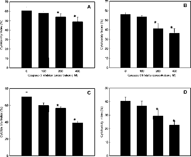
cells. Moreover, the 116-kDa protein was cleaved to give a typical 85-kDa fragment, which demonstrated dose-and time dependences after treatment with 1 and 2 for 12 and 48 h. On other hand, cleavage of pro-caspase 3 was clearly observed after treatment with 1 and 2 in AGS and SNU-1 cells (Figure 4), and 1 showed stronger activity than 2. Pretreatment of AGS and SNU-1 cells with a caspase-3-specific inhibitor in a serial dosage, did slightly reduce cytotoxicity induced by 1 and 2 (Figure 5). We suggest that 1 and 2 can induce apoptosis in AGS and SNU-1 cells, and that activation of caspase-3 may provide a mechanistic explanation for their cytotoxic effects. In conclusion, 1 and 2 were isolated from the active fraction, the D40M of the 70% acetone extracts, and 1 was more cytotoxic than 2 in gastric tumor cells. Therefore, 1 may be a good lead compound for developing an anti-cancer drug.
According to Dr. Newman's review article about natural products as sources of new drugs, major developments of natural products as anticancer drug lead compounds occurred in the period 1981~2002 (Newman et al., 2003). The isolation of 1 and 2 from the leaves of H. angustipetala were first described in this paper. However, 1 was first isolated from Dichroa febrifuga Lour and is abundant in Hydrangea species such as: H. macrophylla var. Otaksa and H. Arten (Chou et al., 1948; Ablondi et al., 1951; Ishih et al., 2003). The structured core of 1 is a 4-quinazolone alkaloid, and it shows strong antimalarial effects against Plasmodium falciparum (Kobayashi et al.,
1999; Hirai et al., 2003; Jiang et al., 2005). Because 1
shows strong liver toxicity, several new derivatives have
|
been synthesized based on febrifugine (Kikuchi et al., 2006; Zhu et al., 2006). In this paper, we found 1 also showed strong cytotoxicity against gastric tumor cells and induced apoptosis via activation of caspase 3. The data indicated that 1 is also a good lead compound from which to develop a new anti-cancer drug.
The other compound, 2, has a phenolic structure and contains hydroxycinnamic acid and coumaroyl groups. Furthermore, constituent 2 has previously been isolated from green coffee beans (Iwai et al., 2004), Cichorium intybus (Norbaek et al., 2002), Onobrychis viciifolia Scop.
(Lu et al., 2000), H. Macrophylla (Takeda et al., 1985),
Loropetalum chinense (Yoshida et al., 1993), and Prunus domestica L. (Nakatani et al., 2000). The antioxidant effects (Sato et al., 1993; Ohmshi et al., 1994; Basnet et al., 1996; Nakatani et al., 2000; Nagaoka et al., 2001), tyrosinase inhibition (Iwai et al., 2004), and human leukemic HL-60 cell inhibition (Chen et al., 2001) of 2 were also previously reported. We clearly demonstrated that 2 induced apoptosis in gastric tumor cells via activation of caspase 3, but its cytotoxicity was less than
that of 1.
Apoptosis is an efficient strategy for cancer chemotherapy. Both 1 and 2 could induce apoptosis in gastric tumor cells and were isolated from the D40M. Moreover, the D40M could prolong the survival time of P-388D1-bearing mice. The results showed that 1 and 2 are the principal antitumor constituents of the D40M and are suitable to serve as bioactivity substance markers for control quality. While many compounds have been shown to inhibit the proliferation of mammalian cells in culture, only a small proportion of these have demonstrated significant selectivity in vivo even in the most chemosensitive animal tumor models. Therefore, we suggested that the D40M of H. angustipetala has the potential for development into chemoprevention products.
Acknowledgment. The authors gratefully acknowledge
the financial support (95TMU-TMUH-14) from Taipei
Medical University Hospital, Taipei, Taiwan.
LITERATURE CITED
Ablondi, F., S. Gordon, and J. Morton. 1951. An antimalarial alkaloid from Hydrangea. J. Org. Chem. 27: 14-18.
Basnet, P., K. Matsushige, K. Hase, S. Kadota, and T. Namba. 1996. Potent antihepatotoxic activity of dicaffeoyl quinic acids from propolis. Biol. Pharm. Bull. 19: 655-657.
Bruggisser, R., K.V. Daeniken, G. Jundt., W. Schaffner, and H. Tullberg-Reinert. 2002. Interference of plant extracts, phy-toestrogens and antioxidants with the MTT tetrazolium assay. Planta Med. 68: 445-448.
Chen, Y.J., M.S. Shiao, M.L. Hsu, T.H. Tsai, and S.Y. Wang.
2001. Effect of caffeic acid phenethyl ester, an antioxidant from propolis, on inducing apoptosis in human leukemic
HL-60 cells. J. Agric. Food Chem. 49: 5615-5619.
|
|||
|
||||
25 50 100 0 50 100 200
Gaspase-3 Inhibitor concentration ( M) Caspase-3 Iribilor concentration ( M)
|
||||
Figure 5. Caspase-3-specific inhibitor pretreatment for 2 h ameliorated the (+)-febrifugine (1 ) and trans-3-p-coumaroylquinic acid (2)-induced cytotoxicity in AGS and SNU-1 cells. 1 induced cytotoxicity in AGS cells at 10 jig/ml for 12 h (A) and in SNU-1 cells at 1 jig/ml for 12 h (B); 2 induced cytotoxicity in AGS cells at 20 j g/ml for 48 h (C) and in SNU-1 cells at 2.5 j g/ml for 48 h (D). Data were used from three separate experiments. * Student's t-test,p <- 0.05.
|
||||
|
||||
|
|||
50
|
Botanical Studies, Vol. 51, 2010
|
||
|
|||
Chou, T.Q., F.Y. Fu, and Y.S. Kao. 1948. Antimalarial constituents of Chinese drug, Ch'ang Shan, Dichvoa febvifuga Lour. J. Am. Chem. Soc. 70: 1765.
Hirai, S., H. Kikuchi, H.S. Kim, K. Begum, Y. Wataya, H. Tasa-
ka, Y. Miyazawa, K. Yamamoto, and Y. Oshima. 2003. Metabolites of febrifugine and its synthetic analogue by mouse liver S9 and their antimalarial activity against Plasmodium malaria parasite. J. Med. Chem. 46: 4351-4359.
Ishih, A., T. Miyase, K. Ohori, and M. Terada. 2003. Different
responses of three rodent plasmodia species, Plasmodium yoelii 17XL, P. berghei NK65 and P. chabaudi AS on treatment with febrifugine and isofebrifugine mixture from Hydrangea macrophylla var. otaksa leaf in ICR mice. Phy-
tother. Res. 17: 650-656.
Iwai, K., N. Kishimoto, Y. Kakino, K. Mochida, and K. Fujita. 2004. In vitro antioxidative effects and tyrosinase inhibitory activities of seven hydroxycinnamoyl derivatives in green
coffee beans. J. Agric. Food Chem. 52: 4893-4898.
Jiang, S., Q. Zeng, M. Gettayacamin, A. Tungtaeng, S. Wannay-ing, A. Lim, P. Hansukjariya, C.O. Okunji, S. Zhu, and D. Fang. 2005. Antimalarial activities and therapeutic properties of febrifugine analogs. Antimicrob. Agents Chemother.
49: 1169-1176.
Kikuchi, H., K. Yamamoto, S. Horoiwa, S. Hirai, R. Kasahara, N. Hariguchi, M. Matsumoto, and Y. Oshima. 2006. Exploration of a new type of antimalarial compounds based
on febrifugine. J. Med. Chem. 49: 4698-4706.
Kobayashi, S., M. Ueno, R. Suzuki, H. Ishitani, H.S. Kim, and Y. Wataya. 1999. Catalytic asymmetric synthesis of anti-malarial alkaloids febrifugine and isofebrifugine and their
biological activity. J. Org. Chem. 64: 6833-6841.
Koh, D.W., T.M. Dawson, and V.L. Dawson. 2005. Mediation of cell death by poly(ADP-ribose) polymerase-1. Pharmacol.
Res. 52: 5-14.
Lazebnik, Y.A., S.H. Kaufmann, S. Desnoyers, G.G. Poirier, and W.C. Earnshaw. 1994. Cleavage of poly(ADP-ribose) polymerase by a proteinase with properties like ICE. Nature
371: 346-347.
Lu, Y., Y. Sun, L.Y. Foo, W.C. McNabb, and A.L. Molan. 2000.
Phenolic glycosides of forage legume Onobrychis viciifolia. Phytochemistry 55: 67-75.
Nakatani, N., S.I. Kayano, H. Kikuzaki, K. Sumino, K. Katagiri, and T. Mitani. 2000. Identification, quantitative determina-
|
tion, and antioxidative activities of chlorogenic acid isomers in prune (Prunus domestica L.). J. Agric. Food Chem. 48:
5512-5516.
Nagaoka, T., A.H. Baskota, Q. Xiong, Y. Tezuka, and S. Kadota. 2001. Synthesis and antihepatotoxic and antiproliferative activities of di- and tri-O-caffeoylquinic acid derivatives.
Wakan Iyakugaku Zassi 18: 183-190. Newman, D.J., G.M. Cragg, and K.M. Snader. 2003. Natural
products as sources of new drugs over the period
1981-2002. J. Nat. Prod. 66: 1022-1037.
Nicotera, P., M. Leist, E. Fava, L. Berliocchi, and C. Volbracht. 2000. Energy requirement for caspase activation and neuro-
nal cell death. Brain Pathol. 10: 276-282.
Norbaek, R., K. Nielsen, and T. Kondo. 2002. Anthocyanins from flowers of Cichorium intybus. Phytochemistry 60:
357-359.
Nunez, G., M.A. Benedict, Y. Hu, and N. Inohara. 1998. Cas-
pases: the proteases of the apoptotic pathway. Oncogene 17:
3237-3245.
Ohnishi, M., H. Morishita, H. Iwahashi, S. Toda, Y. Shirataki, M. Kimura, and R. Kido. 1994. Inhibitory effects of chlo-rogenic acids on linoleic acid peroxidation and haemolysis.
Phytochemistry 36: 579-583.
Pezzuto, J.M. 1997. Plant-derived anticancer agents. Biochem.
Pharmacol. 53: 121-133.
Sato, M., A. Hiraoka, and M. Watanabe. 1993. Inhiition of y-irradiation induced oxidation of chlorogenate by 1,3-di-caffeoylquinate in Xanthium occidentale. Phytochemistry
33: 1357-1360.
Takeda, K., R. Kubota, and C. Yagioka. 1985. Copigments in the blueing of sepal colour of Hydrangea macrophylla. Phyto-
chemistry 24: 1207-1209. Yang, L.L., C.C. Chang, L.G. Chen, and C.C. Wang. 2003. An-
titumor principle constituents of Myrica rubra var. acumi-
nata. J. Agric. Food Chem. 51: 2974-2979. Yoshida, T., S. Tanei, Y.Z. Liu, K. Yuan, C.R. Ji, and T. Okuda.
1993. Hydrolysable tannins from Loropetalum chinense.
Phytochemistry 32: 1287-1292. Zhu, S., L. Meng, Q. Zhang and L. Wei. 2006. Synthesis and
evaluation of febrifugine analogues as potential antimalarial
agents. Bioorg. Med. Chem. Lett. 16: 1854-1858.
|
||
|
|||
|
|||
HSIEH et al. ― Cytotoxic constituents of Hydrangea angustipetala
|
51
|
||
|
|||
狹葉八仙花對胃癌細胞之毒性成分
謝清睿1 游璧如2 陳立耿3 趙淑妙4 張君照1 王靜瓊2
1臺北醫學大學附設醫院消化內科
2臺北醫學大學藥學院藥學系 3嘉義大學生命科學院醫藥科學研究所 4中央研究院生物多樣性研究中心 狹葉八仙花(虎耳草科)生長在北台灣山區,常用於抗瘧疾的民間藥。本硏究採集葉部,利用 70%丙酮均質萃取後'配合細胞毒性分析'追蹤分離其活性成分。以40%甲醇沖提Diaion HP-20管柱
之沖提物(D40M),可抑制P-388D1細胞之生長,並可延長P-388D1擔癌鼠之生命。繼而從D40M沖 提物中'分離得到(+)-febrifugine (1)及fran2s-3-p-coumaroylquinic acid (2)之天然物'並皆具有誘導AGS 及SNU-1胃癌細胞凋亡之作用。而且(+)-febrifugine的細胞毒性比fran2s-3-p-coumaroylquinic acid強' (+)-febrifugine加入AGS及SNU-1胃癌細胞48小時,其抑制50%濃度分別爲0.17及0.05 ig/ml 。此兩 者亦可降解AGS及SNU-1胃癌細胞中PARP及pro-caspase 3 ,當加入caspase-3-specific抑制劑時,此 兩個天然物所引起之細胞毒性將可被緩解。因此推測,此兩個天然物所引起之細胞毒性可能是透過活化 caspase 3之路徑。而(+)-febrifugine將可作爲開發抗癌藥時的起始物。 關鍵詞:凋亡;(+)-febrifugine ;狹葉八仙花;P-388D1-CDF1擔癌鼠;trans-3-p-coumaroylquinic acid;
虎耳草科。
|
|||
|
|||
|
||