Botanical Studies (2010) 51: 75-80.
MICROBIOLOGY
Severe decline of wax apple trees caused by Fusarium solani in northern Taiwan
Pi-Han WANG1, Yun-San CHEN1, Mei-Ju LIN2, Yi-Jung TSOU2, and Wen-Hsiung KO2 *
(Received November 27, 2008; Accepted June 3, 2009)
ABSTRACT. Wax apple (Syzygium samarangense) is an important fruit crop in Taiwan. Severe decline of wax apple trees was noticed in 2003 in northern suburban Taiwan. A fungus consistently isolated from diseased twigs of declining wax apple trees, was identified as Fusarium solani based on morphological characteristics. Fusarium solani from wax apple shared 92.0 to 98.6% and 93.0 to 99.6% intraspecific sequence similarity of ITS and 28S, respectively, with those available in GenBank. Upon inoculation, the isolated F. solani caused twig blight on healthy wax apple trees, and F. solani was reisolated from the diseased twigs, thus fulfilling Koch's postulates. All the control trees remained healthy throughout the experiment. Numerous microconidia of F. solani produced on the cut surfaces of diseased twigs under moist conditions were considered to be the main inoculum source for secondary infection of diseased trees and primary infection of healthy trees.
Keywords: Fusarium solani; ITS; 28S; Sequence similarity; Tree decline.
INTRODUCTION
Wax apple (Syzygium samarangense Merr. et Perry) which is native to Southeast Asia, is an important fruit crop in Taiwan. The main goal of commercial cultivation is the production of fresh fruit for local consumption. Pingtung County in southern Taiwan is the primary production area with more than 6000 ha of wax apple orchards. In this area, flowering and fruiting of wax apple trees are carefully regulated and managed (Wang and Hung, 2005). In contrast, there are about 80 ha of wax apple orchards with unregulated flowering and fruiting and minimum care in Taipei's northern suburbs. Several of these orchards are used for agricultural tourism. Additionally, wax apple trees have been planted around private homes because of their handsome dark evergreen foliage and the production of bright red fruit during the summer.
In 2003, many wax apple trees in suburban Taipei suffered from an unknown ailment. Affected trees displayed different stages of decline (Figure 1). Since then, an increasing number of wax apple trees around private homes, along the road sides, and in the orchards have died. Some of the dead trees were 20 to 30 years old. Several wax apple orchards were abandoned because of the disease. The possibility of the disease being spread to the main wax apple production area in southern Taiwan has become a major concern.
At the beginning, Phellinus noxius (Corner) G. H. Cunn. was suspected to be the causal agent of wax apple tree decline because the pathogen is widespread (Ann et al., 2002) and has caused brown root rot and death among such trees before in Taiwan (Ann et al., 1999). However, no brown root rot or any other root abnormality typical of Phellinus rot was observed when the roots of diseased wax apple trees were exposed and inspected. Field observation also did not show evidence of insect damage (Wen, 2004). The disease appeared to originate from twigs. Most of the twigs of affected trees defoliated and died (Figure 2). The objectives of this study were to identify the causal agent of the wax apple tree decline and to determine the possible source of inoculum for infection in the field.
MATERIALS AND METHODS
Isolation and morphology of pathogen
To isolate the pathogen, sections (ca 5 mm diam., 10 mm long) of diseased twigs showing dark brown discoloration on the scraped surface were surface-sterilized with 0.6% NaOCl for 3 min. After rinsing with sterile distilled water, these tissues were blotted with sterilized paper towels, plated on 2% water agar, V-8 agar containing 10% V-8 juice, 0.02% CaCOg and 2% agar or selective medium for Acremonium spp. consisting of
10% V-8 juice, 0.02% CaCO3, 50 ppm nystatin, 100 ppm
ampicillin and 2% agar (Ko and Kunimoto, 1999). Single-microconidium isolates LW1-1 and LW1-2 obtained from diseased twigs collected from different locations were maintained on potato dextrose agar (PDA) at 24°C under
*Corresponding author: E-mail: kowh@dragon.nchu.edu.tw; Tel: 886-4-22840780; Fax: 886-4-22877585.
76
Botanical Studies, Vol. 51, 2010
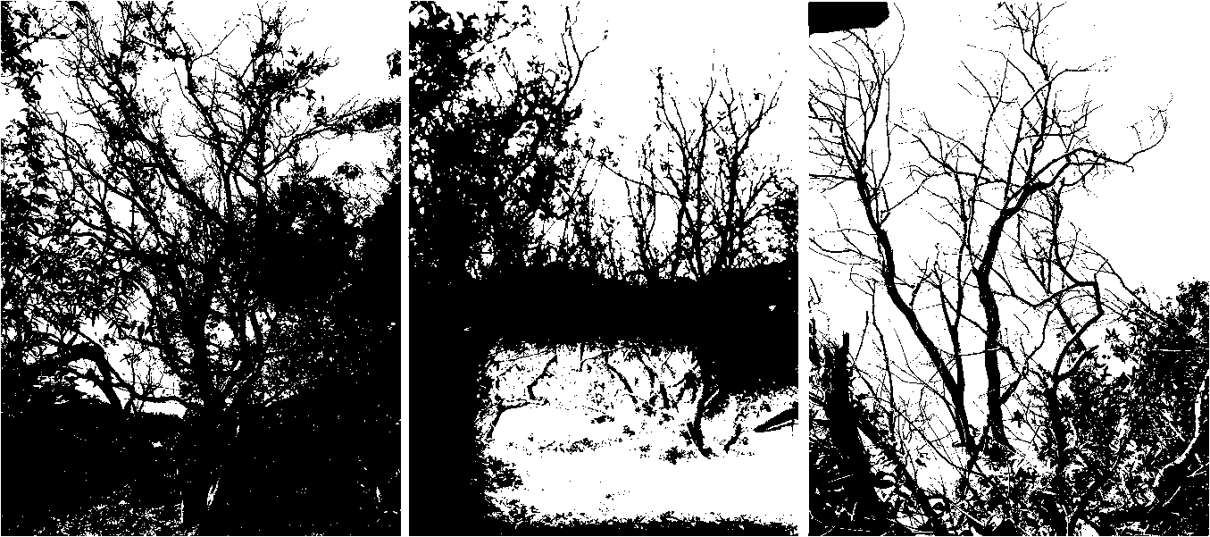
Figure 1. Wax apple trees at intermediate (left), severe (middle) or deceased (right) stage of decline.
placed on the scraped portion of the twig, wrapped with Parafilm, and secured with vinyl tape. Grains were left on until the end of the experiment. Inoculated plants were checked every two days for the first sign of infection. Twigs similarly inoculated with autoclaved grains were used as controls.
Source of inoculum
To determine if diseased twigs may serve as a source of inoculum for secondary or primary infection, sections of disease twigs approximately 5 mm in diameter by 10 mm long were surface-sterilized as described above, and cut into two halves longitudinarily under aseptical conditions. One portion of halfed sections were dipped in sterile distilled water for 10 sec, and then placed on sterilized moistened paper towel in a Petri plate. The other section halves were placed in empty sterile Petri plates and used as the control. Ten diseased and 10 healthy twigs were used for each location.
DNA extraction and polymerase chain reaction (PCR)
The DNA of F. solani isolate LW1-1 from wax apple was extracted from 0.1 g of 3-day-old mycelia grown on cellophane placed on PDA by the plant genomic DNA extraction kit (GeneMark Technology Co., Taichung, Taiwan). The ITS region was amplified with primers ITS1 and ITS4 (White et al., 1990). PCR was performed in a 50 |ul volume reaction containing 2 |ul DNA, 1 pmole of upstream and downstream primers and 2.5 units of SuperTaq polymerase (Protech Technology Enterprise Co., Ltd, Taipei, Taiwan) with buffer system recom­mended by the manufacturer. Cycling conditions of PCR were: initial denaturation at 94C for 2 min, 30 cycles at
94C for 30 sec, 55C for 30 sec, 72C for 1 min, and a
final elongation at 72°C for 6 min. The PCR product was
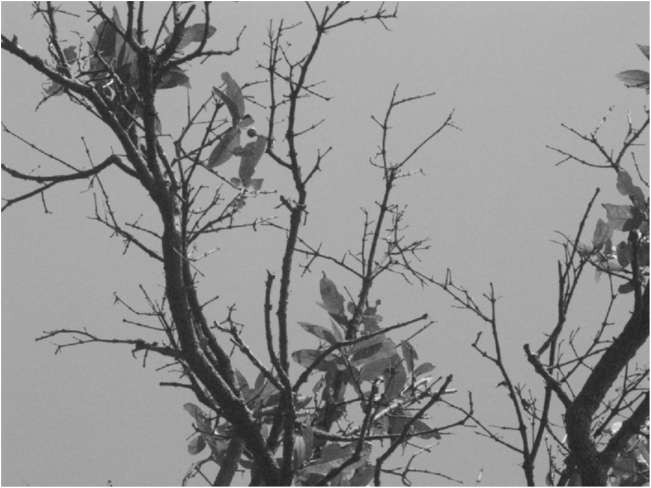
Figure 2. Branches of a declining wax apple tree with may diseased or dead twigs.
light, and used for further study.
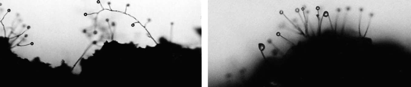
The isolated organisms produced abundant microconidia on PDA. Macroconidia were produced after a piece (ca 10 x 10 x 3 mm) of PDA culture was trans­ferred to water agar and incubated at 24°C under light. Chlamydospores were produced by growing the fungus in celery juice as previously described (Huang et al., 1983).
Pathogenicity tests
For pathogenicity tests, 5-year-old wax apple plants (90-120 cm high) growing in pots were inoculated. The fungus was grown in a wheat-oat medium (10 ml whole wheat grains, 10 ml whole oat grains and 10 ml distilled water) for 2 weeks at 24C (Ko et al., 1986). Wax apple twigs, approximately 5-7 mm in diameter, were scraped gently with a surgical scalpel to remove the epidermis from bark tissue. Four grams of colonized grains were
WANG et al. ― Severe decline of wax apple trees caused by Fusarium solani
77
analyzed by electrophoresis in a 1.2% agarose gel. In the same manner the large ribosomal subunit, 28S, was analyzed with primer pairs LROR and LR7 (Vilgalys and Hester, 1990). The annealing temperature was changed to
50C.
Cloning and sequence analysis
PCR amplified DNA products were cloned into pCRII-TOPO vector (Invitrogen, Carlsbad, CA, USA) according to the manufacturer's instruction. Plasmid clones with expected size DNA inserts were screened and used for sequencing analysis. Sequencing of the target DNA insert was done by an automatic DNA sequencer (ABI
PRISM 377, Perkin-Elmer, CA, USA) with the BigDye
Terminator Cycle Sequencing Ready Reaction Kit (Perkin-Elmer Applied Biosystems, CA, USA). Sequence data were analyzed by Lasergene 7 Software (DNASTAR,
Inc., USA).
RESULTS
Symptoms
On naturally infected wax apple trees, initial symptoms were leaves on the apical portion of infected twigs that turned gray and lost vigor. The color of leaves attached to twig sections that were brown changed to reddish brown and eventually fell off. The interior of the infected twig turned brown. As the disease progressed, increasing number of leaves were browning and abscising (Figure 2). The severity of decline increased with the number of increasing infected twigs and was reflected in the amount of fallen leaves (Figure 1). Eventually some branches were also infected and the whole tree died.
Isolation and identification
When diseased wax apple twigs collected from two different locations on June 5, 2007 were used for isolation, distinctive conidiophores with microconidia in false heads (Figure 3A) were found on cut surface of every section placed on water agar or selective medium (Table 1). However, on V-8 agar the same kind of fruiting bodies
were found on only two of four sections from location 1. Water agar and selective medium, therefore, were chosen for isolation of the fungus from diseased wax apple twigs collected from four other locations on June 24, 2007. Again, the same kind of fruiting bodies were found on the cut surfaces of all the diseased twig sections tested
(Table 1).
On PDA, the fungus formed white colony with dense aerial mycelium and yellowish pigments beneath the colony. Microconidia developed abundantly in spherical false heads on tips of conidiophores which were long and sturdy monophialids. They were oval, ellipsoid, reniform, and fusiform in shape, had none to 1-2 septa and measured 3-16 x 3-5 ^m. Macroconidia developed when a PDA culture block (ca 5 x 5 x 3 mm) was transferred to water agar and incubated at 24C with light for 7 days. They were fusoid with a well-marked foot cell, and 5 to 7 septate measuring 14-46 x 3-5 ^m. Chlamydospores de­veloped abundantly in celery juice after 1-month incuba­tion at 24C in darkness. They were globose to oval, 6-9 x 7- 10 [im, and terminal or intercalary. They also formed chains. The fungus fits the description of Fusarium solani
Table 1. Isolation of the fungus producing distinctive conidiophores with microconidia in moist heads temporar­ily designated as Fx from diseased twigs of wax apple trees at different locations in suburban Taipei (Beitou) of northern Taiwan.
No. of twigs with Fx/No. of twigs tested
Locations
Collection date
Water agar
Selective medium
V-8 agar
1
6/5/07
4/4
4/4
2/4
2
6/5/07
2/2
2/2
2/2
3
6/24/07
5/5
5/5
NTa
4
6/24/07
1/1
1/1
NT
5
6/24/07
2/2
2/2
NT
6
6/24/07
4/4
4/4
NT
a NT = not tested.
Figure 3. Conidiophores of Fusarium solani with microconidia in false heads (100X magnification) produced on the cut surface of diseased twig section placed on 2% water agar (A) or moistened paper towel (B).
78
Botanical Studies, Vol. 51, 2010
(Mart.) Sacc. (Booth, 1971; Huang and Sun, 1997). Isolate
LW1-1 of F. solani used in this study was deposited in the Culture Collection Center of Food Industry Research and Development Institute, Hsinchu, Taiwan with the
accession no. BCRCNO 34274.
Sequence comparison of ITS and 28S
There were hundreds of ITS sequences of F. solani in GenBank. Therefore, only nine randomly selected plant pathogenic isolates were used for comparison with wax apple isolate. The ITS sequence of wax apple isolate shared more than 98% similarity with that of F. solani f. sp. robiniae or F. solani f. sp. pisi, but only 92% similarity with that of F. solani f. sp. glycines (Table 2). Since there were only 10 full length 28S sequences of F solani in GenBank, all of them were used for comparison with that of wax apple isolate. The 28S sequence of wax apple isolate shared 99.6% similarity with that of isolate FRC#S1027 or LCP9.2 of F. solani, but only 93.0% similarity with that of F. solani from tropical forest (Table 3).
Pathogenicity tests
Two weeks after inoculation, some inoculated twigs showed disease symptoms similar to those observed on naturally infected plants. Leaves above inoculation site turned gray initially, became brown to reddish brown, and
fell off eventually. Both isolates of LW1-1 and LW1-2
were pathogenic causing disease incidence ranging from 60 to 100% on healthy wax apple twigs in three separate tests (Table 4). Fusarium solani was recovered from all the diseased twigs thus fulfilling Koch's postulates for proving pathogenicity. All the control twigs remained healthy throughout the experiment.
Source of inoculum for secondary and primary infections
When diseased twigs obtained from the declining wax apple trees in the field were cut into sections and kept under moist conditions, the pathogen F. solani produced microconidia on cut surfaces of most sections tested (Table 5, Figure 3B). Most of the diseased twigs collected
Table 2. The ITS sequence similarity between Fusarium solani isolated from wax apple and nine plant pathogenic isolates of F. solani available in GenBank.
Taxon
Isolate
Associated habitat
GenBank accession no.
Similarity (%)
Fusarium solani f. sp. mori
MAFF 840046
Unknown
AF 129105
97.7
F. solani f. sp. pisi
MAFF 840047
Pisum sativum
AF 130142
98.6
F. solani f. sp. robiniae
NRRL 22161
Unknown
AF 178395
98.5
F. solani f. sp. batata
NRRL 22402
Unknown
AF 178408
94.0
F. solani f. sp. cucurbitae
NRRL 22142
Cucurbita sp.
AF 178411
97.6
F. solani f. sp. glycines
NRRL 22825
Glycine max
AF 178419
92.0
F. solani f. sp. piperis
NRRL 22570
Piper nigrum
AF 178422
94.7
F. solani f. sp. eumartii
Fs 122
Tomato
DQ 164845
94.8
Nectria haematococca
NRRL 22141
Soybean
L 36619
98.0
Table 3. The 28S sequence similarity between Fusarium solani isolated from wax apple and isolates of F. solani available in GenBank.
Taxon
Isolate
Associated habitat
GenBank accession no.
Similarity (%)
Fusarium solani
Unknown
Lotus japonicus
AB 258994
99.0
F. solani
BOL STR 060803
Tropical forest
DQ 139962
93.0
F. solani
NRRL 34123
Unknown
DQ 236687
97.3
F. solani
FRC#s 1027
Unknown
DQ 236813
99.6
F. solani
LCP 9.2
Unknown
EF 579657
99.6
F. solani f. sp. batatas
NRRL 22402
Unknown
AF 178377
98.1
F. solani f. sp. glycines
NRRL 22823
Glycine max
AF 178387
96.6
F. solani f. sp. glycines
NRRL 22825
Glycine max
AF 178388
96.6
F. solani f. sp. piperis
NRRL 22570
Piper nigram
AF 178391
96.4
F. solani f. sp. radicicola
Unknown
Unknown
AY 819046
99.2
WANG et al. ― Severe decline of wax apple trees caused by Fusarium solani
79
Table 4. Incidence of twig blight on wax apple trees after inoculation with isolates LW1-1 and LW1-2 of Fusarium solani.
southern Taiwan are currently under investigation. How the disease started and where this pathogen came from deserve further study. Considerable specialization in pathogenicity has been demonstrated in F. solani and a number of formae speciales have been proposed in this species (Booth, 1971). Two formae speciales, F. solani f. sp. cucurbitae and F. solani f. sp. pisi, have been reported previously from Taiwan (Huang and Sun, 1997). It is not known if the F. solani pathogenic to wax apple trees is host specific.
Although F. solani is generally considered a soil-borne plant pathogen (Booth, 1971), the twig blight of wax apple trees caused by F. solani documented in the present report is apparently an air-borne disease. Die-back of American holly (Ilex opaca Ait) caused by Fusarium martii Appel & Wollenw, currently Haematonectria haematococca (Berk. & Broome) Samuels & Rossman 1999 was reported as early as 1941 (Bender, 1941). Recently, die-back of Indian rosewood (Dalbergia sissoo Roxb. ex DC) caused by F. solani has also been reported from Nepal (Shakya and Lakhey, 2007). Root infection of trees usually results in slow decline, while trunk infection frequently causes quick decline (Ko, 2009). Tree decline caused by twig infection as shown in this study is gradual and is similar to the decline resulting from root infection. However, twig blight usually spreads faster than root rot.
This study also revealed the production of abundant microconidia on the cut surfaces of diseased wax apple twigs under moist conditions. There were many diseased twigs remaining on the affected wax apple trees in the field. These diseased tissues are likely to serve as the main inoculum source for secondary infection of the diseased trees and for primary infection of the healthy trees. Therefore, removal of diseased twigs and branches appears to be very important in the control of the disease.
The ITS sequence of F. solani from wax apple shared 92.0 to 98.6% similarity with those of the same species available in GenBank (Table 2). Although Phytophthora palmivora (Butler) displayed high intraspecific ITS sequence similarity ranging from 97.8 to 100%, variable intraspecific ITS sequence similarity has also been reported for Phytophthora capsici Leonian ranging from 92.2 to 100% and Peronospora parasitica (Pers. Ex Fr.) Fr. ranging from 75.4 to 99.6% (Zhang et al., 2007). The 28S sequence of F. solani from wax apple shared 93.0 to 99.6% similarity with those of the same species available in GenBank (Table 3). This is in conformity with the report of Bremia lactucae Regel which showed intraspecific 28S sequence similarity of 92.1 to 99.4%
(Zhang et al., 2007).
Acknowledgements. This study was supported in part by a grant from the National Science Council of Taiwan (NSC96-2313-B-055-001). We thank H. F. Cheng for manuscript typing, P. J. Ann for supplying wax apple plants used in this study, and J. W. Huang for assistance in pathogen identification.
Isolate
No. diseased / No. inoculateda
Exp. 1
Exp. 2
Exp. 3
LW1-1
4/5
3/5
5/5
LW1-2
5/5
5/5
5/5
Control
0/5
0/5
0/5
aData were recorded one month after inoculation.
Table 5. Formation of conidiophores with microconidia in moist heads of Fusarium solani on sections of diseased wax apple twigs collected from various locations.
No. of sections with F. solani microconidia /No. of sections tested
Location
Treatment
4
8 (days after treatment)
1
Moistened
10/10
10/10
Control
0/10
0/10
2
Moistened
5/10
7/10
Control
0/10
0/10
3
Moistened
5/10
6/10
Control
0/10
0/10
4
Moistened
8/10
9/10
Control
0/10
0/10
5
Moistened
8/10
10/10
Control
0/10
0/10
6
Moistened
4/10
9/10
Control
0/10
0/10
7
Moistened
3/10
6/10
Control
0/10
0/10
from seven different locations surveyed produced F. solani microconidia on cut surfaces under moist conditions but not under dry conditions. The amounts of diseased twigs showing F. solani microconidia under moist conditions ranged from 60% at locations 3 and 7 to 100% at locations 1 and 5.
DISCUSSION
The results indicate that wax apple tree decline in the northern suburbs of Taipei is actually twig blight caused by F. solani which also invades branches during the later stage of disease development. To our knowledge, this is the first report of a wax apple disease caused by F solani, and is also the first observation of an epiphytotic disease of wax apple trees. Measures for preventing the spread of the disease to the major wax apple production area in
80
Botanical Studies, Vol. 51, 2010
LITERATURE CITED
Ann, P.J., H.L. Lee, and T.C. Huang. 1999. Brown root rot of 10 species of fruit trees caused by Phellinus noxius in Taiwan. Plant Dis. 83: 746-750.
Ann, P.J., T.T. Chang, and W.H. Ko. 2002. Phellinus noxius brown root rot of fruit and ornamental trees in Taiwan. Plant Dis. 86: 820-826.
Bender, T.R. 1941. Furarium die-back of American holly. Plant
Dis. Reptr. 225: 403-406.
Booth, C. 1971. The Genus Fusarium. CMI, Kew, Surrey, Eng­land.
Huang, J.W. and S.K. Sun. 1991. The Genus Fusarium from Tai­wan. Shih Way Publishers, Taichung, Taiwan.
Huang, J.W., S.K. Sun, and W.H. Ko. 1983. A medium for chla-
mydospore formation in Fusarium. Ann. Phytopathol. Soc. Jpn. 49: 704-708.
Ko, W.H. 2009. Nature of slow and quick decline of macadamia trees. Bot. Stud. 50: 1-10.
Ko, W.H. and R.K. Kunimoto. 1999. Acremonium recifei a new causal agent of macadamia quick decline. Can. J. Plant
Pathol. 21: 42-44.
Ko, W.H., J. Tomita, and R.L. Short. 1986. Two natural hosts of Kretzschmaria clavus in Hawaiian forests. Plant Pathol. 35:
254-255.
Shakya, D.D. and P.B. Lakhey. 2007. Confirmation of Fusarium solani as the causal agent of die-back of Dalbergia sissoo in
Nepal. Plant Pathol. 56: 1041.
Vilgalys, R. and M. Hester. 1990. Rapid genetic identification and mapping of enzymatically amplified DNA from several Cryptococcus species. Bacteriology 172: 4238-4246.
Wang, D.N. and J.J. Hung. 2005. Wax apple. In J.H. Hung, T.D. Fan, and L.N. Lin (eds.), Taiwan Agriculture Encyclopedia, Crop Edition 2. Council of Agriculture, Taipei, Taiwan, pp.
109-120.
Wen, H.C. 2004. Insect pests. In K.C. Kuo, F.J. Tsou, and J.F.
Su (eds.), Protection of Wax Apple. Council of Agriculture, Taipei, Taiwan, pp. 14-52.
White, T.J., T. Bruns, S. Lee, and J. Taylor. 1990. Amplification and direct sequencing of ribosomal RNA genes for phylo-genetics. In M.A. Innis, D.H. Gelfand, J.J. Sninsky, and T.J. White (eds.), PCR Protocols: A Guide to Methods and Ap­plications. Academic Press, San Diego. pp. 315-322.
Zhang, Z.G., X.B. Zheng, C.Y. Wang, and W.H. Ko. 2007. Evaluation of the rearrangement of taxonomic position of Peronophythora litchii based on partial DNA sequences.
Bot. Stud. 48: 79-89.
台灣北部由 Fusarium solani 所引起的蓮霧才封嚴重衰亡
汪碧涵1 陳永三1 林玫珠2 鄒依蓉2 柯文雄2
1東海大學生命科學及熱帶生態學與生物多樣性研究中心
2國立中興大學植物病理學系
蓮霧是台灣重要果樹之一 ,2003年在台灣北部郊區發現蓮霧樹有嚴重衰亡現象。由罹病樹枝常
分離到的真菌,根據其型態鑑定爲 Fusarium solani 。此菌的ITS序列與基因庫同種菌的序列有92.0
98.6%的相似度,其 28S 序列則有 93.099.6% 的相似度,此菌接種到健康蓮霧樹枝,可使之產生與
自然界一樣的病徵。由得病的樹枝又可分離到相同的菌,因而通過柯霍氏法則證明蓮霧樹枝死亡是由 F.
solani 所弓|起,因此而造成蓮霧樹的衰亡。罹病樹枝剖開,放在潮濕的環境會產生許多微分生孢子,因
此罹病樹枝是病樹第二次感染的主要病源,也是健康樹第一次感染的主要病源。
關鍵詞:蓮霧;樹枝枯萎;茄鐮孢菌;樹衰亡。