|
|||||
Botanical Studies (2010) 51: 209-216.
|
reproductive biology
|
|
|||
|
|||||
|
|||||
Reproductive traits of Pteris cadieri and P. grevilleana in Taiwan: Implications for their hybrid origin
Yi-Shan CHAO1, Ho-Yih LIU1, Yao-Moan HUANG2, and Wen-Liang CHIOU3 *
1Department of Biological Sciences, National Sun Yat-Sen University, Kaohsiung 804, Taiwan 2Division of Silviculture, Taiwan Forestry Research Institute, Taipei 100, Taiwan
3Division of Botanical Garden, Taiwan Forestry Research Institute, 53 Nanhai Rd., Taipei 100, Taiwan (Received May 4, 2009; Accepted November 23, 2009)
ABSTRACT. Pteris cadieri (Pteridaceae) exhibits significant morphological variation, its fronds range from simply pinnate to bipinnatifid. The morphological characteristics of P. cadieri and P. grevilleana vary along a continuum and suggest that P. cadieri is a hybrid species and P. grevilleana is a potential parent species. However, previous studies do not provide evidence to support or refute this hypothesis. To explore this hypothesis of hybrid origin, the reproductive biology of P. cadieri and P. grevilleana was studied in Taiwan. There were diploid and triploid P. cadieri, but P. grevilleana was diploid. All plants of both species were apogamous. In both species, spore shape varied (tetrahedral, globose, ellipsoidal) and germination rate varied, the first frond of some juvenile sporophytes had a midrib, and polyembryony occasionally occurred, the latter being the first report of polyembryony in apogmous ferns. Variation in spore number per sporangium, spore size and spore shape indicated abnormal sporogenesis. All these traits suggested that P. cadieri and P. grevilleana are of hybrid origins. However, the hypothesis that P. grevilleana is a parent of P. cadieri should be reconsidered.
Keywords: Apogamy; Hybridization; Morphological variation; Polyploids; Pteris cadieri; Pteris grevilleana.
|
|||||
|
|||||
introduction
Hybridization and polyploidy are the primary mechanisms of speciation and evolution in ferns (Haufler, 2008). Hybridization and polyploidy within a taxon is thought to cause morphological variation (Wagner, 1954; Walker, 1958; Smedmark et al., 2003; Bennert et al., 2005; Ebihara et al., 2005).
Since the research of Manton (1950), more and more cases of interspecific and intraspecific hybridization and polyploidy in ferns have been clarified by studying their reproductive biology. For example, reproductive biology traits clearly delimited the sexual diploids and apogamous triploids of Pteris fauriei Hieron. (Huang et al., 2006). Traits indicating hybrid origin included the reproductive system, variation in spore size and shape, spore number per sporangium and spore germination rate (Hickok and Klekowski, 1974; Nakato, 1975; Haufler and Windham, 1985; Bennert et al., 2005).
Pteris cadieri Christ is distributed throughout eastern Asia, from Japan, China, and Taiwan, to Indochina and Malaysia (Wu, 1990; Iwatsuki, 1995). Marked morphological variation exists among individuals and
|
even among fronds of a single plant. Because of the continuous variation in frond morphology, from simply pinnate to bipinnatifid fronds, there are several synonyms of P. cadieri, including P. dimorpha Copel. and P. plumbea var. sintenensis Masam. (Shieh, 1975; Wu, 1990). This variation in frond morphology has led some researchers to infer that Pteris cadieri arose from hybridization (Wagner, 1978; Kuo and Yu, 1986). Although cytological data were reported for some diploids (P. plumbea var. sintenensis; Walker, 1962) and triploids (P. cadieri; Nakaike, 1992), the relationship between frond morphology and ploidy level never has been studied.
Pteris grevilleana Wall. ex J. Agardh, with regularly bipinnatifid fronds, is distributed in eastern and southern Asia (Wu, 1990; Iwatsuki, 1995). Because the frond morphology of P. cadieri and P. grevilleana varies along a continuum, P. grevilleana was thought to be a parent of P. cadieri (Wagner, 1978; Kuo and Yu, 1986; Nakaike, 1992). However, these inferences were based on morphological observations only.
In this study, we sampled P. cadieri and P. grevilleana with different morphological characters to determine reproductive traits, including spore and gametophyte characteristics, reproductive systems, and the chromosome number of sporophytes. Based on these results, we examined the hypotheses that P. cadieri is of hybrid origin and that P. grevilleana is one parental species of P. cadieri.
|
||||
*Corresponding author: E-mail: chiou@tfri.gov.tw; Tel: 886-2-23039978 ext. 2701; Fax: 886-2-23076220.
|
|||||
|
|||||
|
|||
210
|
Botanical Studies, Vol. 51, 2010
|
||
|
|||
materials and methods
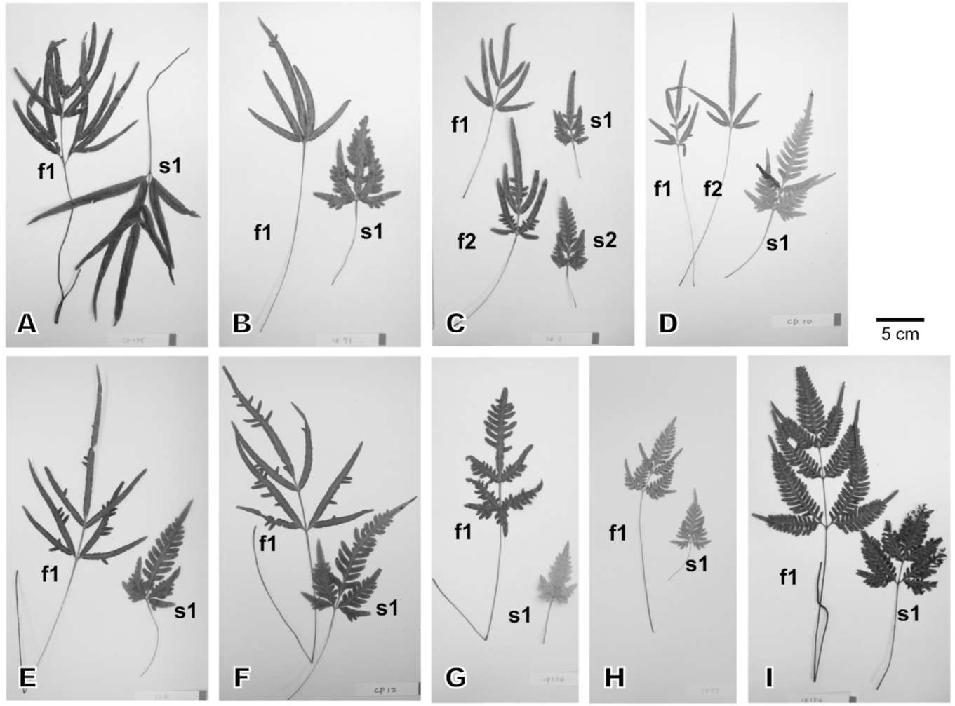
We collected seven Pteris cadieri (Figure 1A-G) and two P. grevilleana (Figure 1H-I) individuals with different frond morphology from five locations in Taiwan (Table 1). The living ferns were cultivated in the Taipei Botanical Garden greenhouse to examine their morphological variation. Voucher specimens were deposited in the Taiwan Forestry Research Institute herbarium (TAIF).
To count chromosomes, root tips were pretreated with 70 ppm cycloheximide and 250 ppm 8-hydroxyquinoline (1:1) at 18-20°C for 8 h. They were fixed sequentially in 45% acetic acid and absolute ethanol (1:3) overnight and preserved in 70% ethanol at 4°C. Then they were macerated in 1 N HCl at 60°C for 10 s, and digested in 4% pectinase for 1-2 h (Sharma, 1982; Huang et al., 2006).
To count the number of spores per sporangium, five mature sporangia were picked randomly from each plant. Spores, including shrunk ones, were counted, but debris was excluded. To determine the spore size, fertile fronds were air-dried for seven days to release their spores. Then,
|
100 spores per plant, excluding shrunk ones, were chosen randomly. The shape and size (equatorial diameter, or length of the long axis) of each spore were recorded. The mean and standard deviation were calculated for each spore shape. One-way Analysis of Variance (ANOVA) followed by Tukey post-hoc test (p < 0.05) was used to compare the spore sizes of individuals.
Germination rates were determined using the methods of Ko et al. (2004) and Huang et al. (2006). Spores of each plant were sown on two pieces of filter paper (pore size 0.45 fim, 47 mm in diameter, Gelman). Each piece of filter paper was placed in a box on a tile lying on the culture medium (vermiculite : peat : perlite = 4:4:2) for 4 weeks. Two hundred spores were sampled randomly from each piece of filter paper. A total of 400 spores from each plant were classified as germinated or ungerminated. Cultures were kept at 20-28°C under white fluorescent illumination (about 24 fimol m-2 s-1, 12 hd-1). After the germination rate had been determined, filter papers and tiles were removed and the gametophytes were remained and cultivated in the medium. After cultivation for three months, 40
|
||
|
|||
|
|||
|
|||
Figure 1. Photographs of the fronds of the 9 plants studied. A-G, representing plant 1-7, respectively, are Pteris cadieri; H & I, representing plant 8 & 9, respectively, are P. grevilleana. Two to four fronds from each plant were photographed. Fronds on the left are fertile (f1 and f2); those on the right are sterile (s1 and s2). The frond morphology of plants 1-9 varies along a continuum, from simply pinnate, irregularly bipinnatifid, to regular bipinnatifid.
|
|||
|
|||
|
|||||||||||||||||||||||||||||||||||||||||||||||||||||||||||||||||||||||||||||||||
CHAO et al. — Reproductive traits of Pteris csdieri & P. grevilleana 211
|
|||||||||||||||||||||||||||||||||||||||||||||||||||||||||||||||||||||||||||||||||
|
|||||||||||||||||||||||||||||||||||||||||||||||||||||||||||||||||||||||||||||||||
|
|||||||||||||||||||||||||||||||||||||||||||||||||||||||||||||||||||||||||||||||||
|
|||||||||||||||||||||||||||||||||||||||||||||||||||||||||||||||||||||||||||||||||
gametophytes with juvenile sporophytes of each plant (1-9) were selected randomly for examination under the light microscope (Wild M8 and Dialux 20). Gametophyte morphology, reproductive system, number of juvenile sporophytes per gametophyte, and the morphology of the first frond were investigated.
results
Sporophyte morphology and chromosome number
The morphology of the fronds of plants 1-9 ranged from simply pinnate, to irregularly bipinnatifid, to regularly bipinnatifid (Figure 1). Because they were all cultivated in the greenhouse, the environmental effects on morphologies were excluded. Plants 1-7, with simple pinna or irregularly bipinnatifid pinna, were Pteris cadieri. Plants 8 and 9, with regularly bipinnatifid pinna, were P. grevilleana. Plants of the same species growing at the same locality differed morphologically (e.g. plants 3-6; Table 1). Within individual plants frond morphology may vary. Simply pinnate, irregularly bipinnatifid, and regularly bipinnatifid fronds often co-occurred on individual plants (e.g. plants 3, 5, 6 and 7).
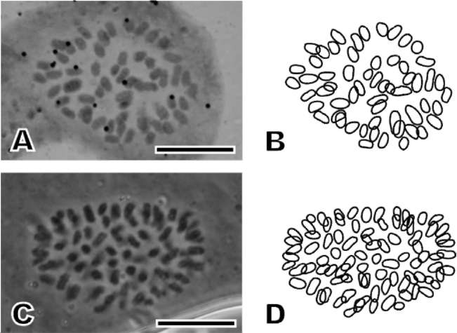
Pteris cadieri had 58 (diploid) or 87 (triploid) chromosomes. Both P. grevilleana had 58 chromosomes and thus were diploid (Figure 2A-D; Table 2).
Spore characteristics and germination
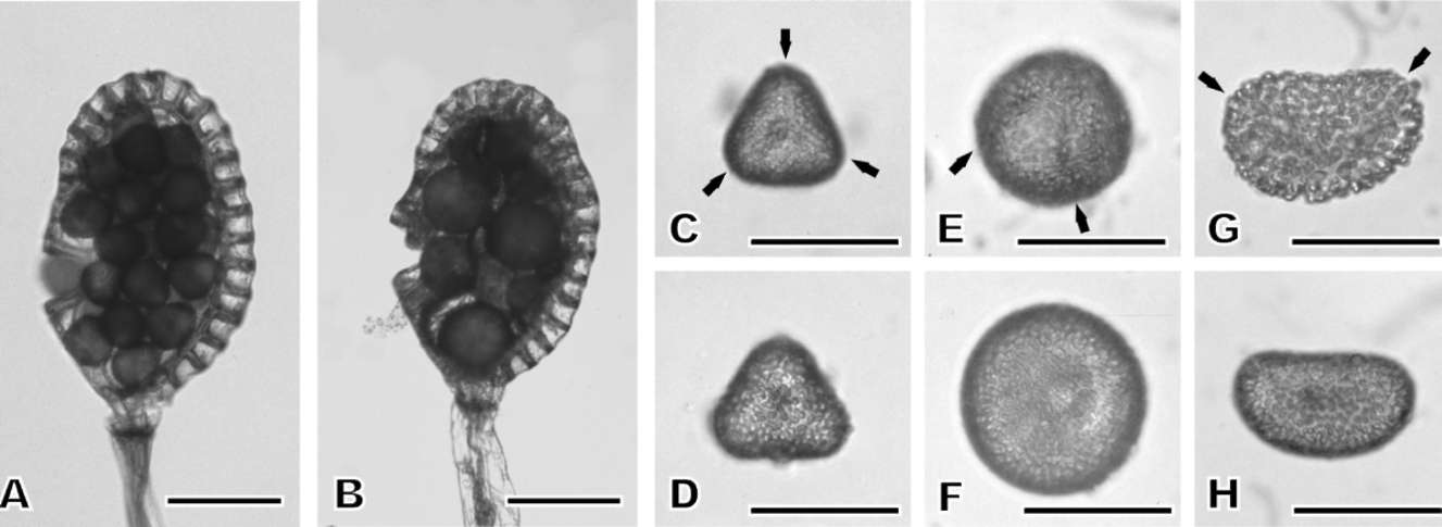
The sporangia of all plants usually contained 32 spores (Table 2, Figure 3A). However, some sporangia contained less than 32 spores (Table 2, Figure 3B), such as 28 (plants 1, 4, 9), 16 (plant 3), or 12 spores (plant 5). Most spores were tetrahedral, but a few were globose or ellipsoidal (Figure 3C-F), and others were shrunk. In sporangia with 16 spores, most spores were globose, some were tetrahedral, and all were larger and more irregularly shaped than the spores from sporangia containing 32 spores.
The mean size of tetrahedral spores ranged from 35.4 fim (plant 9) to 46.9 fim (plant 3). A Tukey-test showed that the spore size of plant 3, a triploid P. cadieri, was
|
|
||||||||||||||||||||||||||||||||||||||||||||||||||||||||||||||||||||||||||||||||
Figure 2. Somatic chromosomes of Pteris grevilleana (A, B, plant 9, diploid with 2n = 58) and P. cadieri (C, D, plant 3, triploid with 2n = 87). B & D, Explanatory drawings of A & C. Scale bars = 10 fim.
|
|||||||||||||||||||||||||||||||||||||||||||||||||||||||||||||||||||||||||||||||||
significantly larger than that of other plants (Table 2). Globose and ellipsoidal spores varied more in size than tetrahedral spores. Within a plant, the mean size of ellipsoidal spores was significantly larger than that of the tetrahedral and globose ones. However, some ellipsoidal spores were smaller than tetrahedral or globose ones. The laesurae of tetrahedral spores were trilete. The laesurae of globose and ellipsoidal spores were neither trilete nor monolete. They looked like a trilete laesura that had lost one radiating branch (Figure 3). Although spore shape varied, the perispore of all spores was tuberculate.
Germination rates ranged from 1 3 . 3 -72. 5%. The germination rates of tetrahedral and globose spores were not counted separately. No ellipsoidal spores germinated.
gametophyte characteristics and reproductive systems
A total of 360 gametophytes (40 from each plant) were examined. Most gametophytes were cordate, and all had a midrib, two wings, and an entire margin. Some gametophytes were irregularly shaped and had a
|
|||||||||||||||||||||||||||||||||||||||||||||||||||||||||||||||||||||||||||||||||
|
|||||||||||||||||||||||||||||||||||||||||||||||||||||||||||||||||||||||||||||||||
|
||||||||||||||||||||||||||||||||||||||||||||||||||||||||||||||||||||||||||||||||||||||||||||||||||||||
212
|
Botanical Studies, Vol. 51, 2010
|
|||||||||||||||||||||||||||||||||||||||||||||||||||||||||||||||||||||||||||||||||||||||||||||||||||||
|
||||||||||||||||||||||||||||||||||||||||||||||||||||||||||||||||||||||||||||||||||||||||||||||||||||||
Table 2. Germination rate, chromosome number, ploidy level, number of spores per sporangium, and spore size of tetrahedral, globose, and ellipsoidal spores of the 9 plants in this study. Plants 1-7 = Pteris cadieri, plants 8-9 = P. grevilleana.
|
||||||||||||||||||||||||||||||||||||||||||||||||||||||||||||||||||||||||||||||||||||||||||||||||||||||
|
||||||||||||||||||||||||||||||||||||||||||||||||||||||||||||||||||||||||||||||||||||||||||||||||||||||
|
||||||||||||||||||||||||||||||||||||||||||||||||||||||||||||||||||||||||||||||||||||||||||||||||||||||
|
||||||||||||||||||||||||||||||||||||||||||||||||||||||||||||||||||||||||||||||||||||||||||||||||||||||
|
||||||||||||||||||||||||||||||||||||||||||||||||||||||||||||||||||||||||||||||||||||||||||||||||||||||
|
||||||||||||||||||||||||||||||||||||||||||||||||||||||||||||||||||||||||||||||||||||||||||||||||||||||
a-fThe same letters indicate the mean sizes of tetrahedral spores are not significantly different according to ANOVA followed by Tukey post-hoc test (p <0.05).
|
||||||||||||||||||||||||||||||||||||||||||||||||||||||||||||||||||||||||||||||||||||||||||||||||||||||
|
||||||||||||||||||||||||||||||||||||||||||||||||||||||||||||||||||||||||||||||||||||||||||||||||||||||
|
||||||||||||||||||||||||||||||||||||||||||||||||||||||||||||||||||||||||||||||||||||||||||||||||||||||
|
||||||||||||||||||||||||||||||||||||||||||||||||||||||||||||||||||||||||||||||||||||||||||||||||||||||
Figure 3. Sporangia and different spore types. A, sporangium with 32, regular spores; B, sporangium with less than 32 spores, of which some are irregular (both from triploid Pteris cadieri plant 3); C & D, Proximal (C) and distal face (D) of a tetrahedral spore (diploid Pteris cadieri plant 1); E & F, Proximal and distal face of a globose spore (plant 3); G & H, Proximal face (G, diploid Pteris cadieri plant 5) and lateral view (H, triploid Pteris cadieri plant 3) of an ellipsoidal spores. The perispore of all spores was tuberculate. Arrows indicate spore laesurae. The laesurae of globose and ellipsoidal spores were neither trilete nor monolete (see text). Scale bars =
100 fm (A, B) and 50 fm (C-H).
|
||||||||||||||||||||||||||||||||||||||||||||||||||||||||||||||||||||||||||||||||||||||||||||||||||||||
|
||||||||||||||||||||||||||||||||||||||||||||||||||||||||||||||||||||||||||||||||||||||||||||||||||||||
slightly notched margin. We found several secondary gametophytes, which arose from primary gametophytes, and most of them were irregularly shaped (Figure 4).
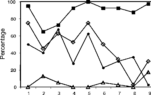
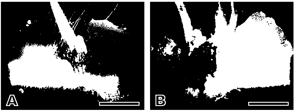
Antheridia of both species were of the common, leptosporangiate type (following the typification of Nayar and Kaur, 1971). They had a cap cell, a ring cell, and a basal cell (Figure 5) and produced active spermatozoid. Antheridia were produced on either the ventral side, the dorsal side, or both sides of the gametophytes. Of the gametophytes with juvenile sporophytes, 47% were male and 53% were asexual (lacking sex organs). We found neither female nor bisexual gametophytes indicateing that all sporophytes were produced apogamously.
Juvenile sporophytes first appeared 45-60 days after spores sowing. All embryos were covered with brown scales. Most gametophytes produced a single embryo
|
|
|||||||||||||||||||||||||||||||||||||||||||||||||||||||||||||||||||||||||||||||||||||||||||||||||||||
Plant
Figure 4. Frequency of gametophyte characters in Pteris cadieri and P. grevilleana (n = 40 for each plant). The numbers 1-9 on the X axis represent the plants 1-9, respectively, listed in Table 1. ●: Gametophyte with secondary gametophyte; male gametophytes; ■: first frond with a midrib; A: polyembryony.
|
||||||||||||||||||||||||||||||||||||||||||||||||||||||||||||||||||||||||||||||||||||||||||||||||||||||
|
||||||||||||||||||||||||||||||||||||||||||||||||||||||||||||||||||||||||||||||||||||||||||||||||||||||
|
||||
CHAO et al. — Reproductive traits of Pteris cadieri & P grevilleana
|
213
|
|||
|
||||
|
||||
|
||||
Figure 5. Antheridia of Pteris cadieri. A, spermatids inside the antheridia (arrows) (plant 3); B, an antheridium including a cap cell (c), a ring cell (r), and a basal cell (b) (plant 3); C, empty antheridia after the spermatids were released (plant 1). Scale bars = 100 fim.
|
||||
|
||||
on the midrib near the apical meristem. In this case, the juvenile sporophyte arose directly from the ventral s ide of gametophyte. However, s ome gametophytes produced two or three embryos (plants 2, 3, 6, 7, and 9). Each embryo had an independent root and grew into a sporophyte (Figure 6). Plant 9 had the highest number of polyembryonate gametophytes. The additional embryos occurred mainly on the midrib of the gametophyte. However, some grew on the wings of the gametophytes (plants 2 and 3) or on secondary gametophytes (plant 2). Most of the first fronds of juvenile sporophytes had midrids and triparted type (88%) (Figure 4); the others were midribless and bifurcate.
discussion
Reproductive biology traits
We found that both Pteris cadieri and P. grevilleana were apogamous, i.e., their next genereation sporophytes were born out of gemetophytic cells without fertilization. Differences were found in spore shape, germination rate, morphology of the first frond of juvenile sporophytes (midrib lacking or present), the occurrence of secondary gametophytes, male gametophytes, and polyembryony. However, the occurrence tendencies of these traits were not consistent with the variations of frond morphology
|
(from simply pinnate, irregular bipinnatifid to regular bipinnatifid) (Figure 4).
In Cornopteris christenseniana, variation in spore germination rates was attributed to interspecific hybridization (Park and Kato, 2003). The fitness of spores from hybrids is usually variable, resulting in high variation in spore germination rates (Quintanilla and Escudero, 2006). The variability of Pteris cadieri and P. grevilleana spore germination rates supports the hypothesis that these species are hybrid origin.
Generally, spore size and ploidy are correlated because an increase in genomic content usually enlarges cell size (Nakato, 1976, 1981; Barrington et al., 1986; Hou and Wang, 2000; Quintanilla and Escudero, 2006). However, based on the size of the tetrahedral spores, this association did not hold in P. cadieri. The spores of one triploid (plant 3) were larger than the spores from the diploid plants, but spores from the other triploid (plant 2) were not. Recently, it was proposed that cell size was determined by cell cycle genes rather than by genome size (Cavalier-Smith, 2005). In P. cadieri, the matter of polypoidy may be not correlated to the increase of spore size.
Embryos with scales and a midrib on the first frond of juvenile sporophytes are indicators of apogamy (Steil, 1939; Kanamori, 1972; Moore et al., 2002; Huang et al., 2006). The gametophytes of P. cadieri and P. grevilleana
|
|||
|
||||
|
||||
|
||||
Figure 6. Polyembryony in Pteris cadieri and P. grevilleana. A, two embryos growing close together (P. grevilleana, plant 9); B, two distinct, separate embryos (P. cadieri plant 2). Scale bars = 1 mm.
|
||||
|
||||
|
|||
214
|
Botanical Studies, Vol. 51, 2010
|
||
|
|||
were apogamous, and all embryos were covered with scales. However, some first fronds lacked a midrib. Thus, an embryo covered with scales appears to be a more reliable indicator of apogamy than venation of the first frond.
Polyembryony has been documented in sexual ferns and is usually attributed to multiple fertilizations (Mo ttier, 1 9 25 ; Klekows ki, 1 9 7 0, 1 9 7 2). The occurrence of polyembryos may be not caused by multi-gametophyte growth. Even in cultures of density experiments, polyembryos occurred in the lowest density condition (Cousens, 1 979). Therefore, polyembryos may arise from intergametophytic or intragametophytic mating in sexual ferns. It is thought that polyembryony would increase the probability of intergametophytic mating in sexual ferns through the neighboring sporophytes and subsequently adjacent gametophytes (Klekowski, 1970, 1972; Lloyd, 1974). Our discovery of polyembryony in P. cadieri and P. grevilleana is the first report of this phenomenon in apogamous ferns. No mating occurs in these apogamous ferns, and the meaning of their polyembryony is not clear yet.
Spore traits indicating abnormal sporogenesis
Abnormal sporogenesis is a common phenomenon in certain polyploids and hybrids (Hickok and Klekowski, 1974; Haufler and Windham, 1985; Bennert et al., 2005). In most sexual, leptosporangiate ferns, including the genus Pteris, an archesporial cell undergoes 4 mitoses and 1 meiosis (two divisions) to produce 64 haplospores per sporangium (Walker, 1979; Huang et al., 2003). Sporogenesis is considered abnormal if there are fewer than 64 spores per sporangium. This results from abnormal reductional meiosis (or nonreductional meiosis) and yields 32, 16, or 8 spores per sporangium (Morzenti, 1962; Hickok and Klekowski, 1973; Wang, 1989). For example, in the Dopp-Manton scheme (Manton, 1950) one mitosis is lost, and in the Braithwaite scheme (Braithwaite, 1964) (which is the same as the scheme of Mehra-Singh, 1957), one meiotic division is lost, both resulting in 32 spores per sporangium. In this study, the "abnormal sporogenesis" of P. cadieri and P. grevilleana produced sporangia with 32, 28, 16 or 12 spores, indicating the deviations from normal sporogenesis are more complicated and varied than those described in the schemes above. Under certain circumstances, lacking or incomplete chromosome pairing during meiosis may yield a number of spores per sporangium being not a multiple of eight with the spores being irregularly shaped (Morzenti, 1962; Lin, Kato and Iwatsuki, 1992; Rabe and Haufler, 1992).
Apogamy and hybrid origins
Pteris cadieri and P. grevilleana, both with 32-spore sporangia, were apogamous. These results concur with those from previous studies that found most leptosporangiate ferns producing 32 spores per sporangium are apogamous (Manton 1950; Hickok and Klekowski,
|
1974; Nakato, 1975; Lovis, 1977; Walker, 1979). In
this study, apogamous diploid and triploid P. cadieri and diploid P. grevilleana were sympatric. Apogamous diploids may result from genetic change in a sexual diploid species, or from hybridization between two sexual diploid species (Manton, 1950). Apogamous triploids originate by crossing between a sexual diploid and a sexual tetraploid species, or by hybridization of an apogamous diploid (unreduced diploid gametes, functionally male) and with a sexual diploid (Manton, 1950; Walker, 1962, 1979; Lovis, 1977; Haufler and Windham, 1985; Suzuki and Iwatsuki, 1990; Lin et al., 1995). Tetraploid P. grevilleana were found in Malaysia (Walker, 1962) and further research is needed to clarify its lineage.
Apogamous diploids and triploids may also be formed by "ploidy reduction". "Ploidy reduction" has been reported in triploid Dryopteris pacifica, which produced both irregular and regular spores. The spores of triploid D. pacifica are capable of developing into apogamous diploid or triploid sporophytes (Lin et al., 1992). The diverse spore sizes and shapes of triploid P. cadieri might be an indicator of with ploidy reduction.
Apogamous taxa commonly are believed to have limited genetic variation. Thus, it is intriguing that P. cadieri exhibits great morphological variation among plants growing at the same location (plants 3-6). The mechanism for producing morphological and genetic diversity in an apogamous species is multiple hybridization between two or more taxa or species. Each hybridization event produces a unique and independent genetic lineage, which will persist unchanged, due to the reproduction barriers arising from apogamy. Furthermore, because gene expression varies in hybrids, the same parents could produce apogamous offspring exhibiting different phenotypes (Guo et al., 2004; Springer and Stupar, 2007; Zhuang and Adams, 2007).
Pteris grevilleana is considered a good species owing to its symmetrical and regular pinnae. In contrast, P. cadieri is thought to be a hybrid because its pinnae are irregular. Pteris grevilleana has been proposed to be a possible parent of P. cadieri (Wagner, 1978). However, this study demonstrats that the spore size and the number of spores per sporangium of P. grevilleana are variable, and that this species is also apogamous. Therefore, the hypothesis that Pteris grevilleana is a parent of P. cadieri has to be reconsidered.
Pteris grevilleana and P. cadieri may share a common ancestor, but arose from independent hybridization events. However, there are two scenarios in which P. grevilleana could still be a parent of P. cadieri. In the first, the antheridia of P. grevilleana supply active sperm that interact with the eggs of another, unidentified, sexual species, and a triploid hybrid is arisen. In the second, P. grevilleana was once a sexual taxon, but it became apogamous after the hybridization events that gave rise to P. cadieri. For example, Pteris cretica, an apogamous diploid species with wide gerographical range, was
|
||
|
|||
|
||||
CHAO et al. — Reproductive traits of Pteris cadieri & P. grevilleana
|
215
|
|||
|
||||
supposed to come from the hybrids between sexual diploids than acquired apogamy (Manton, 1950).
Acknowledgments. We wish to thank Drs. Wilfried Bennert, Donald R. Farrar, Luis G. Quintanilla, and Tom A. Ranker for their valuable comments to improve the manuscript; Pi-Fong Lu and Chun-Ming Chen for their assistance in collecting plant samples; Dr. Alan Warneke for editorial assistance.
literature cited
Barrington, D.S., C.D. Paris, and T.A. Ranker. 1986. Systematic inferences from spore and stomate size in the ferns. Amer. Fern J. 76: 149-159.
Bennert, W., M. Lubienski, S. Korner, M. Steinberg. 2005. Triploidy in Equisetum subgenus Hippochaete (Equisetaceae, Pteridophyta). Ann. Bot. (Oxford) 95:
807-815.
Braithwaite, A.F. 1964. A new type of apogamy in ferns. New Phytol. 63: 293-305.
Cavalier-Smith T. 2005. Economy, speed and size matter: evolutionary forces driving nuclear genome miniaturization and expansion. Ann. Bot. (Oxford) 95: 14-175.
Cousens, M.I. 1979. Gametophyte ontogeny, sex expression, and genetic load as measures of population divergence in Blechnum spicant. Amer. J. Bot. 66: 116-132.
Ebihara, A., H. Ishikawa, S. Matsumoto, S.J. Lin, K. Iwatsuki, M. Takamiya, Y. Watano, and M. Ito. 2005. Nuclear DNA, chloroplast DNA, and ploidy analysis clarified biological complexity of the Vandenboschia radicans complex (Hymenophyllaceae) in Japan and adjacent areas. Amer. J.
Bot. 92: 1535-1547.
Guo, M., M.A. Rupe, C. Zinselmeier, J. Habben, B.A. Bowen, and O.S. Smith. 2004. Allelic variation of gene expression in maize hybrids. Pl. Cell 16: 1707-1716.
Haufler, C.H. 2008. Species and speciation. In T.A. Ranker and C.H. Haufler (eds.), The Biology and Evolution of Ferns and Lycophytes. Cambridge University Press, Cambridge, pp. 303-331.
Haufler, C.H. and M.D. Windham. 1985. Triploidy and its
evolutionary significance in Cystopteris protrusa. Canad. J.
Bot. 63: 1855-1863.
Hickok, L.G. and E.J. Jr. Klekowski. 1973. Abnormal reductional and nonreductional meiosis in Ceratopteris: Alternatives to homozygosity and hybrid sterility in homosporous ferns. Amer. J. Bot. 60: 1010-1022.
Hickok, L.G. and E.J. Jr. Klekowski. 1974. Inchoate speciation in Ceratopteris: An analysis of the synthesized hybrid C. richardii x C. pteridoides. Evolution 28: 439-446.
Hou, X. and Z.R. Wang. 2000. A subspecific taxonomic study on Asplenium trichomanes L. from China. Acta Phytotax. Sin.
38: 242-255.
Huang, Y.M., C.Y. Kuo, and W.L. Chiou. 2003. Morphology of
gametophytes and young sporophytes of Cibotium Kaulf.
|
native to Taiwan. Taiwan J. For. Sci. 18: 163-170.
Huang, Y.M., H.M. Chou, T.H. Hsieh, J.C. Wang, and W.L.
Chiou. 2006. Cryptic characteristics distinguish diploid and triploid varieties of Pteris fauriei (Pteridaceae). Canad. J. Bot. 84: 261-268.
Iwatsuki, K. 1995. Pteridaceae. In K. Iwatsuki, T. Yamazaki, D.E. Boufford, and H. Ohba (eds.), Flora of Japan. Vol. 1. Kodansha, Tokyo, pp. 89-97.
Kanamori, K. 1972. Apogamy in ferns with special reference to the apogamous embryogenesis. Sci. Rep. Tokyo Kyoiku Daigaku., Section B 15: 111-131.
Klekowski, E.J. Jr. 1970. Populational and genetic studies of a homosporous fern-Osmunda regalis. Amer. J. Bot. 57: 1122-1138.
Klekowski, E. J. Jr. 1972. Evidence against genetic self-incompatibility in the homosporous fern Pteridium aquilinum. Evolution 26: 66-73.
Ko, J.W., Y.M. Huang, and W.L. Chiou. 2004. Reproductive
biology of Aglaomorpha meyeniana (Polypodiaceae; Pteridophyta). BioFormosa 39: 83-91.
Kuo, C.M. and H.T. Yu. 1986. Investigation on the Pteridophyta of Kenting National Park. Kenting National Park Administration Office, Hengchun Township, 114 pp.
Lin, S.J., M. Kato, and K. Iwatsuki. 1992. Diploid and triploid
offspring of triploid agamosporous fern Dryopteris pacifica.
Bot. Mag. (Tokyo) 105: 443-452.
Lin, S.J., M. Kato, and K. Iwatsuki.1995. Electrophoretic variation of apogamous Dryopteris varia group
(Drypteridaceae). J. Pl. Res. 108: 451-456.
Lloyd, R.M. 1974. Reproductive biology and evolution in the Pteridophyta. Ann. Missouri Bot. Gard. 61: 318-331.
Lovis, J.D. 1977. Evolutionary patterns and processes in ferns.
Advances Bot. Res. 4: 229-414.
Manton, I. 1950. Problems of Cytology and Evolution in the Pteridophyta. Cambridge University Press, New York, 316
Me hra, P. N. and G. S ing h. 1 9 5 7 . C y to lo g y o f
Hymenophyllaceae. J. Genet. 55: 379-393.
Moore, S.J., T.H. Hsieh, Y.M. Huang, and W.L. Chiou. 2002.
Diplazium maonense Ching. A poorly known species of the Athyriaceae (Pteridophyta) in Taiwan. Taiwan J. Forest Sci. 17: 113-118.
Morzenti, V. M. 1 962. A first report on pseudomeiotic sporogenesis, a type of spore reproduction by which "sterile" ferns produce gametophytes. Amer. Fern J. 52: 69-78.
Mottier, D.M. 1925. Polyembryony in certain Polypodiaceae and Osmundaceae. Bot. Gaz. 80: 331-336.
Nakaike, T. 1992. New flora of Japan Pteridophyta-Revised & Enlarged. Shibundo Company, Tokyo, 868 pp.
Nakato, N. 1975. A cytological study on an intermediate form between Pteris multifida and P. cretica. J. Jap. Bot. 50:
110-125.
|
|||
|
||||
|
|||
216
|
Botanical Studies, Vol. 51, 2010
|
||
|
|||
Nakato, N. 1976. A cytogeographic study on the Japanese Pteris excelsa complex. J. Jap. Bot. 51: 59-64.
Nakato, N. 1981. A cytogeographic study of Pteris dispar Kunze. J. Jap. Bot. 56: 200-205. (In Japanese).
Nayar, B.K. and S. Kaur. 1971. Gametophytes of homosporous
ferns. Bot. Rev. 37: 295-396.
Park, C.H. and M. Kato. 2003. Apomixis in the interspecific triploid hybrid fern Cornopteris christenseniana (Woodsiaceae). J. Pl. Res. 116: 93-103.
Quintanilla, L.G. and A. Escudero. 2006. Spore fitness components do not differ between diploid and allotetraploid species of Dryopteris (Dryopteridaceae). Ann. Bot. (Oxford) 98: 609-618.
Rabe, E.W. and C. H. Haufler. 1 992. Incipient polyploid speciation in the maidenhair fern (Adiantum pedatum; Adiantaceae)? Amer. J. Bot. 79: 701-707.
Sharma, H.C. 1982. A technique for somatic counts from root tips of cereal seedlings raised by embryo culture. Curr. Sci.
51: 143-144.
Shieh, W.C. 1975. Pteridaceae. In H.L. Li, T.C. Huang, T.S. Liu,
T. Koyama, and C.E. DeVol (eds.), Flora of Taiwan, Vol. 1. Epoch Publish Co., Taipei, pp. 281-301.
Smedmark, J.E.E., T. Eriksson, R.C. Evans, and C.S. Campbell. 2003. Ancient allopolyploid speciation in Geinae (Rosaceae): Evidence from nuclear granule-bound starch
synthase (GBSSI) gene sequences. Syst. Biol. 52: 374-385.
Springer, N.M. and R.M. Stupar. 2007. Allele-specific expression
|
patterns reveal biases and embryo-specific parent-of-origin effects in hybrid maize. Pl. Cell 19: 2391-2402.
Steil, W.N. 1939. Apogamy, apospory, and parthenogenesis in
the pteridophytes. Bot. Rev. 5: 433-453.
Suzuki, T. and K. Iwatsuki. 1 990. Genetic variation in apogamous fern Pteris cretica L. in Japan. Heredity 65: 221-227.
Wagner, W.H., Jr. 1954. Reticulate evolution in the Appalachian Aspleniums. Evolution 8: 103-118.
Wagner, W.H., Jr. 1978. Venuloid idioblasts in Pteris and their systematic implication. Acta Phytotax. Geobot. 29: 33-40.
Walker, T.G. 1958. Hybridization in some species of Pteris L. Evolution 12: 82-92.
Walker, T.G. 1962. Cytology and evolution in the fern genus Pteris L. Evolution 16: 17-43.
Walker, T.G. 1979. The cytogenetics of ferns. In A. F. Dyer (ed.), The experimental biology of ferns. Academic Press, London, pp. 87-132.
Wang, Z.R. 1989. A preliminary study on cytology of Chinese
Pteris. Acta Phytotax. Sin. 27: 421-438. Wu, S.W. 1990. Pteridaceae. In R.C. Ching and K.H. Shing
(eds.), Flora Reipublicae Popularis Sinicae, Vol. 3(1). Science Press, Beijing, pp. 15-91.
Zhuang, Y.K. and L. Adams. 2007. Extensive allelic variation in gene expression in Populus F1 hybrids. Genetics 177: 1987-1996.
|
||
|
|||
由二形鳳尾蕨及翅柄鳳尾蕨的生殖特徵推論其雜交起源
趙怡姍1 劉和義1 黃曜謀2 邱文良3
1國立中山大學生物科學系
2農委會林業試驗所育林組 3農委會林業試驗所植物園組 二形鳳尾蕨具有明顯的形態變異,葉形由單回羽片至二回羽裂。由於其形態與翅柄鳳尾蕨成一連
續變異,故過去研究認為二形鳳尾蕨是一個雜交種而翅柄鳳尾蕨為其親本之一,但並無提供進一步證據 或反證。本篇研究以台灣產的二形鳳尾蕨與翅柄鳳尾蕨的生殖特徵,探討此雜交起源的假設。研究發現 二形鳳尾蕨具二倍體與三倍體,翅柄鳳尾蕨為二倍體,但兩種均行無配生殖。兩種在生殖特徵相似:孢 子形狀具變異(四面體形,圓形,豆形),孢子發芽率具變異,幼孢子體的第一片葉具有中脈,配子體 具偶發的多胚現象。生殖特徵的研究結果支持二形鳳尾蕨與翅柄鳳尾蕨同為雜交起源,而建議重新考慮 翅柄鳳尾蕨為二形鳳尾蕨親本的假說。此外,本篇為無配生殖蕨類具多胚性的首次報導。 |
|||
|
|||
關鍵詞:無配生殖;雜交;形態變異;多倍體;二形鳳尾蕨;翅柄鳳尾蕨。
|
|||
|
|||