Botanical Studies (2010) 51: 311-315.
BIOCHEMISTRY
Chemical constituents from fermented broth and
mycelium of the basidiomycete Lacrymaria velutina
Yu-Ming JU1, Guei-Jane WANG2, Chun-Yao CHEN3, Ya-Jing TSAU4, Chang-Hung CHOU5, and
Tzong-Huei LEE5,6 *
1Institute of Plant and Microbial Biology, Academia Sinica, Taipei 115, Taiwan
2
National Research Institute of Chinese Medicine, Taipei 112, Taiwan
3
Department of Life Science, Tzu-Chi University, Hualien 970, Taiwan
4
Institute of Fisheries Science, National Taiwan University, Taipei 106, Taiwan
5
Graduate Institute of Ecology and Evolutionary Biology, China Medical University, Taichung 404, Taiwan
6
Graduate Institute of Pharmacognosy, Taipei Medical University, Taipei 110, Taiwan
(Received August 21, 2009; Accepted January 20, 2010)
ABSTRACT. Lacrymarone (1), a novel diterpenoid, together with seven known compounds 2-8 were isolated from the fermented broth and mycelium of Lacrymaria velutina, an edible fungus collected in Taiwan. The structures of 1-8 were elucidated by analyzing their spectroscopic data. Further biological tests revealed that the co-crystal of 7 and 8 exhibited a moderate inducible nitric oxide synthase (iNOS) inhibitory activity, and with an IC50 value of 59.54 3.74 |iM.
Keywords: Basidiomycete; Inducible nitric oxide synthase (iNOS); Lacrymaria velutina; Lacrymarone.
INTRODUCTION
Fungal natural products have long been an important source of compounds for new drug development (Singh et al., 2000). During our preliminary bioactivity screening of fungal fermentation extracts, Lacrymaria velutina, a basidiomycete, was found to exhibit a significant inducible nitric oxide synthase (iNOS) inhibitory activity without any cytotoxicity at a concentration of 100 fig/ml, the inhibition being 97.9 0.8%. This finding prompted us to investigate the active principles from this fungus, and led to the isolation and identification of a novel diterpenoid along with seven known chemical entities. This paper deals with the characterizations of these compounds and their iNOS inhibitory activities on RAW 264.7 cell line.
MATERIALS AND METHODS
General experimental procedures
Optical rotations were measured on a JASCO DIP-1000 digital polarimeter (JASCO, Kyoto, Japan). 1H-and 13C-NMR were acquired on a Bruker DMX-500 SB spectrometer (Bruker, Ettlingen, Germany). High resolu­tion and low resolution mass spectra were obtained us­ing a ThermoQuest Finnigan MAT 95XL spectrometer (ThermoQuest Finnigan, Bremen, Germany) and VG Platform Electrospray ESI/MS (VG Organic, Altrincham,
*Corresponding author: E-mail: thlee@tmu.edu.tw; Phone: +886-2-2736-1661 ext. 6156.

UK), respectively. Infrared spectra were recorded on a JASCO FT/IR 4100 spectrometer (JASCO, Tokyo, Japan). Ultraviolet spectra were measured on a Helios a spec­trophotometer (Thermo Scientific, Waltham, MA, USA). Column chromatography was carried out with Sephadex LH-20 gel (Amersham Biosciences, Uppsala, Sweden). Pre-coated Si gel plates (Si 60 F254, 0.2 mm, Merck, Darm­stadt, Germany) were used for analytical TLC.
Fermentation of Lacrymaria velutina
Lacrymaria velutina (strain No. 96110901 from Taipei, Taiwan) collected, isolated, and identified by one of us (YMJ), was inoculated into 1-1 Erlenmeyer flasks containing 10 g BactoTM Malt Extract (Becton, Dickinson and Company, Sparks, MD, USA) and 500 ml deionized water. The fermentation was conducted at 25-30°C for 30 days.
Extraction and isolation
The filtered fermented broth (153 l) of L. velutina was partitioned three times with 50 l recycled ethyl acetate, then concentrated in vacuum to dryness (1.7 g). Subsequently, this residue was re-dissolved in 25 ml of MeOH, and applied onto a Sephadex LH-20 column (3 cm i.d. x 65 cm) eluted by MeOH with a flow rate of 2.5 ml/min. Each subfraction (18 ml) collected was checked for its compositions by TLC using isopropanol/isooctane
(2:8, v/v) or EtOAc/acetic acid/H2O (85:10:10, v/v/v) for
development, and observation under UV 254 nm was used in the detection of compounds with similar chromophores.
312
Botanical Studies, Vol. 51, 2010
Subsequently, subfractions were combined into seven portions IVII. The portion II (subfr. 11~13) was purified by HPLC on a semi-preparative reversed-phase column (BDS Hypersil C", 10 x 250 mm, Thermo Hypersil-Keystone, Bellefonte, CA, USA) with acetonitrile/H2O (3:2, v/v) as eluent, 2 ml/min, afforded 1 (3.2 mg). HPLC of the same portion on the same column with acetonitrile/ H2O (1:9, v/v) as eluent, 2 ml/min, gave 2 (11.4 mg). The portion III (subfr. 14~15) was purified by HPLC on the same column with MeOH/H2O (1:4, v/v) as eluent, 2 ml/ min, obtained 3 (21.2 mg). The portion V (subfr. 18~19) was purified by HPLC on a semi-preparative reversed-phase column (BiOSIL PRO-ODS-U, 10 x 250 mm, Biotic
Chemical Co., Taipei, Taiwan) with MeOH/H2O (9:1, v/v)
containing 0.1% trifluoroacetic acid as eluent, 2 ml/min, afforded 4 (12.5 mg), 5 (17.2 mg) and 6 (17.2 mg). The
dried mycelium (20.5 g) of L. velutina was extracted three times with 2 l MeOH, then partitioned using n-hexane to obtain a yellow residue (1.6 g), which was re-crystallized from n-hexane to afford a mixture of 7 and 8 (1:1, 105.8
mg).
Lacrymarone (1)
Rf= 0.45 on Si-60 F254 plate using isopropanol/isooctane (2:8, v/v) for development; Light yellow oil; [ot]18d: +10.4 (c 0.55, pyridine); UV (MeOH): Imax (log s) = 208 (3.7), 251 (3.7) nm; IR (ZnSe): v=3370 (-OH), 2929, 1715 (C=O), 1638 (C=C), 1458, 1255, 1181, 1035 cm-1; 1H-
NMR data: see Table 1; 13C-NMR data: see Table 1; ESI-
MS: m/z = 339 [M + Na]+; HR-ESI-MS: m/z = 339.1933 [M + Na]+; calcd for C20H28O3N+ 339.1936.
Table 1. 1H- and 13C-NMR data of 1 (pyridine, 500 MHz for 1H and 125 MHz for 13C).
No.
13C
1H
HMBC (H—C)
COSY (H^H)
NOESY (H^H)
1
203.7
2a
41.9
2.14 (1H, dd, J = 11.8, 18.1 Hz)
1, 3, 4, 18
2b, 3
2b, 3, 19
2b
41.9
2.40 (1H, dd, J = 7.3, 18.1 Hz)
1,3, 4, 5
2a, 3
2a,17, 18, 19
3
46.2
1.59 (1H, m)
18
2a, 2b, 18
2a, 9b
4
46.4
5
145.5
6
134.6
6.85 (1H, s)
1, 4, 7
15a, 15b, 16
7
80.5
8
48.6
9a
40.4
1.66 (1H, dd, J = 8.1, 13.0 Hz)
4, 7, 11
9b, 10
9b, 10, 11b
9b
40.4
1.82 (1H, dd, J = 2.2, 13.0 Hz)
4, 11, 16
9a, 10
3, 9a, 16
10
81.5
4.45 (1H, dd, J = 2.2, 8.1 Hz)
5, 7
9a, 9b
9a, 17, 20
11a
34.4
1.28 (1H, m)
11b
11b, 12b,16
11b
34.4
1.96 (1H, m)
8, 12, 16
11a
9a, 11a
12a
23.6
2.01 (1H, m)
12b, 13
13, 15a, 15b
12b
23.6
2.09 (1H, m)
12a, 13
11a, 13, 16
13
126.3
6.37 (1H, brd, J = 4.1 Hz)
12a, 12b
12a, 12b, 15a, 15b
14
137.1
15a
63.1
4.53 (1H, d, J = 13.7 Hz)
15b
6, 12a, 13
15b
63.1
4.78 (1H, d, J = 13.7 Hz)
15a
6, 12a, 13
16
19.4
0.89 (3H, s)
7, 8, 9, 11
6, 9b, 11a, 12b
17
18.9
1.25 (3H, s)
3, 4, 5, 10
2b, 10, 18, 20
18
29.8
1.54 (1H, m)
3, 4
3, 19, 20
2b, 17, 19, 20
19
22.4
0.81 (3H, d, J = 6.3 Hz)
3, 18, 20
18
2a, 2b
20
22.2
0.78 (3H, d, J = 6.3 Hz)
3, 18, 19
18
10, 17
JU et al. ― Chemical constituents of Lacrymaria velutina
313
Nitrite measurement and cell viability assay
The methods were essentially the same as reported previously (Wang et al., 2007; Lee et al., 2008). To assess the effects on LPS-induced NO production, crude extracts of fermented broth of L. velutina, compounds 1-8, two positive controls-N^-nitro-L-arginine (L-NNA, a non­selective NOS inhibitor) and aminoguanidine (a specific inhibitor of iNOS)—or vehicle (0.1%, DMSO) were added in the presence of LPS (200 ng/ml) to the RAW 264.7 cells. Both inhibitors were purchased from Sigma-Aldrich Chemical Co. and the purity of each compound was more than 98%.
Statistical analyses
Comparisons of the concentration and treatment effects were made using ANOVA, followed by post hoc comparisons using Newman-Keuls test as appropriate. The average IC50 was determined by data fitting with GraFit (Erithacus Software, Staines, UK).
RESULTS AND DISCUSSION
The fermented broth of L. velutina was partitioned initially using EtOAc to give a brown residue, which was subjected to Sephadex LH-20 column separation followed by HPLC purification to obtain one new diterpenoid (1) along with 5-hydroxymethylfurfural (2) (McNelis et al., 1994), ^is-2,5-hydroxymethylfuran (3) (Fawcett et al., 1987), maltol (4) (Sun et al., 1995), succinic acid
(5) (Abdel-Farid et al., 2007), and furan-2,5-diol (6)
(Rappoporta et al., 2001). Meanwhile, the cultured mycelium of the same fungus was extracted with methanol, then partitioned using n-hexane to obtain a yellow residue, which was re-crystallized from n-hexane to afford a mixture of (22E,24S)-24-methyl-27-norcholesta-5,7,22-trien-3p-ol (7) and (22E,24R)-24-methyl-27-norcholesta-5,7,22-trien-3p-ol (8) (Itoh et al., 1983)/
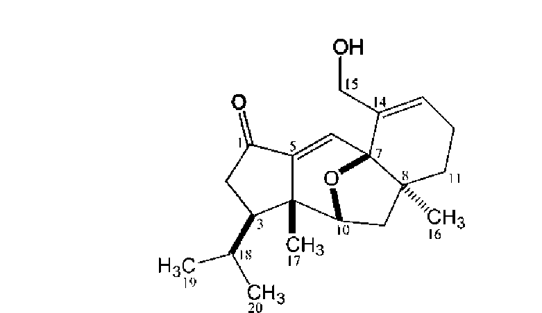
Compound 1 was isolated as light yellow oil, and its molecular formula, C20H28O3, was established through analysis of its 13C-NMR and HR-ESI-MS data. The IR spectrum of 1 exhibited the presence of a carbonyl group
(1,715 cm-1), a double bond (1,638 cm-1) and a hydroxyl group (3,370 cm-1). The interpretation of the 1H-NMR of 1 supported by its HSQC assignments revealed signals for four methyl groups [§h 0.78 (3H, d, J = 6.3 Hz, H3-20), §h 0.81 (3H, d, J = 6.3 Hz, H3-19), §h 0.89 (3H, s, H3-16) and §h 1.25 (3H, s, H3-17)], five methylene groups [§h 1.28 (1H, m, H-11a), §h 1.96 (1H, m, H-11b), §h 1.66 (1H, dd, J = 8.1, 13.0 Hz, H-9a), §h 1.82 (1H, dd, J = 2.2, 13.0 Hz, H-9b), §h 2.01, 2.09 (each 1H, m, H2-12), §h 2.14 (1H, dd, J = 11.8, 18.1 Hz, H-2a), §h 2.40 (1H, dd, J = 7.3, 18.1 Hz, H-2b), §h 4.53, 4.78 (each 1H, d, J = 13.7 Hz, &-15)] and five methine protons [§h 1.54 (1H, m, H-18), §h 1.59 (1H, m, H-3), §h 4.45 (1H, dd, J = 2.2, 8.1 Hz, H-10), §h 6.37 (1H, brd, J = 4.1 Hz, H-13), §h 6.85 (1H, s, H-6)] (Table 1). The 13C-NMR spectrum together with DEPT analysis displayed 20 signals including four methyl carbons, five methylene carbons, three methine carbons and three quaternary carbons in the aliphatic region, two methine carbons and two quaternary carbons in olefinic region (2 double bonds) as well as one carbonyl carbon (Table 1). On account of the molecular formula C20H28O3, the double bond equivalence of 1 was seven including one carbonyl and two olefinic functionalities. Thus, there should be four rings in 1. Analysis of COSY and HSQC spectral data allowed the assignments of three spin systems including an aliphatic chain [(H3-19, -20)-(H-18)-(H-3)-(H2-2)-], two two-resonance units [-(H-10)-(H2-9)- and -(H2-12)-(H-13)-] and five geminal coupled methylene functionalities
[-(H2-2)-, -(H2-9)-, -(H2-11)-, -(H2-12)- and -(H2-15)-].
In the HMBC spectrum, key long range proton-carbon correlations including H3-20 (§h 0.78)/C-19 (§c 22.4), -18 (§c 29.8), -3 (§c 46.2), H3-19 (§h 0.81)/C-20 (§c 22.2), -18 (§c 29.8), -3 (§c 46.2), H3-17 (§h 1.25)/C-10 (§c 81.5), -5 (§c 145.5), -4 (§c 46.4), -3 (§c 46.2), H3-16/C-H (§c 34.4), -9 (§c 40.4), -8 (§c 48.6), -7 (§c 80.5), H-10 (§h 4.45)/C-5 (§c 145.5), H-6 (§h 6.85)/C-7 (§c 80.5), -4 (§c 46.4), -1 (§c 203.7) and H-2b (§h 2.40)/C-5 (§c 145.5), -4 (§c 46.4), -3 (§c 46.2), -1 (§c 203.7) established the major connectivities of the fragments deduced from COSY spectrum. All above assignments suggested that the plane structure of 1 seemed to be analogous to guanacastepenes, unique diterpenoids with a 5/7/6-fused ring system, isolated previously from an unidentified endophytic fungus (Brady et al., 2000, 2001). However, the conspicuous cross peak between carbinoyl H-10 (§h 4.45) and oxygenated quaternary C-7 (S80.5) in the HMBC spectrum of 1 suggested that C-10 and C-7 were bridged by an ether functionality, forming a different 5/6/5/6-fused ring skeleton of 1. In the NOESY spectrum of 1, key mutual correlations were listed as follows: H3-
20/H3-17, H3-17/H-10, H-10/H-9a and H-9b/H3-16, which
were further supported by the three dimensional molecular modeling under minimized energy condition (Figure 1). The relative configurations of the isopropyl group attached at C-3, H3-17, ether linkage between C-7 and C-10, and H3-16 were thus corroborated to be respective p, p, p and a oriented to fit the distinguishing features in NOESY spectrum (Table 1). Accordingly, 1 was characterized as the shown structure (Figure 2), and named lacrymarone.
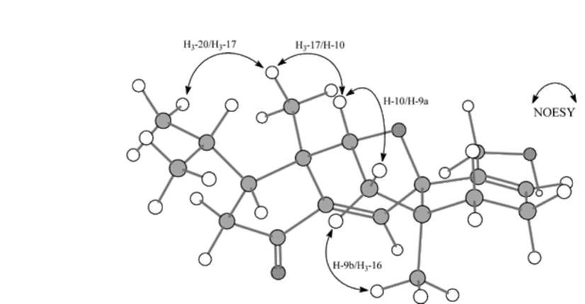
Figure 1. Computer-generated perspective drawing for 1, which accommodate observed mutual key NOESY
314
Botanical Studies, Vol. 51, 2010
Brady, S.F., M.P. Singh, J.E. Janso, and J. Clardy. 2000.
Guanacastepene, a fungal-derived diterpene antibiotic with a new carbon skeleton. J. Am. Chem. Soc. 122: 2116-2117.
Brady, S. F., S.M. Bondi, and J. Clardy. 2001. The
guanacastepenes: a highly diverse family of secondary metabolites produced by an endophytic fungus. J. Am.
Chem. Soc. 123: 9900-9901. Fawcett, A.H., T.F. Yau, J.N. Mulemwa, and C.E. Tan. 1987. The
free-radically prepared copolymers of acrylonitrile with furfuryl alcohol and similar furan derivatives. Brit. Polymer J. 19: 211-221.
Itoh, T., D. Sica, and C. Djerassi. 1983. Isolation and structure elucidation of seventy-four sterols from the sponge Axinella cannabina. J. Chem. Soc. Perkin Trans 1: 147-153.
Lee, T.H., N.K. Huang, T.C. Lai, A.T.Y. Yang, and G.J. Wang.
2008. Anemonin, from Clematis crassifolia, potent and sel e ctive inducible nitri c oxide s y nth ase i nhi bitor. J. Ethnopharmacol. 116: 518-527.
McNelis, B.J., D.D. Sternbach, and A.T. Macphail. 1994.
Synthetic and kinetic studies of substituent effects in the furan intramolecular diels-alder reaction. Tetrahedron 50:
6767-6782.
Rappoporta, Z., Y.X. Leia, and H. Yamatakab. 2001. Observable enols of anhydrides: claimed literature systems, calculations, and pr edictions. Helv. Chim. Acta 84: 1405­1431.
Singh, M.P., J.E. Janso, S.W. Luckman, S.F. Brady, J. Clardy, M.
Greenstein, and W.M. Maiese. 2000. Biological activity of guanacastepene, a novel diterpenoid antibiotic produced by an unidentified fungus CR115. J. Antibiot. 53: 256-261.
Sun, P., W. Ye, J. Zhao, Y. Pei, Z. Wang, Y. Chen, Y. Ogihara,
and T. Takeda. 1 995. Studies on the constituents of Epimedium koreanum. Chem. Pharm. Bull. 43: 703-704.
Wang, G.J., S.M. Chen, W.C. Chen, Y.M. Chang, and T.H. Lee.
2007. Selective inducible nitric oxide synthase suppression by new bracteanolides from Murdannia bracteata. J. Ethnopharmacol. 112: 221-227.
Figure 2. Chemical structure of 1. All the pure isolates 1-8 were examined for their
effects on the LPS-induced NO production in RAW 264.7
cells. All the compounds exhibited no activity even at a concentration of 100 fg/ml except the mixture of 7 and 8. The mixture of 7 and 8 exhibited a moderate iNOS inhibitory activity, and its IC50 value was calculated to be 59.54 ± 3.74 fM. Under the same conditions, the IC50 of aminoguanidine and L-NNA were 27.5 ± 0.4 fM and 145.5 ± 16.7 fM, respectively.
Acknowledgements. This research was supported by a grant from National Science Council, Taipei, Taiwan. We are grateful to Ms. Shwu-Huey Wang and Ms. Shou-Ling Huang for the NMR data acquisition in the Instrumentation Center of Taipei Medical University and Instrumentation Center of the College of Science, National Taiwan University, respectively.
LITERATURE CITED
Abdel-Farid, I.B., H.K. Kim, Y.H. Choi, and R. Verpoorte. 2007. Metabolic characterization of Brassica rapa leaves by NMR Spectroscopy. J. Agric. Food Chem. 55: 7936-7943.
JU et al. ― Chemical constituents of Lacrymaria velutina
315
擔子菌Lacrymaria velutina醱酵液及菌絲體之化學成分探究
朱宇敏1 汪貴真2 陳俊堯3 曹雅菁4 周昌弘5 李宗徽56
1中央硏究院植物暨微生物硏究所
2國立中國醫藥硏究所
3慈濟大學生命科學系
4國立台灣大學漁業科學硏究所
5中國醫藥大學生態學與演化生物學硏究所
6台北醫學大學生藥學硏究所
本硏究自台灣採集的可食真菌株Lacrymaria velutina之醱酵液及菌絲體中'分離、純化出八個化合
物,其中lacrymarone (1)爲一新穎的二萜類化合物。在化學結構上,藉由解析化合物1-8的各種光譜數
據'其化學結構得以加以確認;在生物活性上'化合物78的共結晶(co-crystal)對於誘導型一氧化
氮合成酵素(inducible nitric oxide synthase, iNOS)具中等強度的抑制作用'其半抑制濃度爲59.54 3.74
μM。
關鍵詞:擔子菌;Lacrymaria velutina ;誘導型一氧化氮合成酵素;Lacrymarone 。