Botanical Studies (2011) 52: 197-202.
MICROBIOLOGY
Humicola phialophoroides sp. nov. from soil with potential for biological control of plant diseases
Wen-Hsiung KO, Ching-Hui YANG, Mei-Ju LIN*, Chi-Yu CHEN, and Yi-Jung TSOU
(Received July 2, 2010; Accepted August 24, 2010)
ABSTRACT. A fungus isolated from soil amended with organic matter and with evidence for biological control of plant diseases, produced both Phialophora- and Humicola-like synanamorphs. Based on ITS1-5.8S-ITS2 sequence analyses, this previously undescribed fungus had a closer relationship with the genus Humicola than with Phialophora and was thus named Humicola phialophoroides.
Keywords: Biological control; Humicola phialophoroidesSoil fungus; Synanamorphs; Taxonomy.
INTRODUCTION
Recently, microorganisms capable of utilizing organic vegetable matter for multiplication in soil were selectively isolated and cultured in a broth prepared from this same matter. The liquid cultures that resulted were each tested for their ability to control plant diseases (Ko et al., 2010a). Liquid cultures of six morphologically similar isolates of a fungus obtained from a farm soil collected from southern Taiwan reduced disease incidence of Phytophthora blight of pepper, caused by Phytophthora capsici, and black leaf spot of spoon cabbage (Brassica campestris subsp. chinensis), caused by Alternaria brassicicola, from 100% in the con­trol to less than 10%. During the early stage of growth, the fungus formed branched Phialophora-like conidiophores bearing phialidic cells, later producing Humicola-type aleu-riospores. The fungal characteristics differed from those of recognized species of Phialophora (Schol-Schwarz, 1970; Cole and Kendrick, 1973; Gams and Holubova-Jechova, 1976; de Hoog et al., 1999) or Humicola (Fassatiova, 1964; Fergus, 1964; Ellis, 1971; Nicoli and Russo, 1974; de Ber-toldi, 1976). Molecular phylogenetic study showed that the fungus was phylogenetically closer to the genus Humicola than to Phialophora. The organism was confirmed as a new species of Humicola by Dr. G. J. M. Verkley of CBS Fungal Diversity Center, the Netherlands.
MATERIALS AND METHODS
Isolation and culture of organisms from soil
Soil collected from Tienliau, Kaohsiung, in southern Taiwan, was sifted and moistened to about 65% water-holding capacity. For soil amendment, tomato fruit (Sola-

*Corresponding author: E-mail: jiayi@dragon.nchu.edu.tw; Tel: +886-4-2285-0773; Fax: +886-4-2287-7585.
num lycopersicum), sweet potato tubers (Ipomoea batatas) and greens of spinach (Spinacia oleracea), ong choy (Ipo-moea aquatica) and common purslane (Potulaca oleracea) were purchased at a local market and chopped into small pieces. About 500 g soil was mixed with 20 g of each chopped vegetable in a 1000-ml bottle, and incubated at 24°C for two weeks. To isolate fungi from soil, 1.3 g soil was mixed with 100 ml sterile distilled water in an Omni mixer chamber at 5,000 rpm for 30 s, and the suspension was diluted to 10-4, 10-5 and 10-6. One ml of the diluted soil suspension in a 9-cm Petri plate was mixed with 20 ml of 45°C molten selective medium consisting of 10% soil extract, 0.02% urea, 0.1% Tergitol NP-7 (Sigma-Aldrich, St. Louis, MO) and 1.5% agar (Ko et al., 2010b). Tergitol
NP-7 was added to retard spreading of fast-growing fungi. After autoclaving, the medium was supplemented with 50 ppm chloramphenicol and 50 ppm streptomycin sulfate to inhibit bacteria and actinomycetes. Soil extract was pre­pared by autoclaving 100 g sieved soil, 0.2 g CaCO3 and 1000 ml water for 20 min, filtering the suspension through three layers of cheesecloth and centrifuging the filtrate at 1500 g for 5 min to clarify the extract. Five plates were used for each dilution.
Single spore isolate, KVF-2, was obtained by spreading conidial suspension on 2% water agar and transferring the colony originating from a single conidium to a potato dex­trose agar (PDA) plate as previously described (Ko, 1981). For the study of colony morphology and the effect of tem­perature on growth, an 8-mm culture disc obtained from the 6-day-old colony margin was placed on the center of each PDA plate. The temperatures tested were 4, 8, 12, 16, 20, 24, 28, 32, 36, 40 and 48°C. Three plates were used for each temperature, and the experiment was repeated once.
For spore production, the fungus was grown on PDA at 24°C in darkness or under cool white fluorescent light (2,000 lx). To compare the amount of spores produced, the PDA plate culture was triturated with 100 ml water in
198
Botanical Studies, Vol. 52, 2011
a blender at high speed for 1 min and the spore concentra­tion was determined using a Pipeman microliter pipette (West Coast Scientific Inc., Oakland, California) (Ann et al., 2010 ). Three plates were used for each treatment and the experiments were repeated at least once.
DNA extraction, sequencing and phylogenetic analysis
DNA of KVF-2 was extracted from 0.1 g of 3-day-old mycelium, grown on cellophane and placed on V-8 agar (Wang et al., 2010) using the plant DNA extraction kit (GenMark Technology Co., Taichung, Taiwan). Nucleic acid sequences of the ITS1-5.8S-ITS2 were amplified with primer pairs of ITS1 and ITS4 (White et al., 1990). PCR was performed in a 25 [il reaction solution containing 50 ng DNA, 0.5 pmol each of ITS1 and ITS4 primers and 1.5 units of SuperTaq polymerase (Protech Technology En-texprise Co., Ltd., Taiwan) using the buffer system recom-mended by the manufacturer. Cycling conditions of PCR: initial denaturation at 94°C for 2 min, 30 cycles at 94°C for 30 s, 55°C for 30 s, 72°C for 1 min and the final elon-gation at 72°C for 6 min.
The amplified PCR product was analyzed by electropho-resis in 1.2% agarose gel and cloned into pCR2.1-TOPO vector (Invitrogen, Carlsbad, California) according to manufacturer's instructions. Plasmid clones with expected size DNA inserts were screened and used for sequencing. Sequencing of the target DNA insert was done using an au­tomatic DNA sequencer (ABI PRISM 377, Perkin-Elmer, California) with the BigDye Terminator Cycle Sequencing kit (Perkin-Elmer Applied Biosystem, California).
To examine phylogenetic relationships, the new se­quence and those of species in the genera Humicola and Phialophora, available in GenBank (Table 1), were aligned using the ClustalX Version 1.81 (Thompson, 1997). The multiple alignment was adjusted after visual examina­tion with Sequence Alignment Editor (Se-A1) version 1.0 alpha 1 (Rambaut, 1996). Phylogenetic tree building based on neighbor-joining (NJ) (Saitou and Nei, 1987) and distance matrix for the aligned sequences were performed with Kimura's two-parameter method (Kimura, 1980) of (Felsenstein, 1985).
RESULTS AND DISCUSSION
Humicola phialophoroides sp. nov. formed Phialoph-ora-like conidiospores (Figure 1a, b, c) and Humicola-like aleuriospores (Figure 1d, e). The morphological characteristics of this fungus were different from those of recognized species of Phialophora (Schol-Schwarz, 1970; Cole and Kendrick, 1975; Gams and Holubova-Jechova, 1976; de Hoog et al., 1999) or Humicola (Fasatiova, 1964; Fergus, 1964; Ellis, 1971; Nicoli and Russo, 1974; de Ber-toldi, 1976) in both shape and size of conidiophores and aleuiospores. H. phialophoroides shared only 52.2 to 76.0 % ITS sequence identity with the Phialophora species available in GenBank, but 86.1 to 87.0% with the Humi-cola species (Table 1). The fungus was therefore placed in Humicola. H. phialophoroides was distinct from other spe-cies of Humicola in its production of bottle-like phialides
Table 1. Sequence identity of ITS regions between Humicola phialophoroides and species in the genera of Humicola and Phialoph­ora retrieved from GenBank for phylogenetic analysis.

Species (isolate)

Location

GenBank accession no.

Identity

Humicola fuscoatra (MTCC 6329)

India

EF550969

87.0%
Humicola grisea (DAOM 232586)
Canada
AY706334
86.1%
Humicola grisea (IMI 126329)
Ireland
AJ131856
86.9%
Humicola insolens (IMI 126330)
Ireland
AJ131857
87.0%
Humicola insolens (MTCC 4617)
India
EF550968
87.0%
Phialophora botulispora (DAOM 75261)
USA
AF083198
67.5%
Phialophora brunnescens (A178)
USA
AY249079
72.3%
Phialophora calyciformis (A177)
USA
AY249077
75.4%
Phialophora europaea (CBS 129.96)
Netherlands
EF551553
52.2%
Phialophora lagerbergii (CBS 266.33)
USA
AF083197
67.8%
Phialophora lignicola (WRCF-AW4)
Canada
AY618677
75.5%
Phialophora mustea (DUKE 2314)
USA
AF083194
76.0%
Phialophora sessilis (CBS 243.85)
Netherlands
AY857542
53.1%
Phialophora verrucosa (IFM 5089)
Japan
AB369920
55.8%

KO et al. Humicola phialophoroides sp. nov. from soil
199
Figure 1. Humicola phialophoroides sp. nov. HAST 123948. (a-b) Branched comdiophores; (c) phialides with saucer-shaped top (ar­rows); (d) lateral or terminal aleuriospores; (e) intercalary chlamydospores. Bars, 5 um (a-c), 10 um (d-e).
200
Botanical Studies, Vol. 52, 2011
with saucer-shaped tops, that formed on branched conid-iophores (Figure 1). Phylogenetic relationships inferred from the ITS1-5.8S-ITS2 region sequences of Humicola species and Phialophora species are shown in Figure 2. The phylogenetic tree showed that all Humicola species including H. phialophoroides formed a single clade, while Phialophora species were divided into three clades.
TAXONOMY
Humicola phialophoroides Ko, Yang, Lin, Chen et Tsou
sp. nov.                                                                        Figure 1
Coloniae in PDA albidae, hyphis aeriis abundis, flavi-dae primo, brunneolescens demum. Hyphae hyalinae, sep-tatae, rimosae, 4.0-5.0 fim latae. Conidiophora dichotome rimosa, brunneola basi, 31-72 fim longa. Phialides la-geniformes, 9.5-15.3 fm longae, 2.2-2.7 fim latae basi, at-tenuatae sursum, apicem patelliformem 0.8-1.0 fim latum facientes. Conidia hyalina, sphericus, 1.8-2.6 fim diam, ad apicem phialidis accumulata catervatim. Aleurioconidia atrata, sphericus, unicellularia, 8.8-12.5 im diam. Chla-mydosporae intercalares, 5.6 ^ 9.0-5.6 x 12.1 pim, atra-tae, oblongae vel obovatae.
Etymology. Referring to its Phialophora-like morphol­ogy due to the presence of Phialophora synanamorph.
Holotype. HAST 123948 (dried culture), Herbarium,
Biodiversity Research Center, Academia Sinica, Taipei, Taiwan.
A living culture from the holotype has been deposited at
CBS Fungal Diversity Center, Utrecht, Netherlands (CBS
125784) and at the Culture Collection and Research Cen­ter, Food Industry Research and Development Institute,
Hsinchu, Taiwan (BCRC 34556).
The fungus is relatively slow-growing. It grew 7 mm/ day at the optimum temperature of 32°C in the dark, on PDA. The minimum and maximum temperatures were 12 and 40°C, respectively. Colonies grown on PDA were white with abundant aerial hyphae. Reverse colony color was light yellow initially and later became light brown with irradiation. By the seventh day, conidia were pro­duced on PDA at 24°C with irradiation. Conidiophores fre­quently branched dichotomously, and were pigmented on the basal portion, 31-72 [im long from the basal cell to top of the phialide. Phialides were terminal or lateral, 9.5-15.5 [m long, 2.2-2.7 [m broad at the base with an apical col-
Figure 2. Phylogenetic relationship among species in the genera of Humicola and Phialophora available in GenBank based on rDNA sequence of the ITS1-5.8S-ITS2 region using neighbor-joining method (Saitou and Nei, 1987). The distance matrix for the aligned se­quences was performed with Kimura's two-parameter method (Kimura, 1980) of the program PAUP* 4.0 (Swofferd, 1998). Bootstrap values were generated with 1000 replicate heuristic searches to estimate support for clade stability of the consensus tree (Felsenstein, 1985).
KO et al. Humicola phialophoroides sp. nov. from soil
201
larette and 0.8-1.0 [m broad. Conidia were hyaline, spher­ical, 1.8-2.6 [im in diameter and accumulated in a ball-like mass at the apex of the phialide. After 30 days, the fungus produced a 4.0 x 10⁸ conidia/plate under light but only a 3.2 x 10⁶ conidia/plate in darkness. After incubation on PDA at 24°C for 15 days in darkness, H. phialophoroides produced terminal, lateral and intercalary dark chlamy-dospores. Aleuriospores were spherical, single-celled, 8.8­12.5 [m in diameter, while intercalary chlamydospores were oblong to obovate, 5.6 x 9.0-5.6 x 12.1 [im. After 30 days, the fungus produced 3.2 x10⁵ chlamydospores/plate in darkness, but none under light.
Acknowledgments. We thank Dr. G. J. M. Verkley of CBS for the confirmation of the new species and Dr. Yu-Ming Ju for assistance in Latin diagnosis. This research was supported in part by a grant from the National Science
Council of Taiwan (NSC99-2811-B-005-002).
LITERATURE CITED
Ann, P.J., J.N. Tsai, T.C. Wang, C.H. Chen, M.J. Lin, and W.H. Ko. 2010. Reevaluation of the report of the A2 mating type of Phytophthora infestans on tomato in Taiwan. Bot. Stud. 51: 203-207.
Cole, G.T. and B. Kendrick. 1973. Taxonomic studies of Phia-lophora. Mycologia 65: 661-668.
De Bertoldi, M. 1976. New species of Humicola: an approach to genetic and biochemical classification. Can. J. Bot. 54:
2755-2768.
De Hoog, G.S., X.O. Weenink, and A.H. G Gerrits van den Ende. 1999. Taxonomy of the Phialophora verrucosa com­plex with the description of four new species. Stud. Mycol. 43: 107-142.
Ellis, M.B. 1971. Dematiaceous Hyphomycetes. Kew, England: Commonwealth Mycological Institute.
Fassatiova, O. 1964. Notes on Humicola Traaen. Ceska Mykol. 18: 102-108.
Felsenstein, J. 1985. Confidence limits on phylogenies: An ap­proach using the bootstrap. Evolution 39: 783-791.
Fergus, C.L. 1964. Thermophilic and thermotolerant molds and actinomycetes of mushroom compost during peak heating.
Mycologia 56: 267-284.
Gams, W. and V. Holubova-Jechova. 1976. Chloridium and some other dematiaceous hyphomycetes growing on decay­ing wood. Stud. Mycol. 13: 59-72.
Kimura, M. 1980. A simple method for estimating evolutionary rates of base substitutions through comparative studies of nucleotide sequences. J. Mol. Evol. 16: 111-120.
Ko, W.H. 1981. Reversible change of mating type in Phytoph-thora parasitica. J. Gen. Microbiol. 125: 451-454.
Ko, W.H., Y.J. Tsou, Y.M. Ju, H.M. Hsieh, and P.J. Ann.
2010a. Production of a fungistatic substance by Pseudallescheria boydii isolated from soil amended with vegetable tissues and its significance. Mycopathologia 169: 125-131.
Ko, W.H., Y.J. Tsou, M.J. Lin, and L.L. Chern. 2010b. Activity
and characterization of secondary metabolites produced by a new microorganism for control of plant disease. New Bio-technol. 27 (in press).
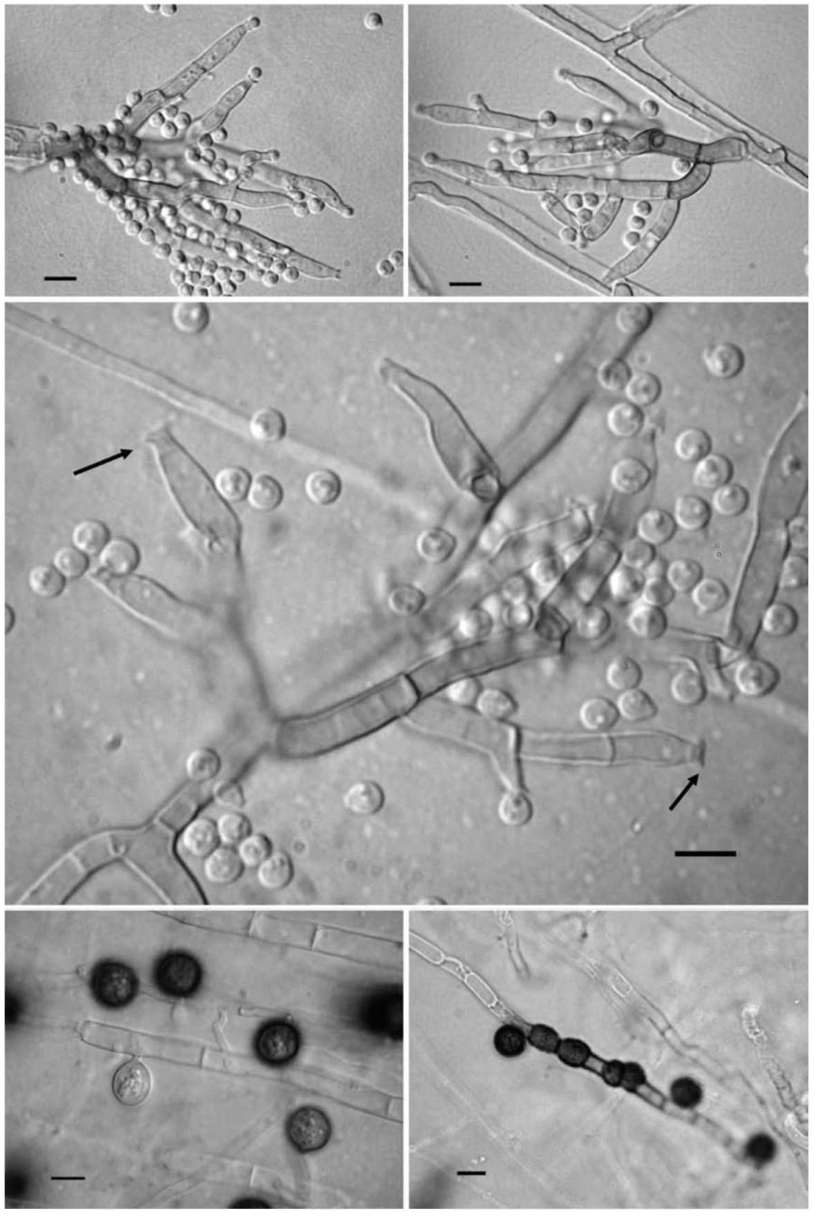
Nicoli, R.M. and A. Russo. 1974. The genus Humicola Traaen and the closely related genera (Hyphomycetes). Nova Hedw 3-4: 737-797.
Rambaut, A. 1996. Se-Al Version 1.0 Alpha 1. Sequence Align­ment Editor. Oxford, UK: Department of Zoology, Univer­sity of Oxford.
Saitou, N. and M. Nei. 1987. The neighbor-joining method: A new method for reconstructing phylogenetic trees. Mol.
Biol. Evol. 4: 406-425.
Schol-Schwarz, M.B. 1970. Revision of the genus Phialophora (Moniliales). Persoonia 6: 59-94.
Swofford, D.L. 1998. PAUP* 4.0: Phylogenetic Analysis Using Parsimony (*and Other Method). Sunderland, Massachu­setts: Sinauer Associates.
Thompson, J.D., T.J. Gibson, F. Plewniak, F. Jeanmougin, and D.G. Higgins. 1997. The CLUSTAL_X windows interface: flexible strategies for multiple sequence alignment aided by quality analysis tools. Nucleic Acids Res. 25: 4876-4882.
Wang, P.H., Y.S. Chen, M.J. Lin, Y.J. Tsou, and W.H. Ko. 2010. Severe decline of wax apple trees caused by Fusari-um solani in northern Taiwan. Bot. Stud. 51: 75-80.
White, T.J., T. Bruns, S. Lee, and J.W. Taylor. 1990. Ampli­fication and direct sequencing of fungal ribosomal RNA genes for phylogenetics. In M. A. Innis, D. H. Gelfand, J. J. Sninsky and T. J. White (eds.), PCR Protocols: A Guide to Methods and Applications. Academic Piers, San Diego, pp. 315-322.
202
Botanical Studies, Vol. 52, 2011
由土壤中分離之新種真菌 Humicola phialophoroides
具植物病害生物防治的潛力
柯文雄 楊靜惠 林玫珠 陳啟予 鄒依蓉
國立中興大學植物病理學系
由添加有機質土壤中分離到一株真菌,具有植物病害生物防治的潛力,且產生的孢子和Humicola
屬及Phialophora屬的真菌相似。此尚未被發表敘述的真菌之ITS1-5.8S-ITS2序列,在演化親緣關係
上,同Humicola屬的親緣比同Phialophora屬接近。所以將此微生物命名為Humicola phialophoroides
關鍵詞:生物防治;Humicola phialophoroides ; 土壤真菌;synanamorphs ;分類學•