|
|||||
Botanical Studies (2011) 52: 257-264.
|
BIOCHEMISTRY
|
|
|||
|
|||||
|
|||||
Sweet potato storage root defensin and its tryptic hydrolysates exhibited angiotensin converting enzyme
|
|||||
|
|||||
Guan-Jhong HUANG1,8 *, Te-Ling LU2 #, Chuan-Sung CHIU1,3, Hsien-Jung CHEN4, Chieh-Hsi WU2,
Ying-Chin LIN5, Wen-Tsong HSIEH6, Jung-Chun LIAO2, Ming-Jyh SHEU2, and Yaw-Huei LIN7 * |
|||||
|
|||||
1School of Chinese Pharmaceutical Sciences and Chinese Medicine Resources, China Medical University, Taichung 404,
Taiwan 2School of Pharmacy, China Medical University, Taichung 404, Taiwan
3Nursing Department, Hsin Sheng College of Medical Care and Management, Taoyuan 325, Taiwan
4Department of Biological Sciences, National Sun Yat-Sen University, Kaohsiung 804, Taiwan 5Nursing and Management, Jen-Teh Junior College of Medicine, Taiwan 6Department of Pharmacology, China Medical University, Taichung 404, Taiwan |
|||||
|
|||||
(Received September 7, 2010; Accepted October 29, 2010)
|
|||||
|
|||||
ABSTRACT. Sweet potato defensin (SPD1) overproduced in E. coli (M15) was purified by Ni2+-chelate affinity chromatography. The molecular mass of SPD1 is about 8,600 Da determined by SDS (sodium dodecyl sulfate)-PAGE (polyacrylamide gel electrophoresis). Our previous paper showed that SPD1 had antimicrobial, dehydroascorbate reductase and monodehydroascorbate reductase activities. The activity of SPD1 to inhibit angiotensin converting enzyme (ACE) was shown using N-[3-(2-furyl) acryloyl]-Phe-Gly-Gly (FAPGG) as substrate in a dose-dependent manner (27.56 ~ 52.58 % inhibition). The 50% inhibition (IC50) of ACE activity required 190.47 [ig/mL SPD1 while that of Captopril was 10 nM (868 ng/mL). Thin layer chromatography (TLC) also identified SPD1 as an ACE inhibitor. SPD1 acted as a mixed type inhibitor against ACE using FAPGG as a substrate. When 200 (ig/mL SPD1 (10 ig) were added, Vmax and Km were 0.01 AA/min and 0.69 mM, respectively; without SPD, 0.03 AA/min and 0.42 mM. Trypsin was used for SPD1 hydrolysis and part of the reaction mixture was removed and analyzed at set times. ACE inhibitory activity increased from 52.47% to about 74.38% after 24 h hydrolysis. The results suggested that small peptides increased by trypsin hydrolysis of the SPD1 ACE inhibitory capacity also increased up to 24 h, then decreased, which may be due to the disappearance of some active ingredients. Six peptides, namely GFR, FK, IMVAEAR, GPCSR, CFCTKPC and MCESASSK, were synthesized based on the simulated trypsin digest of SPD1, then tested for ACE inhibitory activity. IC50 values of individual peptides were 94.25 ± 0.32, 265.43 ± 1.24, 84.12 ± 0.53, 61.67 ± 0.36, 1.31 士 0.07 and 75.93 士 0.64 [iM, respectively, suggesting that CFCTKPC might represent the main domain for the ACE inhibition. The consumption of sweet potatoes may thus help alleviate hypertension and other diseases due to their SPD1 and hydrolysate content.
|
|||||
|
|||||
Keywords: Angiotensin converting enzyme (ACE); Defensin; Inhibition; Sweet Potato.
|
|||||
|
|||||
INTRODUCTION
|
(e.g. 2-9 amino acids) and possession of hydrophobic amino acid residues, including proline, lysine or arginine groups (Lin et al., 2006). Bioactive peptides are among the many functional components identified in foods. These are small protein fragments that have biological effects once they are released during gastrointestinal digestion in the organism or by previous in vitro protein hydrolysis. Recent research has focused on bioactive peptides with immunostimulating (Huang et al., 2010), antioxidant or angiotensin-converting enzyme (ACE) inhibitor (Liu et al., 2007), antithrombotic (Scarborough et al., 1991) and bactericidal (Bellamy et al., 1993) functions.
|
||||
Many bioactive peptides have common structural properties that include a relatively short peptide residue length
|
|||||
8These two authors contributed equally to this work. *Corresponding author: E-mail: boyhlin@gate.sinica.edu.tw, Fax: 886-2-2782-7954, Tel: 886-2-2787-1172 (Yaw-Huei LIN); E-mail: gjhuang@mail.cmu.edu.tw, Tel: +886-42205-3366 ext 5508, Fax: +886-4-2208 3362 (Guan-Jhong HUANG).
|
|||||
|
|||||
|
|||
258
|
Botanical Studies, Vol. 52, 2011
|
||
|
|||
ACE (peptidyldipeptide hydrolase EC 3.4.15.1) is a glycoprotein and a dipeptide-liberating exopeptidase classically associated with the renin-angiotensin system regulating peripheral blood pressure (Mullally et al., 1996). ACE removes a dipeptide from the C-terminus of angiotensin I to form angiotensin II, a very hypertensive compound (Lin et al., 2008). Several endogenous peptides, such as enkephalins, p-endorphin, and substance P, were reported to be competitive substrates and ACE inhibitors. Numerous food-derived peptides from a-lactoalbumin, p-lactoglobulin (Pihlanto-Leppala et al., 1998), mucilage (Huang et al., 2006) and trypsin inhibitors (TI) (Huang et al., 2007) also inhibited ACE, as did a number of antioxidant (reduced glutathione and car-nosine-related peptides) (Hou et al., 2003) and synthetic peptides (Huang et al., 2006).
Plant defensins were originally termed y -thionins because they have a similar size (5 kDa) and the same number of disulfide bridges (four) as a- and ^-thionins. y-Thionins however, are structurally different from a-and ^-thionins and are more similar, both in structure and in function, to insect and mammalian defensins. This class of plant peptides was thus named 'plant defensins' (Haasen and Goring, 2010). A variety of plant defensins, from both monocotyledonous and dicotyledonous plants, have been isolated and characterized (Terras et al., 1992). Plant defensin families have been known as potent growth inhibitors of a broad spectrum of fungi and bacteria, however their antimicrobial activity is quite diverse. They have been divided into two main groups (A and B), whose amino acid sequence shares only 25% similarity (Harrison and Lederberg, 1998). Plant defensins have been detected in the leaves, flowers, seeds, tubers and other parts of plants (Moreno et al., 1994). Furthermore, the expression of some defensin genes is developmentally regulated, whereas that of other genes is greatly elevated in response to external biotic and abiotic stimuli (Epple et al., 1997).
In our previous report, SPD1 exhibited antimicrobial, dehydroascorbate reductase and monodehydroascorbate reductase activities (Huang et al., 2008a). In this work, we report for the first time that SPD1 exhibited dose-dependent ACE inhibitory activity when Captopril was used as a positive control. Commercial bovine serum albumin (BSA), which was frequently found in the literature as the peptide resource of ACE inhibitors, was chosen for comparison. The Ki values of SPD1 against ACE were calculated. We also used trypsin to hydrolyze SPD1, analyzed it at set times, and determined the changes in ACE inhibitory activity. IC50 of ACE inhibitory activities using synthetic peptides were also determined for comparison.
|
(CA, USA); Seeblue prestained markers for SDS-PAGE
including myosin (250 kDa), BSA (98 kDa), glutamic de-hydrogenase (64 kDa), alcohol dehydrogenase (50 kDa), carbonic anhydrase (36 kDa), myoglobin (30 kDa), and lysozyme (16 kDa) were from Invitrogen (Groningen, The Netherlands); FAPGG, ACE (1 unit, rabbit lung); Coo-massie brilliant blue G-250; peptide (GL Biochem, China), and other chemicals and reagents were purchased from
Sigma Chemical Co. (St. Louis, MO, USA).
|
||
Defensin expression in E. coli
|
|||
Defensin with its pre-pro-sequence (SPD1) was expressed in E. coli. The coding sequence was amplified from cDNA SPD1 using an oligonucleotide (5'-A GGAT CCATG GCTTC ATCTC TTCGT TC -3') with a BamHI site (underlined) at the putative initial Met residue, and an oligonucleotide (5'-GCCTT GCTAA TTCAG TCGAC CGCTG T -3') with a SalI site at the 3' end. The PCR fragment was subcloned in a pGEM T-easy vector. The plasmid was then digested with BamHI and SalI and the excised fragments were subcloned in a pQE30 expression vector (QIAexpress expression system, Qiagen). The resulting plasmid, termed pQE-SPD1, was introduced into E. coli (M15). Cultures of the transformed E. coli (M15) overexpressed a protein of expected molecular mass, which was purified by affinity chromatography in Ni-nitrilotriacetic acid (NTA) columns (Qiagen), according to
Huang et al. (2004).
|
|||
ACE inhibitory activity determination
|
|||
The ACE inhibitory activity was measured according to the method of Holmquist et al. (1979) with some modifications. Four microliters (4 microunits) of commercial ACE (1 unit, rabbit lung) was mixed with 50 fiL of different amounts of SPD1 or BSA (50, 100, and 200 (ig/mL), then 200 [iL of 0.5 mM N-[3-(2-fu^l) aciyloyl]-Phe-Gly-Gly [FAPGG, dissolved in 50 mM Tris-HCl buffer (pH 7.5) containing 0.3 M NaCl] was added. The decreased absor-bance at 345 nm (A4 inhibitor) was recorded after 5 min at room temperature. Deionized water was used instead of sample solution for blank experiments (A4 control). Captopril (molecular mass 217.3 Da) was used as a positive control for the ACE inhibitor (1.25, 2.5, 5, 10, 20, 40 and 80 nM). ACE activity was expressed as A4 345 nm, and the ACE percent inhibition was calculated as follows: [1 - (A4 inhibitor / A4 control)] x 100. Means of triplicates were determined. The 50% inhibition (IC50) of ACE activity was defined as the concentrations of samples that inhibited 50% of ACE activity under experimental conditions.
|
|||
|
|||
MATERIALS and METHODS
|
ACE inhibitory activity determination using TLC
|
||
|
|||
Materials
|
The ACE inhibitor activity of SPD1 was also determined using TLC (Holmquist et al., 1979). Each 100 fL of SPD1 or BSA (225 (ig/mL) was premixed with 15 micro-units of ACE for 1 min, and then 200 fL of 0.5 mM FAPGG was added and allowed to react at room tempera-
|
||
Tris, electrophoretic reagents and silica gel 60 F254 were purchased from E. Merck Inc. (Darmstadt, Germany); Captopril was purchased from Calbiochem Co.
|
|||
|
|||
|
||||
HUANG et al. — Defensin with angiotensin converting enzyme inhibitory activity
|
259
|
|||
|
||||
ture for 10 min. Then 800 fL of methanol was added to stop the reaction. The blank experiment contained FAPGG only; in the control experiment, ACE reacted with FAPGG under the same conditions. Each was dried under reduced pressure and redissolved with 400 fL of methanol, and 50 fL was spotted on a silica gel 60 F254. The FAPGG and FAP (ACE hydrolyzed product) were separated by TLC in 1-butanol-acetic acid-water, 4:1:1 (V/V/V), and observed under UV light.
|
|
|||
Kinetic properties of ACE inhibition determination by defensin
|
||||
The kinetic properties of ACE (4 fU) without or with SPD1 (200 (g/mL) in a total volume of 250 [L were determined using different concentrations of FAPGG as substrate (0.1 mM to 0.5 mM). The Km (without SPD1) and Km' (with SPD1) were calculated from Lineweaver-Burk plots, where Km' was the Michaelis constant in the presence of 200 (g/mL SPD1.
|
||||
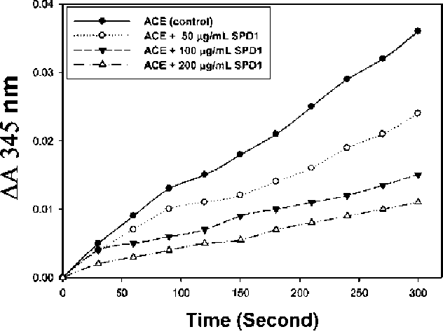
Figure 1. Inhibitory activity of different amounts of defensin (SPD1) (0, 50, 100 and 200 (ig/mL) from sweet potato storage root on ACE activity (AA 345 nm).
|
||||
|
||||
ACE inhibitory activity determination by de-fensin trypsin hydrolysates
|
affinity chromatography, which yielded highly purified His-tagged SPD1. Preparing SDS-PAGE (Huang et al., 2008a) was used as the next step for SPD1 purification.
|
|||
Six mg of SPD1 were dissolved in 1 mL of 50 mM Tris-HCl buffer (pH 7.9). Then 0.1 mL of 12 mg of trypsin was added and hydrolysis was carried out at 37°C for 8, 16, 24 and 32 h. After hydrolysis the solution was heated at 100°C for 5 min to stop enzyme reaction. The trypsin was heated before SPD1 hydrolysis for the 0 h control reaction. Each of the 60 [iL SPD1 hydrolysates was used in spectro-photometry to determine ACE inhibition.
|
||||
Spectrophotometric determination of ACE inhibitor activity of defensin
|
||||
Purified SPD1 was used to determine ACE inhibitory activity. Figure 1 shows time progression for the effect
of the different amounts of SPD1 (0, 50, 100 and 200 (g/
mL) on the ACE activity (AA 345 nm). Compared with the ACE only (control), it was found that the higher the amount of SPD1 added during a 300 sec reaction period, the lower the AA 345 nm would be. Figure 1 shows that purified SPD1 could inhibit ACE activity in a dose-dependent manner.
|
||||
Defensin tryptic hydrolysate chromatograms on a Sephadex G-50 column
|
||||
The unhydrolyzed SPD1 and tryptic SPD1 hydrolysates at 24 h were separated by Sephadex G-50 chromatography (1 x 60 cm). The column was eluted with 20 mM Tris-HCl buffer (pH 7.9). The flow rate was 30 mL/h, and each fraction contained 2 mL, the absorbance of which was determined at 570 nm.
|
||||
Effects of defensin, BSA and captopril on ACE activity shown by spectrophotometry
|
||||
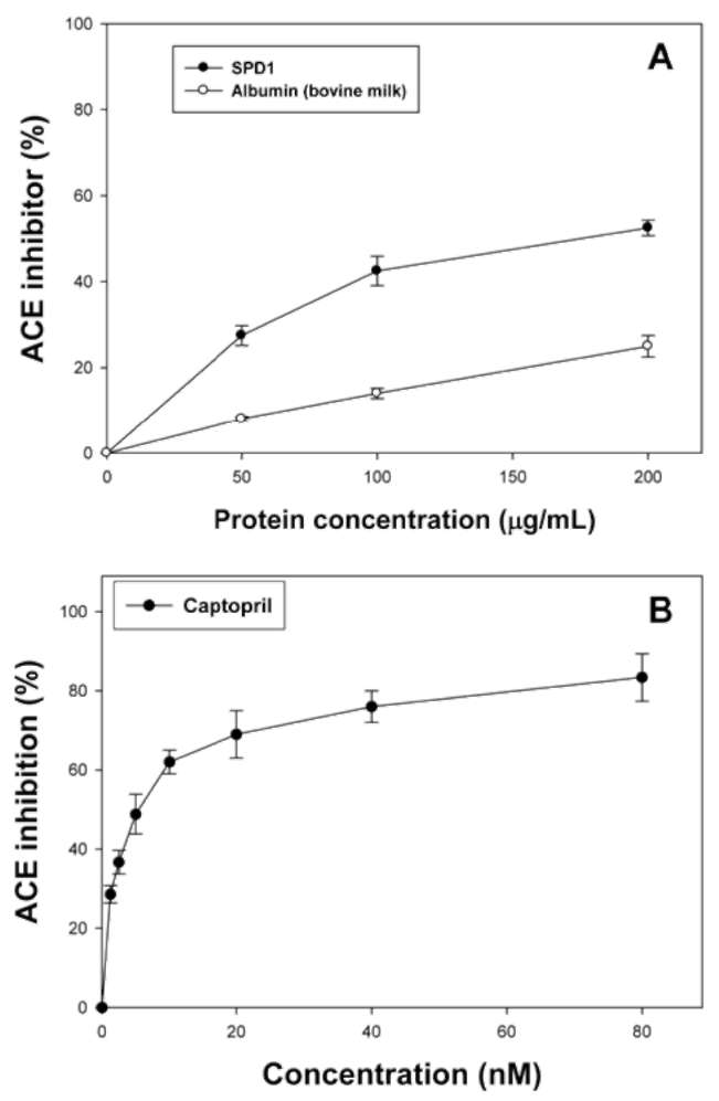
We were interested in learning whether BSA also exhibited ACE inhibitory activity. Figure 2A shows the effects of SPD1 (0, 50, 100, and 200 ig/mL), BSA (0, 50, 100, and 200 (ig/mL) or Captopril (Figure 2B; 0, 1.25, 2.5, 5,
10, 20, 40 and 80 nM; corresponding to 0, 108.5, 217, 434, 868, 1,736, 3,472 and 6,844 ng/mL, respectively) on ACE
activity. BSA showed less ACE inhibitory activity (less than 25% inhibition) without dose-dependent inhibition patterns. However, SPD1 exhibited dose-dependent ACE inhibitory activity (50~200 (ig/mL giving, respectively, 27.56 ~ 52.58% inhibition). From calculations, the 50%
inhibition (IC50) of SPD1 against ACE activity was 190.47
(g/mL compared to that of 10 nM (868 ng/mL) for Cap-topril, which was similar to the report (7 nM) by Pihlanto-Leppala et al. (1998); while the IC50 of yam dioscorin was 250 (ig/mL (Hsu et al., 2002). Both BSA and purified SPD1 were proteins, but only the purified SPD1 showed specific dose-dependent ACE inhibitory activity. In the
|
||||
Statistical analysis
|
||||
Means of triplicates were calculated. Student's t test was used for comparison between two treatments. A difference was considered to be statistically significant when p < 0.05.
|
||||
RESULTS and DISCUSSION
|
||||
Expression of defensin in E. coli
|
||||
SDS-PAGE analysis of SPD1 crude extracts from the transformed E. coli (M15) showed high amounts of a poly-peptide with the expected molecular mass (ca. 8,600 Da). This polypeptide was found as a soluble protein in the supernatant, and was absent in protein extracts obtained from E. coli transformed with pQE-30 vector. The expressed protein was purified from crude extracts by Ni2+-chelate
|
||||
|
||||
|
|||
260
|
Botanical Studies, Vol. 52, 2011
|
||
|
|||
|
Defensin ACE inhibitor activity determination using TLC
|
||
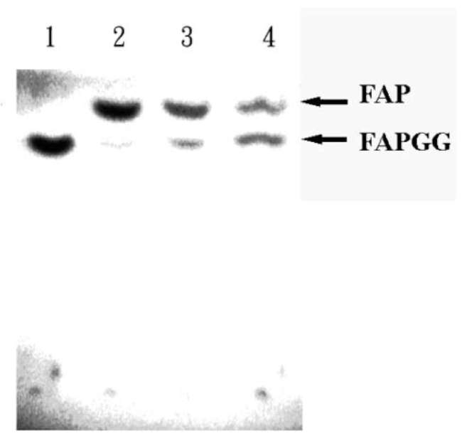
FAPGG and FAP (products of ACE catalyzed hydrolysis reaction) were separated by TLC using water saturated 1-butanol: acetic acid: water, 4:1:1 (V/V/V) as developing solvents according to the method of Hol-mquist et al. (1979). Figure 3 shows the qualitative results of TLC chromatograms of a silica gel 60 F254 about the effects of 225 (g/mL of commercial BSA (lane 3) or SPD1 (lane 4) on 15 microunits of ACE. Compared to
the control test (lane 2), SPD1 (lane 4) inhibited ACE reaction showing less amounts of FAP production observed under UV light. However, similar amounts of FAP were found between the control test (lane 2) and BSA (lane 3). These results demonstrated again that SPD1 exhibited ACE inhibitor activity.
|
|||
Determination of the kinetic properties of ACE Inhibition by defensin
|
|||
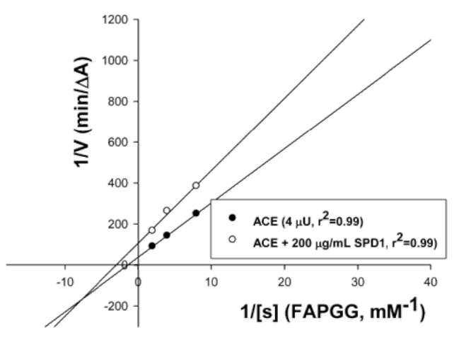
The Lineweaver-Burk plots of ACE (4 fU) with or without purified SPD1 (200 (ig/mL) under different concentrations of FAPGG are shown in Figure 4. The results indicated that purified SPD1 acted as a mixed type inhibitor against ACE using FAPGG as a substrate. When 200 (g/mL SPD1 (10 [g) were added, Vmax and Km were, respectively, 0.01 AA/min and 0.69 mM; while they were
0.03 AA/min and 0.42 mM without SPD1. In conclusion,
|
|||
|
|||
Figure 2. The effects of SPD1, albumin and Captopril on ACE activity determined by spectrophotometry. SPD1 (0, 50, 100 and 200 (ig/mL) or bovine serum albumin (0, 50, 100 and 200 (ig/ mL) was used. ACE inhibition (%) was calculated according to the equation [1-(AA inhibitor + AA control)] x 100%.
|
|||
literature, the protein hydrolysates were used as sources to purify peptides as ACE inhibitors (Mullally et al., 1996). From calculations, the IC50 of SPD1 against ACE activity was 190.47 (g/mL, which was smaller than the synthetic peptide a-lactorphin (YGLF, 322.7 (ig/mL). Several identified peptide fragments exhibited much lower IC50 values than our purified SPD1; for example, Tyr-Pro of whey proteins, 8.1 (g/mL (Yamamoto et al., 1999) and HHL of soybean proteins, 2.2 (g/mL (Shin et al., 2001). Alternately, several peptide fragments exhibited much higher IC50 values than our purified SPD1; for example, hydrolysates of whey proteins (a-lactalbumin and p-lactoglobulin) were effective with IC50 values between 345-1,733 (g/mL (Pihlanto-Leppala et al., 2000), LAHKAL of a-lactalbumin hydrolysates, 406 (ig/mL; GLDIQK of p-lactoglobulin hy-drolysates, 391 ig/mL; and VAGTWY of p-lactoglobulm hydrolysates, 1,171 (g/ mL. In our previous paper (Huang et al., 2008b), the IC50 of ACE activity required 187.96 [ig/ mL TI from sweet potato, which was lower than the IC50 value of purified SPD1.
|
|||
Figure 3. The TLC chromatograms of a silica gel 60 F254 showing the effects of defensin from sweet potato storage root or bovine serum albumin on ACE activity. Lane 1, blank test (FAPGG only); lane 2, control test (ACE reacted with FAPGG to produce FAP); lane 3, 225 (ig/mL bovine serum albumin added; lane 4, 225 (ig/mL SPD1 added. Each solution was dried under reduced pressure and redissolved with 400 fL methanol. Each 50 fL was spotted on a silica gel 60 F254. FAPGG and FAP were separated by water saturated 1-butanol:acetic acid:water, 4:1:1 (V/V/V). Arrows indicate the positions of both FAP and FAPGG.
|
|||
|
|||
|
||||
HUANG et al. — Defensin with angiotensin converting enzyme inhibitory activity
|
261
|
|||
|
||||
|
We used synthetic peptides (according to SPD1 gene sequence) to measure ACE inhibitor activity (Table 1). Kohmura et al. (1989) synthesized some human p-casein peptide fragments and found that their length had an influence on the ACE inhibitory activity. The necessary ACE inhibitors were namely peptides composed of 3-10 amino acids with proline on the C-terminal, and Leu-Arg-Pro, from food protein hydrolysates, is reportedly the most potent natural ACE inhibitor, with an IC50 value of 0.27 (ig/mL or 1.0 (iM. Byun and Kim (2002) studied the ACE inhibitory activity of a series of dipeptides, and indicated that tryptophan, tyrosine, proline, or phenylalanine at the C-terminal and branched-chain aliphatic amino acid at the N-terminal were required for a peptide to bind to ACE.
Synthetic peptides were designed by simulated trypsin cutting sites of SPD1 gene (accession number: AY552546) products from sweet potato (http://www.expasy.org/tools/ peptidecutter/). Six peptides, namely GFR, FK, IMVAEAR, GPCSR, CFCTKPC and MCESASSK, were synthesized based on the simulated trypsin digest of SPD1, then tested for ACE inhibitory activity (Table 1). IC50 values
|
|||
Figure 4. The Lineweaver-Burk plots of ACE (4 fiU) without or with SPD1 (200 (ig/mL) from sweet potato storage root using different concentrations of FAPGG (0.1 to 0.5 mM).
|
||||
SPD1 exhibited dose-dependent ACE inhibitory activity and acted as a mixed type inhibitor with respect to the substrate (FAPGG). A similar work was reported with the calculated Km as 0.25 mM FAPGG for ACE in the presence of purified dioscorin, the calculated Km' was 0.33 mM (Hsu et al., 2002).
The mixed-type inhibition suggests that there might be an inhibitor-binding site (or I-site) for SPD1 on the enzyme (ACE) surface in addition to the substrate-binding site (or S-site). It is noteworthy that SPD1 binds tighter to the I-site on the substrate-bound form of ACE than that on the free form. The Km values in the presence of the inhibitors are also higher than in their absence. In other words, binding of the substrate to the S-site increases the binding affinity of the inhibitor to the I-site, whereas binding of the inhibitor to the I-site decreases the binding affinity of the substrate to the S-site. The inhibitory mode of SPD1 was unique and hardly analyzed with a simple Michaelis-Menten-type interaction between the enzyme and inhibitor. The inhibitory mode of SPD1 must be examined kineti-cally and thermodynamically in the next phase of research.
|
||||
|
||||
Determination of ACE inhibitory activity by de-fensin trypsin hydrolysates and their peptide distributions
|
||||
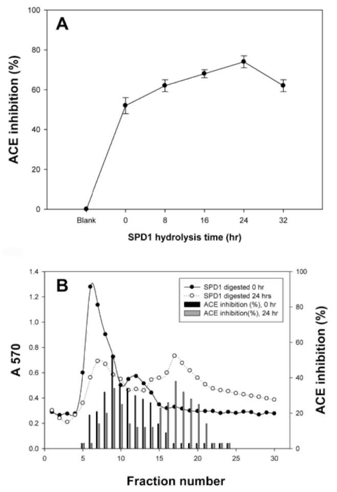
Figure 5 shows the ACE inhibitory activity (AA 345 nm) of tryptic SPD1 hydrolysates. Figure 5A shows the ACE inhibition (percent) of tryptic SPD1 hydrolysates collected at set times. From the results (Figure 5A), ACE inhibitory activity increased from 52.47% (0 h) to about 74.38% (24 h). Figure 5B shows the chromatograms of unhydrolyzed SPD1 and tryptic SPD1 hydrolysates (24 h) on a Sephadex G-50 column. It was found that smaller peptides increased the longer that trypsin was hydrolyzed. The ACE inhibitor activities of tryptic SPD1 hydrolysates decreased after 24 h hydrolysis (Figure 5A), suggesting that some active ingredients got lost after 24 hours.
|
||||
Figure 5. ACE inhibitor activity of tryptic hydrolysates of sweet potato SPD1. The plot shows the ACE inhibition (%) of tryptic SPD1 hydrolysates obtained at different trypsin hydrolysis time (A). The proteins and the inhibition of ACE (%) were shown (B). The inhibition of ACE (%) was calculated according to the equation [1-(AA inhibitor + AA control)] x 100%.
|
||||
|
||||
|
|||||||||||||||||||||||||||||||||||||||
262
|
Botanical Studies, Vol. 52, 2011
|
||||||||||||||||||||||||||||||||||||||
|
|||||||||||||||||||||||||||||||||||||||
Table 1. SPD1 peptides with ACE inhibition activity.
|
LITERATURE CITED
|
||||||||||||||||||||||||||||||||||||||
|
|||||||||||||||||||||||||||||||||||||||
|
Bellamy, W., R.H. Wakabayashi, M. Takase, K. Kawase, S. Shi-mamura, and M. Tomita. 1993. Role of cell-binding in the antibacterial mechanism of Lactoferricin B. J. Appl. Bacte-riol. 75: 478-484.
Byun, H.G. and S.K. Kim. 2002. Structure and activity of angio-tensin I converting enzyme inhibitory peptides derived from Alaskan pollack skin. J. Biochem. Mol. Biol. 35: 239-243.
Epple, P., K. Apel, and H. Bohlmann. 1997. ESTs reveal a mul-tigene family for plant defensins in Arabidopsis thaliana. FEBS Lett. 400: 168-172.
Haasen, K.E. and D.R. Goring. 2010. The recognition and rejection of self-incompatible pollen in the Brassicaceae. Bot. Stud. 51: 1-6.
Harrison, P.F. and J. Lederberg. 1998. Antimicrobial Resistance. National Academies Press.
Holmquist, B., P. Bunning, and J.F. Riordan. 1979. A continuous spectrophotometric assay for angiotensin converting enzyme. Anal. Biochem. 95: 540-548.
Hou, W.C., H.J. Chen, and Y.H. Lin. 2003. Antioxidant
peptides with angiotensin converting enzyme inhibitory activities and applications for angiotensin converting enzyme purification. J. Agric. Food Chem. 51: 1706-1709.
Hsu, F.L., Y.H. Lin, M.H. Lee, C.L. Lin, and W.C. Hou.
2002. Both dioscorin, the tuber storage protein of yam (Dioscorea alata cv. Tainong No. 1), and its peptic hydrolysates exhibited angiotensin converting enzyme inhibitory activities. J Agric Food Chem. 50: 6109-6013.
Huang, D.J., C.D. Lin, H.J. Chen, and Y.H. Lin. 2004. Antioxi-dant and antiproliferative activities of sweet potato (Ipom-oea batatas [L.] Lam. 'Tainong 57') constituents. Bot. Bull. Acad. Sin. 45: 179-186.
Huang, D.J., W.C. Hou, H.J. Chen, and Y.H. Lin. 2006.
Sweet potato (Ipomoea batatas [L.] Lam 'Tainong 57') storage roots mucilage exhibited angiotensin converting enzyme inhibitory activities in vitro. Bot. Stud. 45: 397-402.
Huang, G.J., H.J. Chen, Y.S. Chang, M.J. Sheu, and Y.H.
Lin. 2007. Recombinant Sporamin and its synthesized peptides with Antioxidant Activities in vitro. Bot. Stud. 48: 133-140.
Huang, G.J., H.C. Lai, Y.S. Chang, M.J. Sheu, T.L. Lu, S.S.
Huang, and Y.H. Lin. 2008a. Antimicrobial, dehydroascor-bate reductase and monodehydroascorbate reductase activities of defensin from sweet potato (Ipomoea batatas [L.] Lam. 'Tainong 57') storage roots. J. Agric. Food Chem. 56: 2989-2995.
Huang, G.J., Y.L. Ho, H.J. Chen, Y.S. Chang, S.S. Huang,
H.J. Hung, and Y.H. Lin. 2008b. Sweet potato storage root trypsin inhibitor and their peptichydrolysates exhibited angiotensin converting enzyme inhibitory activity in vitro. Bot. Stud. 49: 101-108.
Huang, C.Y., C.L. Wen, Y.L. Lu, Y.S. Lin, L.G. Chen, and W.C. Hou. 2010. Antihypertensive activities of extracts from tis-sue cultures of Vitis thunbergii var. taiwaniana. Bot. Stud. 51: 317-325.
|
||||||||||||||||||||||||||||||||||||||
Note: The sequence of SPD1 contains pre-pro-sequence. These sequences were retrieved from the NCBI (National Center for Biotechnology Information, http://www.ncbi.nlm.nih.gov) with the following accession numbers AY552546. |
|||||||||||||||||||||||||||||||||||||||
of individual peptides were 94.25± 0.32, 265.43± 1.24,
84.12± 0.53, 61.67 ± 0.36, 1.31 ± 0.07 and 75.93 ± 0.64 [M, suggesting that CFCTKPC might represent the main domain for the ACE inhibition (Table 1). These results demonstrated that simulated synthetic peptides from tryp-tic SPD1 hydrolysates exhibited ACE inhibitory activities. Our work suggests that (1) CFCTKPC might represent the main active site for the ACE inhibition; (2) there are marked structural similarities in peptides with antihyper-tensive, immunomodulatory and antioxidant activities that may be used as criteria in selecting or designing multifunctional foods to control cardiovascular diseases.
In our previous paper, TI inhibited ACE activation in a dose-dependent manner. The IC50 of ACE activity required 187.96 [g/mL TI. And TYCQ was synthesized based on the simulated pepsin digest of TI. The IC50 values of the
TYCQ peptide was 2.30 fM (Huang et al., 2008b). In
this work the IC50 values of the CFCTKPC peptide was 1.31 [M. This value was much better than TI and its hy-drolysates. The CFCTKPC peptide contains seven amino acids with three cysteine residues. Thus far, the structure-activity correlations among the various ACE inhibitory peptides remain ambiguous. On the basis of some common structure patterns, it seems that the most favorable amino-terminal residues are branched amino acids such as Val and Ile and the most preferred carboxyterminus residues are among Trp, Tyr, Pro, or Phe (Zhou et al., 2010).
In summary, SPD1 exhibited dose-dependent ACE inhibitory activity. SPD1 acted as a mixed type inhibitor toward ACE with IC50 of 1.31 ± 0.07 [iM. Its peptic hydrolysates also showed ACE inhibitory activities. Some peptides derived from food proteins demonstrated antihy-pertensive activities against spontaneously hypertensive rats (Yoshii et al., 2001). The role of sweet potato in hypertension control deserves further investigation.
|
|||||||||||||||||||||||||||||||||||||||
Acknowledgements. The authors want to thank the National Science Council (NSC 97-2313-B-039-001-MY3) and China Medical University (CMU) (CMU95-PH-11,
CMU96-113, CMU97-232 and CMU99-S-29) for their financial support.
|
|||||||||||||||||||||||||||||||||||||||
|
|||||||||||||||||||||||||||||||||||||||
|
||||
HUANG et al. — Defensin with angiotensin converting enzyme inhibitory activity
|
263
|
|||
|
||||
Kohmura, M., N. Nio, K. Kubo, Y. Minoshima, E. Munekata, and Y. Ariyoshi. 1989. Inhibition of angiotensin-converting enzyme by synthetic peptide fragments of human p-casein.
Agric. Biol. Chem. 53: 2107-2114. Lin, C.L., S.Y. Lin, Y.H. Lin, and W.C. Hou. 2006. Effects of
tuber storage protein of yam (Dioscorea alata cv. Tainong No. 1) and its peptic hydrolysates on spontaneously hypertensive rats. J. Sci. Food Agric. 86: 1489-1494.
Lin, S.Y., C.C. Wang, Y.L. Lu, W.C. Wu, and W.C. Hou.
2008. Antioxidant, anti-semicarbazide-sensitive amine oxidase, and antihypertensive activities of geraniin isolated from Phyllanthus urinaria. Food Chem. Toxicol. 46: 2485-2492.
Liu, Y.H., M.T. Chuang, and W.C. Hou. 2007. Methanol-
soluble, p-elimination products from preparations of alginic acid hydroxamate exhibited DPPH scavenging and angiotensin converting enzyme inhibitory activities. Bot. Stud. 48: 141146.
Moreno, M., A. Segura, and F. Garcia-Olmedo. 1994. Pseudothi-onin-St1, a potato peptide active against potato pathogens. Eur. J. Biochem. 223: 135-139.
Mullally, M.M., H. Meisel, and R.J. Fitzgerald. 1996.
Synthetic peptides corresponding to a-lactalbumin and p-lactoglobulin sequences with angiotensin-I-converting enzyme inhibitory activity. J. Biol. Chem. 377: 259-260.
Pihlanto-Leppala, A., T. Rokka, and H. Korhonen. 1998. Angio-tensin I converting enzyme inhibitory peptides derived from bovine milk proteins. Int. Dairy J. 8: 325-331.
Pihlanto-Leppala, A., P. Koskinen, K. Piilola, T. Tupasela, and H. Korhonen. 2000. Angiotensin I-converting enzyme inhibi-
|
tory properties of whey protein digest: concentration and characterization of active peptides. J. Dairy Sci. 67: 53-64.
Scarborough, R.H., J.W. Rose, M.H. Hsu, D.R. Phillips, V.A. Fried, A.M. Campbell, L. Manniaai, and I.F. Charo. 1991. Barbourin A GpIIb-IIIa specific integrin antagonist from the venom of sistrurus M. barbouri. J. Biol. Chem. 266: 93599360.
Shin, Z.I., R. Yu, S.A. Park, D.K. Chung, C.W. Ahn, H.S.
Nam, K.S. Kim, and H.J. Lee. 2001. His-His-Leu, an angiotensin I converting enzyme inhibitory peptide derived from Korean soybean paste, exerts antihypertensive activity in vivo. J. Agric. Food Chem. 49: 3004-3009.
Terras, F.R., I.J. Goderis, F. Van Leuven, J. Vanderleyden, B.P. Cammue, and W.F. Broekaert. 1992. In vitro Antifungal Activity of a Radish (Raphanus sativus L.) Seed Protein Homologous to Nonspecific Lipid Transfer Proteins. Plant Physiol. 100: 1055-1058.
Yamamoto, N., M. Maeno, and T. Takano. 1999. Purification and characterization of an antihypertensive peptide from a yogurt-like product fermented by Lactobacillus helveticus CPN4. J. Dairy Sci. 82: 1388-1393.
Yoshii, H., N. Tachi, R. Ohba, O. Sakamura, H. Takeyama, and T. Itani. 2001. Antihypertensive effect of ACE inhibitory oligopeptides from chicken egg yolks. Comp. Biochem. Physiol. C Pharmacol. Toxicol. Endocrinol. 128: 27-33.
Zhou, F., Z. Xue, and J. Wang. 2010. Antihypertensive effects of silk fibroin hydrolysate by alcalase and purification of an ACE inhibitory dipeptide. J. Agric. Food Chem. 58: 67356740.
|
|||
|
||||
|
|||
264
|
Botanical Studies, Vol. 52, 2011
|
||
|
|||
甘藷塊根中防禦素及其合成之胜肽具有血管收縮素
轉化酶抑制活性
|
|||
|
|||
黃冠中1 陸德齡2 邱傳淞1,3 陳顯榮4 吳介信2 林穎志5
謝文聰6 廖容君2 許明志2 林耀輝7 |
|||
|
|||
1中國醫藥大學中國藥學暨中藥資源系
2中國醫藥大學藥學系
3新生醫護管理專科學校護理科
4中山大學生命科學系
5仁德醫護管理專科學校護理科
6中國醫藥大學醫學系藥理學科
7中央研究院植物暨微生物研究所
|
|||
|
|||
在大腸桿菌(M15)中大量表現甘藷塊根之重組蛋白質防禦素(SPD1),利用鎳離子螯合之親和性管
柱純化。SPD1經SDS-PAGE分析其分子量約為8,600 Da 。由以前的研究發現SPD1具有抗微生物活 性,去氫抗壞血酸還原酶,單去氫抗壞血酸還原酶的活性。SPD1以W-[3-(2-furyl) acryloyl]-Phe-Gly-Gly (FAPGG)為受質,利用分光光度計的方法分析抑制血管收縮素轉化酶(angiotensin converting enzyme, ACE)的能力,其效果隨劑量增加而增加(50到200 ig/mL SPD1 ,分別抑制27.56 - 52.58%血管收縮素 轉化酶活性)。SPD1對於血管收縮素轉化酶之50%抑制濃度(IC50)為190.47 ig/mL ,對照組Captopril 為10 nM (868 ng/mL)。另外利用螢光silica TLC偵測FAPGG及其水解產物FAP ,結果也顯示SPD1對 於血管收縮素轉化酶有抑制的效果。SPD1對於血管收縮素轉化酶是屬於混合型抑制。利用胰蛋白晦以 不同時間水解SPD1時,發現反應24小時時其血管收縮素轉化酶活性有抑制的效果可以從52.47 % (0 h) 增加到74.38 % (24 h)。由結果可知小分子的胜肽會隨著水解時間增加且血管收縮素轉化酶活性抑制也 有增加,但水解時間超過24 h時,血管收縮素轉化晦活性抑制會降低,可能是由於一些胜肽的結構被 破壞。利用電腦模擬胰蛋白酶水解SPD1的結果,得到六種人工合成具有抑制血管收縮素轉化酶活性胜 肽:GFR, FK, IMVAEAR, GPCSR, CFCTKPC和MCESASSK ,測定其IC50為94.25 士 0.32, 265.43 士 1.24, 84.12 士 0.53, 61.67 士 0.36, 1.31 士 0.07和75.93 士 0.64 fiM 。結果發現CFCTKPC具有很好的抑制血管 收縮素轉化酶活性。當人們食用甘藷塊根時,SPD1及其胜肽也許對於高血壓和其他疾病的控制是有益 的。 |
|||
|
|||
關鍵詞:甘藷;防禦素;血管收縮素轉化酶;抑制作用。
|
|||
|
|||