Botanical Studies (2011) 52: 417-426.
BIOCHEMISTRY
Characterization of a leaf-type catalase in sweet potato (Ipomoea batatas Lam. (L.))
Hsien-Jung CHEN1*, Mufidah AFIYANTI1, Guan-Jhong HUANG2, Shyh-Shyun HUANG1, and Yaw-Huei LIN3 *
1Department of Biological Sciences, National Sun Yat-sen University, 804 Kaohsiung, Taiwan
2 Graduate Institute of Chinese Pharmaceutical SSciences, China Medical University, 404 Taichung, Taiwan
3Institute of Plant and Microbial Biology, Academia Sinica, Nankang, 115 Taipei, Taiwan
(Received January 21, 2011; Accepted April 12, 2011)
ABSTRACT. In this report a major sweet potato catalase was detected and identified from fully-expanded mature leaves using an in-gel activity staining assay with native- and SDS-PAGEs. The catalase activity was optimal from pH 8 to 12, and was repressed by p-mercaptoethanol and a catalase inhibitor 3-amino-1,2,4-triazole. However, catalase activity was much less affected by temperature within the range of 5 to 45°C. Temporal and spatial expression demonstrated that it was specifically detected in leaves, but not in roots and stems. Its activity increased from the immature L2 leaves, reached the maximum in the fully-expanded mature L3 leaves, slightly decreased in the partly-yellowing senescent L4 leaves, and was almost undetectable in the completely yellow L5 leaves, which were similar to the folded and unopened immature L1 leaves. The catalase levels approximately inversely correlated with the H2O2 amounts in leaves of different developmental stages. Dark and ethephon, an ethylene-releasing compound, also enhanced catalase activities from 6 h to 24 h, however, the enhanced activity decreased from 24 h to 48 h in detached leaves after treatment. The catalase levels also approximately inversely correlated with the H2O2 amounts in treated leaves. These data suggest a possible role for sweet potato catalase in coping with H2O2 scavenging in leaves. Based on these data we conclude that a major leaf-type catalase is identified in sweet potato leaves. Its activity level is maximal in mature leaves and becomes significantly lower in natural and in induced senescent leaves. The leaf-type sweet potato catalase likely functions in H2O2 removal in leaves.
Keywords: 3-Amino-1,2,4-triazole; Catalase; Ethephon; Leaf senescence; Sweet potato.
INTRODUCTION
senescence and cell death (Wang et al., 2002). Examples concerning the role of elevated oxidative stress have been reported in the natural senescence of pea leaves (Pastori and del Rio, 1997), induced senescence by ethylene in sweet potato leaves (Chen et al., 2010), JA (Hung and Kao, 2004a) and ABA (Hung and Kao, 2004b) in rice leaves, and wounding in tomato leaves (Orozco-Cardenas and Ryan, 1999).
Plant catalases have been intensively studied and function mainly in the removal of excessive H2O2 generated during developmental processes or by environmental stimuli into water and oxygen in all aerobic organisms (Mhamdi et al., 2010). Catalases are gener­ally composed of a multigene family and various isoform numbers have been previously reported in different plant species. There was one catalyse in sweet potato storage root (Sakajo and Asahi, 1986), two in Hordeum vulgare (Skadsen et al., 1995), three in maize (Guan and Scanda-lios, 1995), tobacco (Havir et al., 1996) and Arabidopsis (Frugoli et al., 1996), five from two interacting subunits in cottonseed (Ni et al., 1990), and eight in Helianthus ann-uus cotyledons (Eising et al., 1990). Various plant catalase
Leaves are the main organ of photosynthesis and serve as a source of carbohydrates for sink nutrients in plants. Their longevity and senescence thus affect photosynthesis efficiency and crop yield. Leaf senescence is influenced by endogenous and exogenous factors, including plant growth regulators, starvation, wounding, darkness, ozone levels, UVrays, and other environmental stresses (Yoshida, 2003; Lim et al., 2007). Elevated oxidative stresses caused by environmental stimuli, including ozone, UV-B, and wounding have been reported and enhance ethylene pro­duction via ACC synthase and ACC oxidase (Wang et al., 2002). In ozone treatment, ethylene also enhances reactive oxygen species (ROS) generation, which in turn leads to

*Corresponding authors: E-mail: boyhlin@gate.sinica.edu.
tw
; Phone: +886-2-27871172; Fax: +886-2-27827954
(Yaw-Huei Lin); E-mail: hjchen@faculty.nsysu.edu.tw;
Phone: 886-7-5252000 ext. 3630; Fax: 886-7-5253630
(Hsien-Jung Chen).
418
Botanical Studies, Vol. 52, 2011
isoforms are temporally and spatially regulated and may respond differentially to developmental and environmental stimuli (Guan and Scandalios, 1995; Zimmermann et al.,2006; Du et al., 2008).
In Arabidopsis, three major catalase isoforms, CAT1, CAT2 and CAT3, were detected in an activity gel (Zim-mermann et al., 2006; Du et al., 2008). For CAT2, it is`the predominant catalase isoform in Arabidopsis, and its activity increased and reached a maximum in mature leaves. The activity levels of CAT3 and CAT1 were much lower than for CAT2, but increased during leaf senescence (Zimmermann et al., 2006). CAT3 was also induced dur-ing sucrose starvation in direct response to the increase in oxidative stress caused by the rapid activation of alterna-tive catabolic pathways (Orendi et al., 2001; Contento and Bassham, 2010). In tobacco, there were three catalase genes isolated and named as CAT-1, CAT-2, and CAT-3. Gene expression patterns detected with RT-PCR demon-strated that CAT-1 and CAT-2 were detected in veinal and interveinal tissues of non-senescent leaves, however, were found in interveinal but not veinal tissues of senescing leaves. The amount of CAT-2, but not CAT-1, was signifi-cantly reduced in senescent leaves compared to non-senes-cent leaves. CAT-3 was detected in veinal and interveinal tissues of both non-senescent and senescing leaves, how-ever, the veinal tissues of senescing leaves exhibited a sig-nificant increase of CAT-3 (Niewiadomska et al., 2009).
The catalase activity levels were inversely correlated with the cellular H2O2 amounts of plants (Zimermann et al., 2006). Therefore, a light-dependent source of H2O2 via photorespiration in the peroxisomes can act as a signal molecule and its concentration is regulated by catalase (Queval et al., 2007). In Arabidopsis, a catalase 2 knock­out mutant (cat2) was produced and its growth in ambient air caused severely decreased rosette biomass, intracellular redox perturbation and activation of oxidative signaling pathways (Queval et al., 2007). Transgenic tobacco plants expressing CAT-1 antisense construct displayed severely reduced catalase activity and developed chlorosis and necrosis on some of the lower leaves. These same leaves showed high level pathogenesis-related protein 1 accumu­lation and enhanced resistance to the tobacco mosaic virus (Takahashi et al., 1997). These data suggest that catalases play an important role in the regulation of plant cellular homeostasis of reactive oxygen species and are also in­volved in the regulation of particular gene expressions and stress responses.
Sweet potato (Ipomoea batatas (Lam.)) is an important food crop in the tropics and subtropics including Tai­wan. Its storage roots and leaves are the edible portions, and contain plenty of vitamin B complex, vitamin C, p-carotenoids, multiple minerals and high levels of cal­cium (Yang et al., 1975; Hattori et al., 1985). Research on sweet potato catalase, however, was limited. In the sweet potato storage root, a candidate catalase protein band was detected by SDS-PAGE from a root microbody after par­tial purification with ammonium sulfate fractionation and
Sepharose 6B column chromatography (Esaka and Asahi, 1982). Sakajo and Asahi (1986) detected a catalase protein band with an antibody against sweet potato catalase, which was an in vitro translation product with a wheat germ sys­tem from storage root mRNA. In addition, a full-length cDNA encoding putative catalase had also been cloned from sweet potato storage root (Sakajo et al., 1987a), and its mRNA level was induced in wounded root tissue (Sakajo et al., 1987b). Sweet potato catalase, however, was not intensively studied in tissues other than storage roots. In this report, we identified and characterized for the first time a major leaf-type catalase in sweet potato. The pos­sible physiological role and function of the leaf-type cata-lase in association with H2O2 removal and leaf senescence are addressed.
MATERIALS AND METHODS
Plant materials
Sweet potato (Ipomoea batatas (L.) Lam.) storage roots were grown in a growth chamber at 28°C/16 h day and 23°C/8 h night cycle. Plantlets that sprouted from the stor­age roots provided detached mature leaves for darkness and ethephon treatments at 28°C/16 h and 23°C/8 h cycle in the dark, and different developmental stages of leaves for temporal and spatial expression experiments. Leaves were arbitrarily divided into L1 to L5 according to their size and different developmental stages. L1 was the stage with folding, unopened immature leaves. L2 was the stage with unfolding but not fully-expanded immature leaves. L3 was the stage with fully-expanded mature leaves. L4 and L5 were the stages with partial and completely yel­lowing senescent leaves, respectively. Samples collected from different leaf stages were used for catalase activity assays as described below. Detached mature leaves (L3) were also used in darkness and ethephon experiments, re­spectively, as described below.
Detection and characterization of catalases from L3 mature leaves
Mature sweet potato leaves were collected from L3 stage, as described above, for detection and characteriza­tion of catalases. Mature leaves (L3) were ground into powder with a mortar and pestle in liquid nitrogen, then homogenized with two folds volumes (w/v) of extraction buffer (100 mM Tris-HCl pH 8.0, 20% glycerol and 30 mM p-mercaptoethanol) at 4°C. After centrifugation at 13,000 xg for 30 min at 4°C, the supernatant was trans­ferred into a new centrifuge tube. Its protein content was quantified according to the method of Bradford (1976) using bovine serum albumin (BSA) as standard, then, ca. 2 [ig soluble proteins were used for a qualitative in-gel activity assay with native-PAGE and SDS-PAGE, a quantitative catalase activity assay, and characteriza­tion for optimal pH, temperature, and inhibition by p-mercaptoethanol or catalase inhibitor 3-amino-1,2,4-triazole as described below.
CHEN et al. ― Sweet potato catalase
419
Temporal and spatial expression of catalase
on catalase activity, different concentrations of 3-amino-1,2,4-triazole (0, 1, 3, 5 or 10 mM) were added separately to crude extracts, as described above, and incubated at 4°C for 30 min. Each sample containing ca. 2 [ig soluble proteins was then loaded into the sample well for native PAGE. After electrophoresis, the gel was immersed in 100 mM Tris-HCl pH 8.0 buffer for 15 min, then stained for in-gel catalase activity as described below.
In order to analyze sweet potato catalase isoform pat­terns, samples were collected from stems, roots and leaves at different leaf developmental stages (L1 to L5) as de­scribed above. These samples were qualitatively and quan­titatively analyzed for catalase activity as described below for temporal and spatial expression patterns. For different leaf samples (L1 to L5), additional assays were performed, including leaf morphology, chlorophyll content, photo­chemical Fv/Fm, and H2O2 amount as described below.
Leaf morphology
Ethephon and darkness treatments
Leaves from treatments mentioned above were scanned for morphological record and comparison. Each experi­ment was repeated at least three times and a representative one was shown (Chen et al., 2010).
For the darkness treatment, detached mature leaves (L3) were placed on a wet paper towel containing 3 mM 2-(N-morpholino)ethanesulphonic acid (MES) buffer pH 5.8, and kept at 28°C in the dark. Samples were collected individually at 0, 6, 24, and 48 h after treatment. For the ethephon treatment, detached mature leaves (L3) were also placed on a wet paper towel containing 3 mM MES buffer pH 5.8 plus 1 mM ethephon, and kept at 28°C in the dark. Samples were also collected individually at 0, 6, 24, and 48 h, respectively. Samples from both the darkness and ethephon treatments were analyzed for leaf morphology, chlorophyll content, photochemical Fv/Fm, H2O2 amount, and catalase activity as described below.
Measurement of chlorophyll content
Leaves from treatments mentioned above were mea­sured and recorded directly with non-invasive CCM-200 Chlorophyll Content Meter. Each leaf measurement was taken in at least five different parts of the leaf, and each treatment was repeated at least three times. The data were expressed as mean S.E. (Chen et al., 2010).
Measurement of photochemical Fv/Fm
Leaves from treatments mentioned above were mea­sured and recorded with a non-invasive Chlorophyll Fluorometer (WALZ JUNIOR-PAN). The photochemical Fv/Fm is used to determine the maximal quantum efficien­cy of photosystem II primary photochemistry. In healthy leaves, this value is close to 0.8, independent of the plant species. The photochemical Fv/Fm was thus measured, recorded and compared to the control and treated samples. Each leaf measurement was taken in at least five different parts of the leaf, and each treatment was repeated at least three times. The data were expressed as mean S.E. (Chen
et al., 2010).
Effects of p-mercaptoethanol, pH, temperature and 3-amino-1,2,4-triazole, on in vitro catalase activities
To determine the effect of p-mercaptoethanol on cata-lase activity, different concentrations of p-mercaptoethanol (30, 120, 240, 480, and 960 mM) were added separately to crude extracts as described above, incubated at 4°C for 30 min, then each sample containing ca. 2 [g soluble proteins was loaded into the sample well for native PAGE. After electrophoresis, each sample well of the gel was cut into separate strips and immersed in 100 mM Tris-HCl pH 8.0 buffer for 15 min, then stained for in-gel catalase activity as described below.
To study the effects of different pHs (4.0, 6.0, 8.0, 10.0,
and 12.0) on catalase activity, each sample containing ca. 2μg soluble proteins was loaded into the sample well for native PAGE. After electrophoresis, each sample well of the gel was cut into separate strips and immersed in dif­ferent pHs (pH 4.0; 6.0; 8.0; 10.0; 12.0) of 100 mM Tris-HCl buffer, respectively, for 15 min, then stained for in-gel catalase activity as described below.
To determine the effects of different temperatures (5, 15, 25, 35, or 45°C) on catalase activity, each sample containing ca. 2 [g soluble proteins was loaded into the sample well for native PAGE. After electrophoresis, each sample well of the gel was cut into separate strips and immersed in 100 mM Tris-HCl pH 8.0 buffer for 15 min, then stained for in-gel catalase activity at different tem­peratures (5, 15, 25, 35, or 45°C) as described below.
To study the in vitro effects of 3-amino-1,2,4-triazole
Measurement of H2O2 amount
For quantitative measurement of H2O2 amount, leaves from treatments mentioned above were analyzed based primarily on the method reported by Kuzniak et al. (1999). There were about ten leaf discs (diameter 1 cm) incubated in 2 mL reagent mixture (50 mM phosphate buffer pH 7.0, 0.05% guaiacol and horseradish peroxidase (2.5 U mL-1)) for 2 h at room temperature in the dark. Four moles of H2O2 are required in order to form 1 M of tetraguaiacol, which has an extinction coefficient of s = 26.6 cm-1 mM-1 at 470 nm. The absorbance in the reaction mixture was measured immediately at 470 nm and expressed as fimole H2O2 cm-2. Each treatment was repeated at least three times. The data were expressed as mean S.E.
Analysis of catalase activity
Leaves from treatments mentioned above were used for qualitative in-gel and quantitative catalase activity as­says. For the in-gel activity assay with native PAGE, each
420
Botanical Studies, Vol. 52, 2011
sample containing ca. 2 [g soluble proteins was mixed with 5x sample buffer without SDS (60 mM Tris-HCl pH 6.8, 50% glycerol, 28.8 mM p-mercaptoethanol, 0.1% bromophenol blue), then separated into 7.5% native-PAGE gels (0.375 M Tris-HCl pH 8.8) with a 4% stacking gel (0.125 M Tris-HCl pH 6.8) at 4°C, 80 V for 4 h. The in-gel catalase activity staining method is based primarily on the method reported by Chandlee and Scandalios (1983). After electrophoresis, the gels were immersed in 100 mM Tris-HCl pH 8.0 buffer for 15 min, soaked in 0.01% of H2O2 solution for 10 min, washed twice with mini-Q water, and finally incubated in 1% of both FeCl3 and K3[Fe(CN)6] for 10 min. After staining, the gels were washed again with mini-Q water and photographed.
The methods for the in-gel activity assay with SDS-PAGE were basically the same as for the in-gel catalase activity assay with native PAGE, except that the SDS was added to the 5x SDS sample buffer (60 mM Tris-HCl pH 6.8, 50% glycerol, 2% SDS, 28.8 mM β-mercaptoethanol,`0.1% bromophenol blue), the 10% SDS-PAGE gels (0.1% SDS, 0.375 M Tris-HCl pH 8.8) and the 4% stacking gels (0.05% SDS, 0.125 M Tris-HCl pH 6.8). After electro-phoresis, the gel was cut into two halves. The half that contained the molecular weight marker was stained with Coomassie Brilliant Blue R-250 dye, and the other half was used for the in-gel catalase activity assay. The half gel was first immersed in 10 mM Tris-HCl buffer pH 8.0 containing 25% (v/v) isopropanol and shaken for 10 min twice, in order to remove SDS from the gel, then equili-brated in 100 mM Tris-HCl buffer pH 8.0 for 15 min be-fore in-gel catalase activity staining based on the method reported by Chandlee and Scandalios (1983).
For the quantitative catalase activity assay, we used about the same amount of soluble proteins for each sample as we did for the assay described above. The catalase ac­tivity was monitored spectrophotometrically, based on the method reported by Aebi (1984), by measuring the rate of decrease in H2O2 absorbance at 240 nm at room tempera­ture. A 50 μl sample of the supernatant was mixed with 950 μl of 50 mM potassium phosphate buffer pH 8.0 and 100 μl 100 mM H2O2, and the change in A240 was mea­sured for 1 min. This value was used to determine the rate of decomposition of hydrogen peroxide by catalase. The final units for the assay were famole H2O2 consumed min-1 mg protein-1. To normalize data for all assays, soluble pro­tein was determined using a Bradford assay, following the manufacturer's instructions (BioRad, Hercules, CA). Each treatment was repeated at least three times. The data were expressed as mean S.E.
for catalase isoform analysis with native- and SDS-PAGE gels. A major activity band was detected and identified in both native- and SDS-PAGE gels with a molecular mass near 72 kDa (Figure 1A).
The effects of pH, p-mercaptoethanol, temperature and 3-amino-1,2,4-triazole on catalase activity are also shown in Figure 1. For pH, the catalase activity was almost un-detectable at pH 4 and pH 6, then the activity significantly increased from pH 8 until pH 10 and pH 12 (Figure 1B). For p-mercaptoethanol, the catalase activity was higher when the exogenous amount was less than 120 mM, how­ever, it gradually decreased when exogenous amount was over 120 mM. Its activity was almost completely repressed at exogenous 960 mM p-mercaptoethanol (Figure 1C). These data demonstrate that sweet potato catalase activity was significantly repressed by pH and p-mercaptoethanol. With regards to temperature, however, the catalase activity was much less affected when temperatures ranged from 5 to 45°C (Figure 1D). This is consistent with the report that Mesembryanthemum crystallinum catalase activity from leaf extract remained most active (ca. 77 to 88% of control) at 50°C for 10 min (Niewiadomska and Miszalski, 2008). These data demonstrate that sweet potato catalase activity was less sensitive to temperature fluctuations be­tween 5 and 45°C. For catalase inhibitor 3-amino-1,2,4-tri-azole, the in-gel activity band was significantly repressed when exogenous concentrations of the catalase inhibitor were greater than 3 mM (Figure 1E). These data clearly demonstrate that the activity band was a catalase, and that sweet potato leaves contained a major catalase isoform.
Temporal and spatial expression of sweet po­tato catalase
There were five different leaf stages (L1 to L5) arbi­trarily divided according to leaf size and developmental stage (Figure 2A), chlorophyll content (Figure 2A), and photochemical Fv/Fm (Figure 2B). L1 was the leaf stage with folding, not fully open immature leaves. L2 was the leaf stage with unfolding but not fully-expanded immature leaves. L3 was the leaf stage with fully-expanded mature leaves. L4 and L5 were the leaf stages with partial and completely yellowing senescent leaves, respectively. The chlorophyll content increased gradually from L1 leaves and reached maximum at L3 leaves, then decreased gradu­ally from partial yellowing senescent L4 leaves until com­pletely yellowing senescent L5 leaves (Figure 2A). The photochemical Fv/Fm showed little difference between L1, L2 and L3 leaves, decreased slightly in L4 leaves, and decreased continuously until it reached the L5 stage of completely yellow senescence (Figure 2B). These data demonstrate that the fully-expanded mature L3 leaves contain the highest chlorophyll content and photochemical Fv/Fm.
Quantitative H2O2 amounts were also measured in leaves at different developmental stages. The results showed that the H2O2 amount was highest in L1 leaves, decreased gradually to the lowest amount in leaves from
RESULTS
Identification and characterization of sweet potato catalase in leaves
Catalase is one of the major H2O2 scavenging enzymes and catalyzes H2O2 into water and oxygen. In sweet pota­to, the fully-expanded mature leaves (L3 stage) were used
CHEN et al. ― Sweet potato catalase
421
L2 to L3, then increased again from L4 partial and L5 completely yellow senescent leaves, which had an amount close to that of L1 leaves (Figure 2C). Temporal and spa­tial patterns showed that the sweet potato catalase isoform was specifically expressed in leaves. Its activity was first detected in immature L2 leaves, increased and reached its maximum in mature L3 leaves, gradually decreased in par­tially yellow senescent L4 leaves until it was almost unde-tectable in completely yellow senescent L5 leaves (Figure 2D). No significant catalase activity was detected in sweet potato stems and roots (Figure 2E). The quantitative cata-lase activity assay matched the qualitative in-gel activity assay (Figure 2D and 2E). These data conclude that sweet potato contains a major leaf-type catalase isoform with maximum activity in mature L3 leaves. The leaf-type
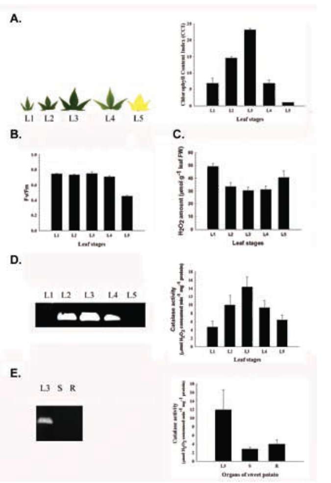
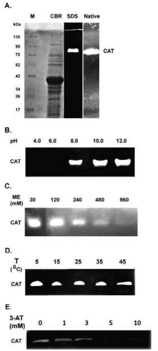
Figure 1. Detection and analysis of catalase activity from sweet potato mature L3 leaves. (A) Detection of catalase activity in native- and SDS-PAGE. CBR, Native and SDS denote Coo-massie Brilliant Blue R250 staining, native- and SDS-PAGE, respectively. M and CAT denote molecular mass and catalase, respectively; (B) Effect of pH on catalase activity; (C) Effect of p-mercaptoethanol on catalase activity; (D) Effect of temperature on catalase activity; (E) Effect of 3-amino-1,2,4-triazole on cata-lase activity. The ME, T, and 3-AT denote p-mercaptoethanol, temperature, and 3-amino-1,2,4-triazole, respectively. The ex­periments were performed three times and a representative one was shown.
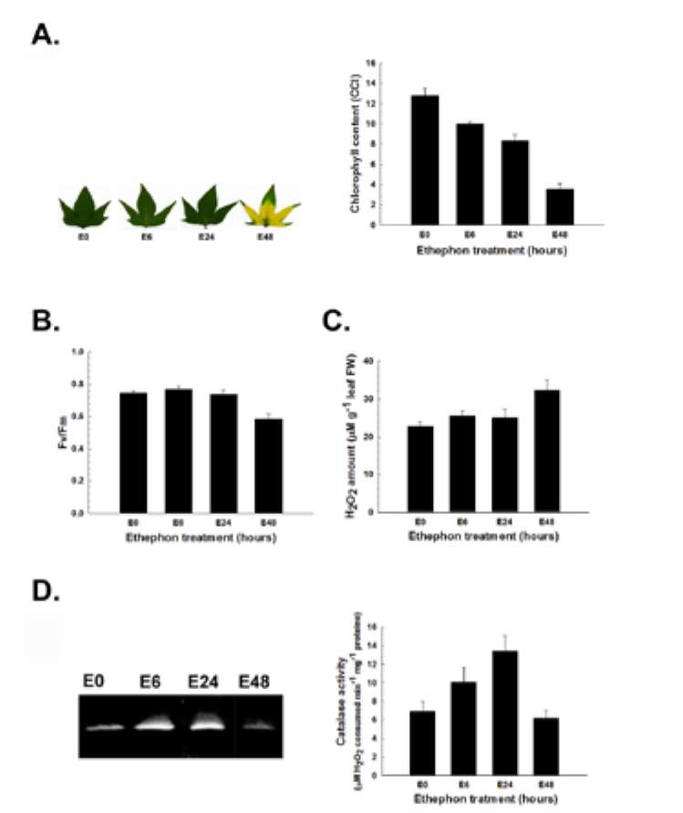
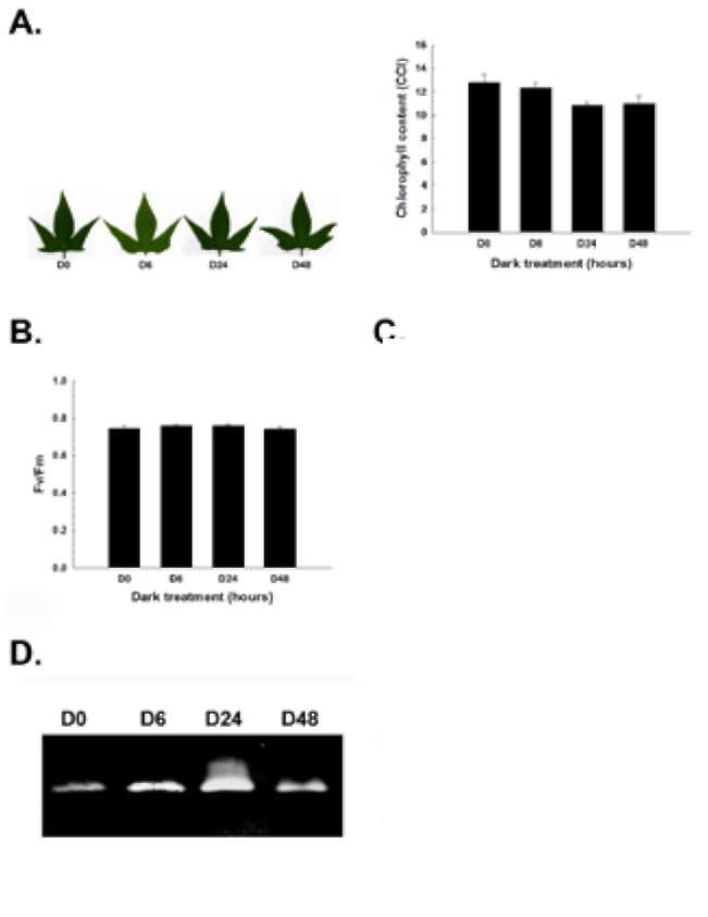
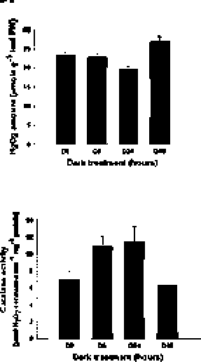
Figure 2. Temporal and spatial catalase expression in sweet potato. (A) Leaf morphology and chlorophyll content; (B) Photochemical Fv/Fm; (C) H2O2 amount; (D) Temporal cata-lase activity assay; (E) Spatial catalase activity assay. The leaf morphology, chlorophyll content, photochemical Fv/Fm, H2O2 amount, and in-gel and spectrophotometric catalase activity were analyzed in different leaf stages (L1, L2, L3, L4 and L5) and organs (mature L3 leaf, stem and root) of sweet potato. Stages of leaves were described in "Materials and Methods". L, S and R denote leaf, stem and root, respectively. The data were shown as mean ± S.E.. The experiments were performed at least three times and a representative one was shown.
422
Botanical Studies, Vol. 52, 2011
catalase activities also inversely correlated with the H2O2 amounts in leaves. These data suggest the possible physi­ological role of the leaf-type catalase in homeostasis regu­lation of reactive oxygen species H2O2 amounts in leaves.
The major leaf-type catalase activity was af­fected by darkness
Effects of darkness on leaf senescence, chlorophyll content, photochemical Fv/Fm, H2O2 amount, and catalase activity were studied. Darkness did not promote signifi­cant leaf senescence (Figure 3A), decrease in chlorophyll content (Figure 3A), or reduction of photochemical Fv/Fm (Figure 3B) within 48 h treatment. These data suggest that darkness may not be a key regulator in leaf senescence promotion. However, darkness did affect the H2O2 amount and the catalase activity in treated detached leaves. The H2O2 level decreased gradually from 0 h to 24 h, was the lowest at 24 h after treatment, then increased gradually un­til 48 h after the darkness treatment (Figure 3C). However, the leaf-type catalase activity increased gradually from 0 h to 24 h, was the highest at 24 h after treatment, then
Figure 4. Effects of ethephon on catalase activity. (A) Leaf mor­phology and chlorophyll content; (B) Photochemical Fv/Fm; (C) H2O2 amount; (D) Catalase activity assay. The leaf morphology, chlorophyll content, photochemical Fv/Fm, H2O2 amount and in-gel and spectrophotometric catalase activity were analyzed in mature L3 leaves. Detached leaves were treated with 1 mM ethephon for 0, 6, 24 and 48 h, respectively. E denotes ethephon treatment. The data were shown as mean ± S.E.. The experi­ments were performed at least three times and a representative one was shown.
decreased gradually until 48 h after the darkness treatment (Figure 3D). The H2O2 levels were inversely correlated with catalase activities in dark-treated detached leaves (Figure 3C and 3D). These data provide more evidence to support the possible physiological role of the leaf-type catalase in homeostasis regulation of H2O2 in leaves.
The major leaf-type catalase activity was af­fected by ethephon
Effects of ethephon on leaf senescence, chlorophyll content, photochemical Fv/Fm, H2O2 amount, and catalase activity were also studied. Ethephon did cause significant promotion of leaf senescence (Figure 4A), a decrease in chlorophyll content (Figure 4A), and reduction of pho­tochemical Fv/Fm (Figure 4B) at 48 h after treatment. These data are consistent with those showing the key role of ethylene in leaf senescence promotion. Ethephon also affected the H2O2 amount and catalase activity in treated detached leaves. Quantitatively, there was a slight increase
Figure 3. Effects of darkness on catalase activity. (A) Leaf mor­phology and chlorophyll content; (B) Photochemical Fv/Fm; (C) H2O2 amount; (D) Catalase activity assay. The leaf morphology, chlorophyll content, photochemical Fv/Fm, H2O2 amount and catalase activity were analyzed in mature L3 leaves. Detached leaves were treated with darkness for 0, 6 h, 24 h and 48 h, re­spectively. D denotes darkness treatment. The data were shown as mean ± S.E.. The experiments were performed at least three times and a representative one was shown.
CHEN et al. ― Sweet potato catalase
423
in the H2O2 level from 0 h to 24 h, but it was the highest at 48 h after treatment (Figure 4C). The leaf-type catalase ac­tivity, however, increased from 0 h to 24 h, then decreased gradually and was the lowest at 48 h after treatment (Fig­ure 4D). The H2O2 levels were approximately inversely correlated with catalase activities in ethephon-treated detached leaves (Figure 4C and 4D). These data provide more evidence to support the possible physiological role of the leaf-type catalase in homeostasis regulation of H2O2 in leaves.
not clear. In Mesembryanthemum crystallinum leaves, the active catalase dimer, with ca. 160 kDa molecular mass, can be separated under mild-denaturing conditions into three subunits with molecular masses of 79, 74 and 62 kDa using p-mercaptoethanol on SDS-PAGE. In samples incubated with 10% p-mercaptoethanol for 15 min prior to electrophoresis, the catalase activity was significantly re­pressed, compared to the untreated control in native PAGE, due to the reduction of active oligomeric and dimeric catalase forms into inactive monomeric catalase forms (Niewiadomska and Miszalski, 2008). Our data agree with these reports and suggest that the repression of sweet po­tato catalase activity by p-mercaptoethanol is likely due to a similar mechanism. Sweet potato catalase activity was also affected by pH. Its activity was significantly repressed at a pH lower than 8.0 (Figure 1B). The reasons are not clear. However, a mechanism similar to p-mercaptoethanol effect, that renders the catalase to a less or inactive mono-meric form (Niewiadomska and Miszalski, 2008), is sug­gested. It is possible that most of the catalase was reduced to a less or inactive form at a pH lower than 8. At a pH higher than 8, most of the catalase oxidized into an active form.
There was one catalase activity band detected in sweet potato leaves but not in stem or root tissues (Figure 2D and E). It is not clear whether there is only one catalase isoform in sweet potato leaves. However, a catalase pro­tein band was detected with SDS-PAGE from a storage root microbody after partial purification with ammonium sulfate fractionation and Sepharose 6B column chroma-tography from a sweet potato storage root (Esaka and Asahi, 1982). Sakajo and Asahi (1986) also detected one catalase protein band on SDS-PAGE with an antibody against sweet potato catalase from an in vitro translation product of storage root mRNA using a wheat germ system. In tobacco, low-peroxidatic (LP-CAT) isoforms consist of CAT-1 and CAT-2 catalases, and enhanced-peroxidatic (EP-CAT) isoforms composed of CAT-3 catalase. Dis­tribution of the LP-CAT and EP-CAT catalase isoforms were studied with antibodies raised specifically against LP-CAT and EP-CAT, respectively. Total catalase activity was about three-fold higher in leaves than in green stems. In leaves, ca. 85-90% of the total LP-CAT and EP-CAT activity was of a minor form. Whereas in the stem, EP-CAT was the predominant form and LP-CAT was less than 15% of the total activity (Havir et al., 1996). These reports suggest that different tissues contain different predominant catalase isoforms. Our data are consistent with these re-ports and suggest that the identified sweet potato catalase is a predominant leaf-type isoform.
In Arabidopsis leaves, CAT2 is the major catalase iso-form. Its activity increased and reached its maximum in mature leaves, then gradually decreased after vegetative to reproductive phase transition and senescence of the plant (Zimmermann et al., 2006). CAT3 and CAT1 activ­ity levels were much lower than those of CAT2, and were detected and steadily increased during leaf senescence
DISCUSSION
A major leaf-type catalase was identified in sweet po­tato and its activity was significantly repressed by catalase inhibitor 3-amino-1,2,4-triazole at exogenous concentra­tions 5 and 10 mM (Figure 1A and E). In tobacco, activi­ties of different catalase isoforms (CAT-1, CAT-2, and CAT-3) were also inhibited by 3-amino-1,2,4-triazole at concentrations between 0.03 to 20 mM. It took between 2.5 to 5 min for 50% inhibition of CAT-1 and CAT-3 at 5 mM and 10 mM 3-amino-1,2,4-triazole (Havir, 1992). Our data agree with the report and suggest that the detected ac­tivity band was a sweet potato catalase.
The estimated molecular mass of sweet potato catalase detected in leaves with SDS-PAGE was close to 72 kDa (Figure 1A). However, the catalase protein band reported by Sakajo and Asahi (1986), with an antibody against sweet potato catalase from in vitro translation product of storage root mRNA using a wheat germ in vitro translation system, had a molecular mass between 43 kDa and 67 kDa on SDS-PAGE. The reason for the difference are not clear. In Phaseolus vulgaris nodules and Medicago sativa leaves, partially-purified catalases from immobilized metal ion affinity chromatography had respective molecular masses of 42 kDa and 46 kDa on SDS-PAGE. Native forms on sephacryl S-300 columns however, exhibited molecular masses of 59 and 48 kDa for Phaseolus vulgaris, and 88 and 53 kDa for Medicago sativa (Tejera Garcia et al., 2007). In Mesembryanthemum crystallinum, the active catalase isolated from leaves had a high molecular mass of about 320 kDa on native PAGE, and was broken down into the less active, putative dimer of 160 kDa molecular mass on SDS-PAGE. Three subunits with respective mo-lecular masses of 79, 74 and 62 kDa were resolved after p-mercaptoethanol treatment under mild-denaturing condi-tions on SDS-PAGE (Niewiadomska and Miszalski, 2008). The existence of different tissue-specific catalase isoforms may provide one of the possible explanations. Post-trans-lational modification and assay method variations, such as mixing the sample with SDS sample buffer and keeping it at 4°C overnight without boiling at 95°C, before loading it into SDS-PAGE gel for an activity assay in our system, may provide other possibilities.
Sweet potato catalase activity was significantly re­pressed by exogenous β-mercaptoethanol at concentra­tions higher than 120 mM (Figure 1C). The reasons are
424
Botanical Studies, Vol. 52, 2011
(Zimmermann et al., 2006). CAT-1 was predominant in to­bacco, detectable just 8 days post-germination and steadily increased to its maximum at 21 days post-germination (Havir and McHale, 1987; Havir et al., 1996). Tobacco CAT-1 exhibited higher thermal stability and was more sensitive to 3-amino-1,2,4-triazole inhibition (Havir and McHale, 1987; Havir, 1992). Our data agree with these reports and suggest that the sweet potato major leaf-type catalase exhibits biochemical and physiological properties similar to Arabidopsis CAT2 and tobacco CAT-1.
The relationship between catalase activity and H2O2 amount was also studied, and an inverse correlation was observed during leaf development (Figure 2A, C and D). Although the catalase activities were much lower in fold­ing, un-opened immature L1 leaves and completely yellow senescent L5 leaves, their H2O2 amounts were significantly higher than those of the other leaf stages. In Arabidop-sis, the major catalase CAT2 activity also increased and reached its maximum in mature leaves, then gradually decreased after vegetative to reproductive phase transition and senescence of the plant. Changes in H2O2 amounts also increased significantly during vegetative to reproduc­tive phase transition and senescence of the plant (Zimmer-mann et al., 2006). Our data are consistent with the report and suggest the role of sweet potato leaf-type catalase in homeostasis regulation of reactive oxygen species H2O2. Therefore, when sweet potato leaf-type catalase activ­ity decreased, the H2O2 amounts increased, which in turn promoted leaf senescence. In Arabidopsis, a catalase 2 knock-out mutant (cat2) caused severe decrease of rosette leaf biomass, perturbed intracellular redox homeostasis, and activated oxidative signaling pathways (Queval et al., 2007). Transgenic tobacco plants ectopically expressing CAT-1 antisense constructs also displayed severely re­duced catalase activity and developed chlorosis and necro­sis on some of the lower leaves (Takahashi et al., 1997). These data provide more evidence to support the role of catalases in the regulation of plant cellular homeostasis of reactive oxygen species H2O2, which in turn affect plant physiological and developmental processes, including leaf senescence. Whether sweet potato leaf-type catalase plays a role similar to Arabidopsis CAT2 and tobacco CAT-1 awaits further investigation.
How sweet potato leaf-type catalase was induced by darkness and ethephon treatments (Figures 3 and 4) is not clear. However, an ethephon- and dark-inducible full-length cDNA encoding putative catalase (GenBank acces­sion no. GU230147) has been recently cloned from sweet potato leaves in our laboratory (unpublished data). These results support the inducibility of sweet potato leaf-type catalase by ethephon and darkness. When the leaf-type catalase activity decreased drastically, the H2O2 levels in leaves 48 hours after darkness or ethephon treatments in­creased significantly (Figures 3 and 4). These data provide further evidence to support the role of leaf-type catalase in the assistance of homeostasis of reactive oxygen spe­cies H2O2 in sweet potato leaves. Based on these data, we
conclude for the first time that a major leaf-type catalase isoform is detected and identified in a gel activity stain­ing assay. Its activity is maximal in mature leaves, and decreases significantly in natural and induced senescent leaves. A possible physiological role and function of the major leaf-type catalase in the assistance of homeostasis of reactive oxygen species H2O2 in sweet potato leaves is also suggested.
Acknowledgment. The authors thank the financial sup­port (NSC97-2313-B-110-001-MY3) from the National Science Council and National Sun Yat-sen University, Tai­wan.
LITERATURE CITED
Aebi, H. 1984. Catalase in vitro. Methods Enzymol. 105: 121­126.
Bradford, M.M. 1976. A rapid and sensitive method for the quantification of microgram quantities of protein utilizing the principle of protein-dye binding. Anal. Biochem. 72:
248-254.
Chandlee, J.M. and J.C. Scandalios. 1983. Gene expression dur­ing early kernel development in Zea mays. Dev. Genet. 4:
99-115.
Chen, H丄,Y.J. Tsai, W.S. Chen, G.J. Huang, S.S. Huang, and Y.H. Lin. 2010. Ethephon-mediated effects on leaf senes­cence are affected by reduced glutathione and EGTA in sweet potato detached leaves. Bot. Stud. 51: 171-181.
Contento, A.L. and D.C. Bassham. 2010. Increase in catalase-3 activity as a response to use of alternative catabolic sub­strates during sucrose starvation. Plant Physiol. Biochem. (On line in advance).
Du, Y.Y., P.-C. Wang, J. Chen, and C.-P. Song. 2008. Compre­hensive functional analysis of the catalase gene family in Arabidopsis thaliana. J. Integr. Plant Biol. 50: 1318-1326.
Eising, R., R.N. Trelease, and W. Ni. 1990. Biogenesis of cata-lase in glyoxysomes and leaf-type peroxisomes of sunflower cotyledons. Arch. Biochem. Biophys. 278: 258-264.
Esaka, M. and T. Asahi. 1982. Purification and properties of catalase from sweet potato root microbodies. Plant Cell
Physiol. 23: 315-322.
Frugoli, J.A., H.H. Zhong, M.L. Nuccio, P. McCourt, M.A. McPeek, T.L. Thomas, and C.R. McClung. 1996. Catalase
is encoded by a multigene family in Arabidopsis thaliana (L.) Heynh. Plant Physiol. 112: 327-336.
Guan, L. and J.G. Scandalios. 1995. Developmentally related re­sponses of maize catalase genes to salicylic acid. Proc. Natl. Acad. Sci. USA 92: 5930-5934.
Hattori, T., T. Nakagawa, M. Maeshima, K. Nakamura, and T. Asahi. 1985. Molecular cloning and nucleotide sequence of cDNA for sporamin, the major soluble protein of sweet potato tuberous roots. Plant Mol. Biol. 5: 313-320.
Havir, E.A. 1992. The in vivo and in vitro Inhibition of Catalase from Leaves of Nicotiana sylvestris by 3-Amino-1,2,4-
CHEN et al. ― Sweet potato catalase
425
Triazole. Plant Physiol. 99: 533-537.
Havir, E.A. and N.A. McHale. 1987. Biochemical and devel­opmental characterization of multiple forms of catalase in tobacco leaves. Plant Physiol. 84: 450-455.
Havir, E.A., L.F. Brissonf, and I. Zelitch. 1996. Distribution of catalase isoforms in Nicotiana tabacum. Phytochemistry 41:
699-702.
Hung, K.T. and C.H. Kao. 2004a. Nitric oxide acts as antioxi-dant and delays methyl jasmonate-induced senescence of rice leaves. J. Plant Physiol. 161: 43-52.
Hung, K.T. and C.H. Kao. 2004b. Hydrogen peroxide is neces­sary for abscisic acid-induced senescence of rice leaves. J. Plant Physiol. 161: 1347-1357.
Ni, W., R.N. Trelease, and R. Eising. 1990. Two temporally syn­thesized charge subunits interact to form the five isoforms of cottonseed (Gossypium hirsutum) catalase. Biochem. J. 269: 233-238.
Kuzniak, E., J. Patykowsky, and H. Urbanek. 1999. Involvement of the antioxidative system in tobacco response to fusaric acid treatment. J. Phytopathol. 147: 385-390.
Lim, P.O., H.J. Kim, and H.G. Nam. 2007. Leaf senescence.
Annu. Rev. Plant Biol. 58: 115-136.
Mhamdi, A., G. Queval, S. Chaouch, S. Vanderauwera, F. Van Breusegem, and G. Noctor. 2010. Catalase function in plants: a focus on Arabidopsis mutants as stress-mimic models. J. Exp. Bot. (on line in advance)
Niewiadomska, E., L. Polzienb, C. Deselb, P. Rozpadeka, Z. Miszalskia, and K. Krupinskab. 2009. Spatial patterns of se­nescence and development-dependent distribution of reac­tive oxygen species in tobacco (Nicotiana tabacum) leaves. J. Plant Physiol. 166: 1057-1068.
Niewiadomska, E. and Z. Miszalski. 2008. Partial characteriza-tion and expression of leaf catalase in the CAM-inducible halophyte Mesembryanthemum crystallinum L. Plant Physi-ol. Biochem. 46: 421-427.
Orendi, G., P. Zimmermann, C. Baar, and U. Zentgraf. 2001. Loss of stress-induced expression of catalase3 during leaf senescence in Arabidopsis thaliana is restricted to oxidative
stress. Plant Sci. 161: 301-314.
Orozco-Cardenas, M. and C.A. Ryan. 1999. Hydrogen peroxide is generated systemically in plant leaves by wounding and systemin via the octadecanoid pathway. Proc. Natl. Acad.
Sci. USA 96: 6553-6557.
Pastori, C.M. and L.A. de1 Rio. 1997. Natural senescence of pea leaves - an activated oxygen-mediated function for peroxi-
somes. Plant Physiol. 113: 411-418.
Queval, G., E. Issakidis-Bourguet, F.A. Hoeberichts, M. Van-dorpe, B. Gakiere, H. Vanacker, M. Miginiac-Maslow, F. Van Breusegem, and G. Noctor. 2007. Conditional oxida-tive stress responses in the Arabidopsis photorespiratory mutant cat2 demonstrate that redox state is a key modulator of daylength-dependent gene expression, and define photo-period as a crucial factor in the regulation of H2O2-induced
cell death. Plant J. 52: 640-657.
Sakajo, S. and T. Asahi. 1986. In vitro synthesis of catalase protein in sweet potato root microbodies. FEBS Lett. 205:
337-340.
Sakajo, S., K. Nakamura, and T. Asahi. 1987a. Molecular clon­ing and nucleotide sequence of full-length cDNA for sweet potato catalase mRNA. Eur. J. Biochem. 165: 437-442.
Sakajo, S., K. Nakamura, and T. Asahi. 1987b. Increase in cata-lase mRNA in wounded sweet potato tuberous root tissue.
Plant Cell Physiol. 28: 919-924.
Skadsen, R.W., P. Schulze-Lefert, and J.M. Herbst. 1995. Mo­lecular cloning, characterization and expression analysis of two catalase isozyme genes in barley. Plant Mol. Biol. 29:1005-1014.
Takahashi, H., Z. Chen, D. He, Y. Liu, and D.F. Klessig. 1997.
Development of necrosis and activation of disease resis­tance in transgenic tobacco plants with severely reduced catalase levels. Plant J. 11: 993-1005.
Tejera Garcia, N.A., C. Iribarne, F. Palma, and C. Lluch. 2007. Inhibition of the catalase activity from Phaseolus vulgaris and Medicago sativa by sodium chloride. Plant Physiol.
Biochem. 45: 535-541.
Wang, K.L.C., H. Li, and J.R. Ecker. 2002. Ethylene Biosynthe­sis and Signaling Networks. Plant Cell 14: S131-S151.
Yang, T.H., Y.C. Tsai, C.T. Hseu, H.S. Ko, S.W. Chen, and
R.Q. Blackwell. 1975. Protein content and its amino acid distri­bution of locally produced rice and sweet potato in Taiwan. J. Chin. Agri. Chem. Soc. 13: 132-138.
Yoshida, S. 2003. Molecular regulation of leaf senescence. Curr.
Opin. Plant Biol. 6: 79-84.
Zimmermann, P., C. Heinlein, G. Orendi, and U. Zentgraf. 2006. Senescence-specific regulation of catalases in Arabidopsis thaliana (L.) Heynh. Plant Cell Envir. 29: 1049-1060.
426
Botanical Studies, Vol. 52, 2011
定性分析一個甘藷葉型的過氧化氫酶
陳顯榮1  Mufidah AFIYANTI1  黃冠中2  黃世勳1  林耀輝3
1中山大學生物科學系
2中國醫藥大學中國藥學研究所
3中央研究院植物暨微生物研究所
本硏究利用SDS-PAGE膠體活性染色方法從甘藷完全展開的成熟葉片中辨識出一個分子量接近72
kDa
的主要過氧化氫酶。其活性於pH8-12間較pH小於8時爲高,且受p-mercaptoethanol3-amino-
1,2,4-triazole所抑制。然而其活性於5-45°C間受溫度的影響較小。組織專一性表現顯示其活性主要於葉
中被偵測到,且活性由未完全成熟的L2發育階段葉片開始增加,至完全展開成熟的L3發育階段葉片
時具有最高的活性,然後於部份黃化的L4發育階段的老化葉片梢微減少,至完全黃化的L5發育階段
的老化葉片幾乎偵測不到,類似於未展開未完全成熟的L1發育階段葉片一樣具有最低的活性。葉片中
過氧化氫酶的活性與H2O2含量適成反比關聯性。黑暗及ethephon (釋放乙烯的化合物)處理624
時亦會誘導增加此過氧化氫酶的活性,於處理2448小時後此過氧化氫酶的活性又漸漸減少;處理葉
片中過氧化氫酶的活性亦與H2O2含量適成反比關聯性。依據這些實驗數據我們結論甘藷葉片具有一個
主要的過氧化氫酶,其活性於成熟葉片時爲最高,於自然及誘導的老化葉片則顯著下降,此過氧化氫酶
的活性與葉片清除活化氧族H2O2可能有關。
關鍵詞:3-胺基-1,2,4-三氮唑;過氧化氫酶;Ethephon ;葉片老化;甘藷。