Botanical Studies (2012) 53: 243-254.
MORPHOLOGY
Stipules and colleters of the mangrove Rhizophoraceae: morphology, structure and comparative significance
Chiou-Rong SHEUE1,*, Ying-Ju CHEN1, and Yuen-Po YANG2,3
1Department of Life Sciences, National Chung Hsing University, 250, Kuo Kuang Rd., Taichung 402, Taiwan
2Department of Biological Sciences, National Sun Yat-sen University, Kaohsiung 804, Taiwan
3Department of Bioresources, Dayeh University, 168 University Rd., Dacun, Changhua 515, Taiwan
(Received October 11, 2010; Accepted December 1, 2011)
ABSTRACT. One characteristic possessed by the entire mangrove Rhizophoraceae is the strong enclosure of its young shoots by conspicuous rounded or flattened stipules. Young leaves are conspicuously immersed in resinous to milky exudates from colleters (multicellular external secretory emergences) located at the adaxial stipule bases. We systematically studied and compared morphological and structural features of stipules and their colleters from 18 taxa of the mangrove Rhizophoraceae. Three types of sclereid idioblasts (Ceriops and Rhizophora), collenchymas, and thick cuticles, were found to provide a structural basis for the mechanical support of stipules. Several to hundreds (35-580) of finger-like colleters aggregate into genus-specific shapes: rectangular to trapezoidal (Bruguiera), triangular (Ceriops and Kandelia), or as a band (Rhizophora). Number of rows, total number of colleters per stipule, and their individual size, also vary by taxon, and have taxo-nomic value. All colleters of this subfamily are considered anatomically 'standard', regardless of whether they appear as short-stalked rods (Bruguiera), long-stalked rods (Ceriops, Kandelia) or acuminate rods (Rhizophora, no stalk). Colleters in these taxa are comprised of a central axis of slender, elongated cells and an outer pali­sade-like epidermis, with a secretory function, perpendicular to the axis. Based on stipule and colleter charac­teristics, Ceriops and Kandelia are closely related, but Rhizophora shows more derived features. The structural and mechanical protection provided by stipules and colleter exudates may help shield the young shoots of these mangrove plants from their harsh environments.
Keywords: Anatomy; Bruguiera; Ceriops; Colleter; Kandelia; Mangroves; Rhizophora; Sclereid; Standard type; Stipule.
INTRODUCTION
the naked eye at the adaxial base of stipules in the species of mangrove Rhizophoraceae (Tomlinson, 1986; Sheue et al., 2003a, b; Sheue et al., 2005). Except for the genus Bruguiera, colleters, although few in number, are also present within the bracteoles of mangrove Rhizophoraceae inflorescences (Sheue, 2003; Sheue et al., 2003a).
Rhizophoraceae is well known as the richest mangrove family, having four exclusively mangrove genera (Bru-guiera, Ceriops, Kandelia and Rhizophora) (Tomlinson, 1986). About 21 mangrove species of this family are currently recognized, including new taxa from Kandelia (Sheue et al., 2003b) and Ceriops (Sheue et al., 2009a, b; Sheue et al., 2010). The family is diversified both ecologi-cally (habitat) and morphologically (trait). In addition to the mangroves mentioned above, the family has 15 inland genera with 135 species occurring in inland forests (Jun-cosa and Tomlinson, 1988a). All members of the Rhizo-phoraceae s. s. (excluding Anisophyllea) have large and conspicuous interpetiolar, glabrous and caducous stipules on both vegetative and reproductive shoots, strongly ensheathing the young leaves and inflorescences (Hou, 1958). Several to hundreds of aggregated finger-like col-leters mixed with milky mucilage can be observed with
Colleters are external secretory structures (Fahn, 1990), characterized by the production of a viscous substance that covers, protects from desiccation, and lubricates the bud and young leaves of plants (Kronestedt-Robards and Robards, 1991; Evert, 2007). Hou (1958) mentioned that colleters in Rhizophoraceae were first observed by Al-ston in African Cassipourea, and later by Metcalfe and Chalk in Carallia (both inland genera). These glandular emergences inside the adaxial regions of stipules appear to occur in all Malaysian genera, both mangrove and in-land (Hou, 1858). Colleter exudates from this family are assumed to be composed of phenolic compounds (Rosh-china and Roshchina, 1993).

*Corresponding author: E-mail: crsheue@nchu.edu.tw; Tel/ Fax: +886-4-22857395.
Colleters have long been associated with a number of flowering plants, notably Apocynaceae and Rubiaceae. The color and consistency of colleter exudates are variable in
244
Botanical Studies, Vol. 53, 2012
a somewhat diagnostic manner from species to species of the mangrove Rhizophoraceae (Tomlinson, 1986). How­ever, the knowledge of colleters of Rhizophoraceae is still under-reported and remains obscure. The presence of col-leters in R. mangle was noticed by Warming in 1883 and briefly described (Gill and Tomlinson, 1969): "a feature of some physiological significance in the bud of Rhizophora is a double row of yellow, glandular scales on the side of each stipule. The primodia enclosed by the stipules are always bathed by a viscous fluid which seems to originate from these glands." It is the only species in this family to have the colleters anatomically studied (Lersten and Cur­tis, 1974). The main objectives of the present work are to provide a systematic study of stipules and colleters of the mangrove species of this family in order to understand their morphology, structure, taxonomic value, and com­parative significance.
MATERIALS AND METHODS
Plant materials containing stipules with colleters were collected from 18 mangrove Rhizophoraceae taxa (includ­ing two hybrids of Rhizophora) from Asia and Australia from 1999 to 2009 (Table 1). The voucher specimens were deposited in the Herbarium of National Chung Hsing Uni­versity (TCB), Taichung, Taiwan.
Three individual plants were sampled from a popula­tion, and from each individual, three stipules were care­fully detached from the shoot apices when the young leaves were about to emerge from the stipules. Stipules were stored in 70% ethanol in the field, observed and pho­tographed with a Zeiss dissecting microscope to determine stipule morphology, number of colleters, and their forms of aggregation within the stipule. A portion of the materi­als was observed with a S-2400 Scanning Electron Micro-
Table 1. Taxa and collection information of the mangrove species of Rhizophoraceae in this study.

Species

Collection locality

Habitat

Bruguiera cylindrica (L.) Blume

Pichavarum, Tamil Nadu, India

Back or middle mangroves
Sungei Buloh Wetland Reservoir, Singapore
Bruguiera exaristata Ding Hou
Cairns, Queensland, Australia
Back or middle mangroves
Bruguiera hainesii C. G. Rogers
Pulau Ubin, Singapore
Within mangroves
Bruguiera gymnorhiza (L.) Savigny
Sungei Buloh Wetland Reservoir, Singapore
Middle mangroves
Cardwell, Queensland, Australia
Bruguiera parviflora (Roxb.) W. & A. ex
Pasir Ris Nature Park, Singapore
Back or middle mangroves
Griff.
Cardwell, Queensland, Australia
Bruguiera sexangula (Lour.) Poir.
Benut, Johor, Malaysia
Middle mangroves
Ceriops australis (C. T. White) Ballment, T.
Cardwell, Queensland, Australia
Back mangroves, landward
J. Sm. & J. A. Stoddart
Moreton Bay, Queensland, Australia
Ceriops decandra (Griff.) Ding Hou
Pichavarum, Tamil Nadu, India
Back mangroves
Ceriops pseudodecandra Sheue, H. Y. Liu,
Darwin, Northern Territory, Australia
Middle mangroves, along river bank
C. C. Tsai and Yuen P. Yang
Cairns, Queesland, Australia
Back mangroves
Ceriops tagal (Perr.) C. B. Rob.
Sungei Buloh Wetland Reservoir, Singapore
Within mangroves
Cairns/ Cardwell, Queensland, Australia
Back mangroves
Ceriops zippeliana Blume
Pasir Ris Nature Park, Singapore
Back mangroves
Kandelia candel (L.) Druce
Benut, Johor, Malaysia Malaysia
Along river bank
Kandelia obovata Sheue, H. Y. Liu & J. W.
Tanshui, Taipei, Taiwan
Along river bank, pure stand
H. Yong
Tongshi, Chaiyi, Taiwan
River bank
Rhizophora apiculata Blume
Sungei Buloh Wetland Reservoir, Singapore
Seaward mangroves
Pichavarum, Tamil Nadu, India
Cairns/ Cardwell, Queensland, Australia
Rhizophora x annamalayana Kathiresan
Pichavarum, Tamil Nadu, India
Back mangroves
Rhizophora x lamarckii Montr.
Cairns/ cardwell, Queensland, Australia
Back mangroves
Rhizophora mucronata Lam.
Pichavarum, Tamil Nadu, India
Seaward mangroves
Sungei Buloh Wetland Reservoir, Singapore
Rhizophora stylosa Griff.
Tainan, Taiwan
Along salty canal
Cairns/ Moreton Bay, Queensland, Australia
Seaward mangroves

SHEUE et al. ― Stipules and colleters of the mangrove Rhizophoraceae
245
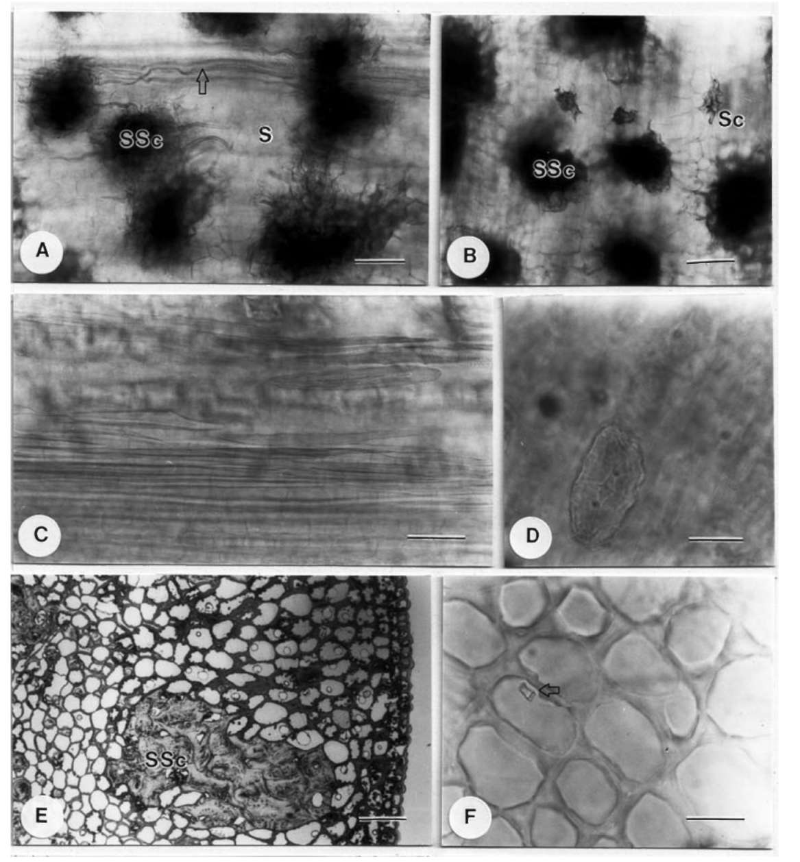
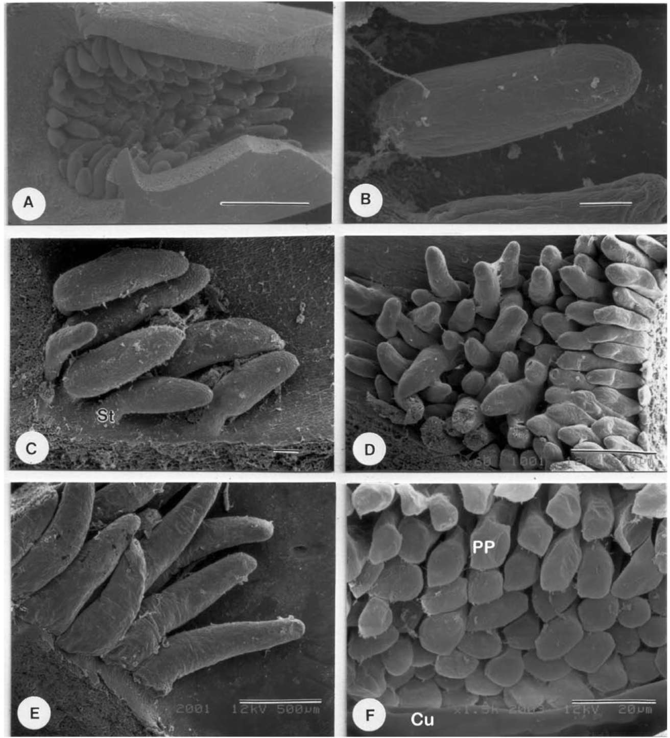
scope (Hitachi, Tokyo, Japan), after treating with ethanol series dehydration, critical point drying and gold coat­ing, allowing shape and size determination in particular. These stipules were also treated with the clearing method (Chiang, 1990) to investigate features such as sclereid id-iobalsts and crystals.
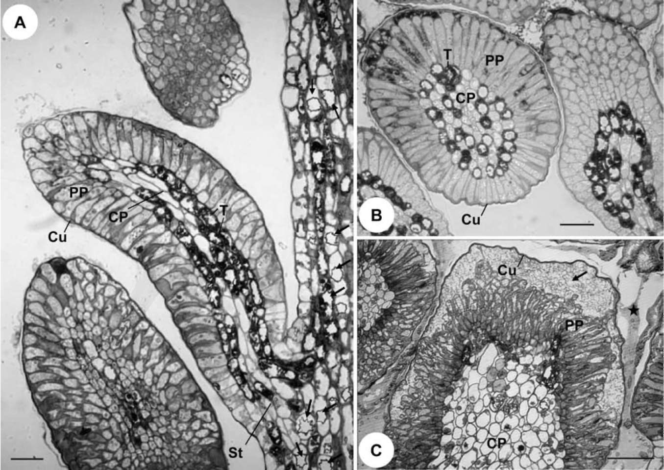
For field fixation for anatomical study, the base of each stipule with colleters was cut into several small pieces and put into 1.25-1.5% glutaraldehyde in 0.1 M phosphate buf­fer with 5% sucrose. Materials were stored in this fixative for 5-7 days during collection abroad. For the second fixa­tion, materials were subsequently transferred to 1% OsO4 in 0.1 M phosphate buffer for about 4 hours. After dehy­dration through an ethanol series, materials were infiltrated for 3 days and embedded within Spurr's-resin. The embed­ded materials were polymerized in an oven at 70°C for 12 hours. Semi-thin sections (1 μm) were made by an Ul-tracut E (Leica, Wetzlar, Germany) or a MTX Microtome (RMC, Tucson, USA), stained with 0.1% Toluidine blue for 1 minute or so, and then observed and photographed with a BH-2 Light Microscope (Olympus, Tokyo, Japan).
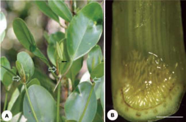
Figure 1. The mangrove Rhizophoraceae with shoots and stipules. (A) The shoot apices and top nodes of Ceriops australis with stipules (broad arrows), which are interpetiolar and cadu­cous. Primodia enclosed by the stipules are always bathed by a viscous fluid. Young expanded leaves coated with resin-like mu­cilage (thin arrow); (B) A fresh stipule of Bruguiera exaristata showing yellowish aggregated finger-like colleters with viscous exudation at the adaxial base. Scale bar = 1 mm.
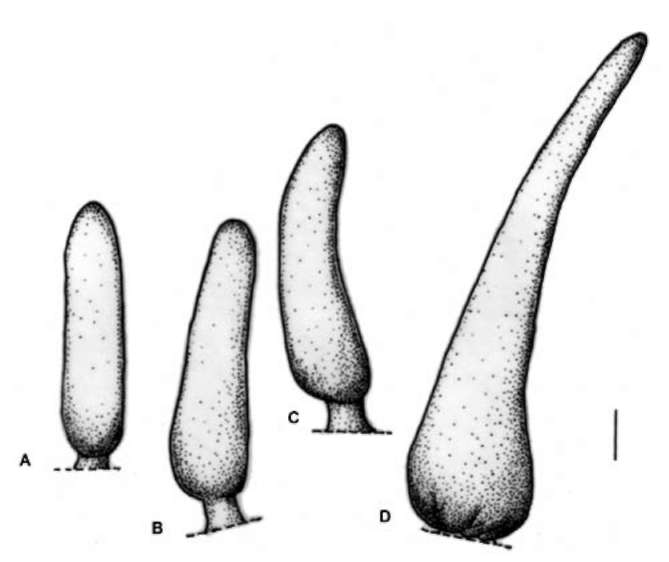
Table 2. Morphological and structural characters of stipules and colleters of the mangrove Rhizophoraceae, including aggregated form, number of rows and total number of colleters in each stipule, and individual shape and color of colleters.

Taxon

Stipule

Colleter


Morphology/ length (cm)/ Sc

Aggregated form/ row no./ total no.

Shape/ color
Bruguiera cylindrica
Round/ 3.0-4.0/ no Sc
1/ 4-7/ 70-90
SRs/ MW
Bruguiera exaristata
Round/ 2.0-3.0/ no Sc
/ 9-14/ 120-135
SRs/ MW-MY
Bruguiera hainesii
Round/ 3.5-4.2/ no Sc
/ 12-15/ 100-146
SRs/ MW
Bruguiera gymnorhiza
Round/ 4.0-5.0/ no Sc
1/ 9-13/ 200-220
SRs/ MW
Bruguiera parviflora
Round/ 4.5-6.5/ no Sc
國晨/ 6-8/ 160-170
SRs/ MW
Bruguiera sexangula
Round/ 4.5-5.5/ no Sc
•/ 3-4 / 35-40
SRs/ MW
Ceriops australis
Flattened/ 1.0-1.2/ Sc 2 types
▲ /9-11/ 90-140
SRl/ MW
Ceriops decandra
Flattened/ 1.2-2.4/ Sc 2 types
▲ /7-8/ 50-70
SRl/ MW
Ceriops pseudodecandra
Flattened/ 2.0-3.0/ Sc 2 types
▲ /8-12/ 80-100
SRl/ MW
Ceriops tagal
Flattened/ 1.5-2.8/ Sc 2 types
▲ /24-26/ 165-205
SRl/ MW
Ceriops zippeliana
Flattened/ 2.5-3.6/ Sc 2 types
▲ / 18-20/ 280-310
SRl/ MW
Kandelia candel
Flattened/ 3.0-4.0/ no Sc
▲ /6-7/ 75-96
SRl/ MY
Kandelia obovata
Flattened/ 2.5-3.2/ no Sc
▲ /8-9/ 95-110
SRl/ MY
Rhizophora apiculata
Round/ 5.0-8.5/ Sc 3 types
―/ 2-4/ 160-200
ARse/ Y
Rhizophora mucronata
Round/ 4.5-6.5/ Sc 3 types
―/ 6-9/ 460-580
ARse/ MY
Rhizophora stylosa
Round/ 4.5-5.5/ Sc 3 types
―/ 4- 6/ 180-300
ARse/ MY
Rhizophora x annamalayana
Round/ 4.5-6.0/ Sc 3 types
―/6-7/130-160
ARse/ MY
Rhizophora x lamarckii
Round/ 4.5-5.5/ Sc 3 types
― / 2-3/ 75-90
ARse/ MY

Symbols: aggregated form: broad band —narrow band —rectangle I; trapezoid A; semicircle triangle Abbreviations: ARse-Sessile acuminate rod; MW-milky white; MY-milky yellow; Y-yellow; Sc: sclereid idioblast; SRl-long stalked rod; SRs-short stalked rod.
246
Botanical Studies, Vol. 53, 2012
Figure 2. Morphological features of stipules and colleters in mangrove Rhizophoraceae. Abbreviations: C: colleter, E: exudates, L: leaf, S: stipule. (A) A shoot of Rhizophora apiculata with stipules, annular stipule scars and leaf scars; (B-C) Cross-section of a Ce-riops zippeliana shoot showing a pair of clasping stipules appearing as flattened type, with thickest part in the folding region and its thickness gradually thinning out toward its margin and apex; (C) A position closer to the base of the stipule with colleters. The con­volute young leaves, colleters and their exudates appear between the space of stipules; (D) Cross-section of a R. apiculata shoot with a pair of clasping stipules appearing as rounded type; (E-F) A Bruguiera gymnorhiza stipule with aggregated colleters forming a rect­angle at the adaxial base. Removing some stipule tissues reveals colleters in (F); (G) Aggregated colleters in B. cylindrica. Note that colleters are less compact at the distal end; (H) A broad band of aggregated colleters in R. mucronata; (I) Part of a colleter aggregation in R. apiculata. Scale bars: (A) and (D) = 5 mm; (B), (C), (E), (F), (G) and (H) = 1 mm; (I) = 500 μm.
SHEUE et al. ― Stipules and colleters of the mangrove Rhizophoraceae
247
RESULTS
(Bruguiera and Rhizophora) (Figure 2A, 2D) and flattened (Ceriops and Kandelia, Figure 2B-C). The adaxial base of each stipule contains a space filled with aggregated col-leters and a slightly sticky resin-like substance, which can be observed from detached stipules (Figures 1B, 2E-I) or stipule cross-sections (Figure 2B-C). Despite the morpho­logical difference, clasping stipule vernation is present in all species of the mangrove Rhizophoraceae, whereby one margin is free and is thickest in the middle region and gradually thins out towards its edge (Figure 2B-D).
Stipule morphology
All the members of mangrove Rhizophoraceae have distinct stipules paired outside young leaves (Figures 1A and 2A). Most of the stipules are green to yellow- green; however, some species (e.g. R. apiculata, R. stylosa, B. gymnorhiza) may have both red-purple and green stipules. The morphology of stipules is consistent within a genus, although size may vary interspecifically (Table 2). Two types of stipule morphology can be recognized: rounded
Figure 3. Anatomical features of mangrove Rhizophoraceae stipules. Abbreviations: S: stipule, Sc: small sclereid idioblast, SSc: strands of aggregated sclereid idioblast. (A-B) Sclereid idioblasts in Rhizophora apiculata, several strands of aggregated sclereid idio-blasts (multibranched) and elongated rod idioblasts (arrow) parallel to the long axis of the stipules in (A); three isolated small sclereid idioblasts and aggregated sclereid idioblasts in (B); (C-D) Sclereid idioblasts in Ceriops australis, elongated rod idioblasts in (C) and an isolated small sclereid idioblast in (D); (E) Cross-section of R. stylosa stipule showing a strand of aggregated sclereid idioblasts; (F) The collenchyma in the base part of a stipule in Bruguiera parviflora, with a prismatic crystal inside a cell (arrow). Scale bars: (A), (B) and (E) = 100μm (C), (D) and (F) = 30μm.
248
Botanical Studies, Vol. 53, 2012
Stipule structure
The clearing method and cross-sections reveal that two genera of the mangrove Rhizophoraceae, Ceriops and Rhizophora, have sclereid idioblasts inside the stipules. Rhizophora has three types of sclereid idioblasts: strands of aggregated sclereid idioblasts (Figure 3A-B), isolated small
idioblasts (Figure 3B) and elongated rod idioblasts, which are mostly parallel to the long axis of stipules (Figure 3A). In Ceriops, two kinds of sclereid idioblasts are observed: elongated rod idioblasts along the thickest mid region (Fig­ure 3C) and isolated small idioblasts (Figure 3D). In con­trast, Bruguiera and Kandelia have no sclereid idioblast in
Figure 4. SEM view of mangrove Rhizophoraceae colleters. Abbreviations: Cu: cuticle, PP: peripheral parenchyma, St: stalked rod colleter. (A- B) Bruguiera exaristata, colleters aggregate as a trapezoid form in (A) and an individual colleter appears as a short stalked rod in (B); (C) Ceriops decandra, with long stalked rod colleters; (D) Kandelia obovata, a part of the aggregated colleters in a trian­gular form; (E-F) Rhizophora apiculata; (E) Colleters appear as sessile acuminate rods; (F) A surface view of a colleter showing the detached cuticle and inner peripheral parenchyma cells. Scale bars: (A) = 1 mm; (B) and (C) = 100 μm; (D) and (E) = 500 μm; (F) = 20
SHEUE et al. ― Stipules and colleters of the mangrove Rhizophoraceae
249
their stipules, both instead possess collenchyma tissue near the abaxial side of their stipules (Figure 3F).
Figure 2H-I). Rhizophora apiculata colleters are bright yellow, but those of other taxa of this genus are milky white. This feature could distinguish R. apiculata from other species of Rhizophora in the field. In addition, row numbers of colleters differ between species of this genus. Rhizophora apiculata has just 2-3 rows of colleters, while R. stylosa has 4-6, and R. mucronata has 6-9 (460-580 col-leters).
Stipule structure among mangrove Rhizophoraceae species is similar, except for the presence of sclereid idio-blasts (Table 2, Figure 3E). Unlike in normal leaves, there is no differentiated palisade and spongy parenchyma in these stipules. A thick cuticle covers the epidermis of both sides. The ground tissue consists mostly of collenchyma, but parenchyma is found near the adaxial side. If pres­ent, sclereid idioblasts mostly occur within collenchyma. Stipule tissues of mangrove Rhizophoraceae are also abun­dant in tannins (Figure 6A) and form druse (Figure 6A), or occasionally prismatic (Figure 3F), calcium oxalate crys­tals.
Individual shape and size of colleter
Colleter attachment comes in both sessile and stalked forms in the mangrove Rhizophoraceae (Table 2; Figures 4-5). Only the genus Rhizophora has sessile colleters (Fig­ures 4E, 5D), while the colleters of Bruguiera, Ceriops and Kandelia have stalks near the base. Colleters of the species of Bruguiera appear as short stalked rods (stalk less than 30 fim long) with similar width from the base to the tip (Figures 4B, 5A). Colleters of Ceriops and Kande-lia have long stalked rods, with stalks about or longer than 100 μm in length (Table 3; Figures 4C, 5B-C). Colleters of Rhizophora are best described as sessile acuminate rods due to the uneven width (Figures 4E, 5D).
Colleter
Several to numerous milky-white to yellowish colleters of various shapes aggregated in multiple rows were found attached to the base of the adaxial side of the stipule (Fig­ure 1B, Table 2). These colleters remain attached to the stipule, but shrink and gradually lose their fresh color as the stipule drops from the parent tree. However, colleters may attach to the corresponding side of the stipule scar along with the basal residue of the stipule if the stipule is removed while it is still strongly enclosing a shoot. Obvi­ous secretions often glue the colleters together in all man­grove taxa observed in this study.
The size (most conveniently measured by length) of individual colleters may vary within the same aggregated group within a stipule. For example, smaller colleters are often observed near the base, but these smaller colleters are relatively few in number. There are larger numbers of larger similarly-sized colleters within a stipule. These colleters are the basis of the comparative measurements in this study. It is clear that the genus Rhizophora has the largest colleters in these four genera of mangrove Rhizo-phoraceae. The colleter size of six selected species of mangrove Rhizophoraceae is compared in Table 3 (stalks included if stalks present). The colleters of R. stylosa reach 1008.9 270 fim in length with a broad base (up to
Aggregated form, rows and numbers of col-leters
A mass of colleters occurring at the adaxial base of each stipule aggregates as a band, rectangle, trapezoid or triangle. It is clear that these aggregated forms are generi-cally consistent (Table 2).
In Bruguiera, multirow colleters aggregate as a rect­angle or trapezoid (Figure 4A), and colleters located at the distal end of a stipule may appear more or less loosely organized (Figure 2G). There are from 3-14 rows, and c. 40-210 colleters in each stipule of the various Bruguiera species (Table 2). It is apparent that row number and total colleter number are species specific, and this could be a di­agnostic character for species differentiation. In the genus Bruguiera, B. gymnorhiza has the greatest number of col-leters (9-12 rows, 200-210 colleters) (Figure 2E-F), while B. sexangula has the least (3-4 rows, 35-40 colleters) (Table 2), although their stipules are nearly the same size.
The stipules of Kandelia and Ceriops are flattened (Figure 2B-C) and both colleters are aggregated into a tri­angular shape (Figure 4D). Due to the tightly overlapping margins of the stipule, the colleters of these two genera are difficult to observe unless the stipule is cut into two halves. It is noteworthy that K. candel, with a larger stipule (3-4 cm long), has fewer colleters (75-96) than K. obovata, with a smaller stipule (2.5-3.2 cm long, 95-110 colleters) (Table 2).
Figure 5. Diagram of the individual shapes of colleters in man­grove Rhizophoraceae stipules. (A) Bruguiera, short stalked rod; (B) Ceriops, long stalked rod; (C) Kandelia, long stalked rod; (D) Rhizophora, sessile acuminate rod. Scale bar = 100 fim.
In the genus Rhizophora, colleters are aggregated either as narrow or broad bands, comprising 2-9 rows (Table 2;
250 Botanical Studies, Vol. 53, 2012
Figure 6. Anatomical structure of colleters in mangrove Rhizophoraceae stipules. Abbreviations: Cu: cuticle, CP: central core of parenchyma, PP: peripheral parenchyma, St: stalked rod colleter; T: Tanninferous cells. (A-B) Kandelia obovata; (C) Rhizophora stylosa. (A) A longitudinal section of a colleter showing the central core of elongated parenchyma, peripheral parenchyma and a stalk attached to a stipule. Abundant drused crystals (arrows) appear in the stipule tissue, but are absent from colleter cells. Tanninferous cells commonly occur in colleter and stipule core parenchyma, but not in the colleter epidermis; (B) Cross-section of a colleter showing central parenchyma surrounded by radically elongated peripheral epidermis; (C) A terminal part of a colleter showing an apparent space filled with exudates (arrow) between peripheral epidermis and the cuticle. Some exudates (starred) that drench young shoots are released after the rupture of the cuticle. All scale bars = 50 μm.
Table 3. Morphometric characters of colleters of six selected species of the mangrove Rhizophoraceae.

Species

N

Length1 (fm) (min) avg (max) std

Width (fm)
(min) avg (max) + std

Stalk length (fm)

Bruguiera exaristata

12

(238) 406.6 (540) 106.6

(119) 153.3 (200) + 27.7

Less than 30
Ceriops australis
10
(204) 371.2 (530) + 100.8
(82) 97.9 (102) 9.1
160 + 24
Ceriops decandra
10
(269) 516.0 (672) + 119.9
(77) 157.2 (211) + 46.1
92.7 + 5.8
Ceriops tagal
10
(510) 674.8 (856) 115.2
(102) 130.6 (142) 18.2
Not measured
Kandelia obovata
10
(275) 500.5 (680) + 129.2
(100) 155.8 (220) 39.2
119 23.9
Rhizophora stylosa
10
(713) 1008.9 (1490) + 270.0
Base2: 300.0 28.2
No stalk
Middle: 222.5 + 11.6
Tip: 100.1 18.1

1Stalks included if stalks present.
2Because colleter is an acuminate rod, widths were measured from the base, middle and tip.
SHEUE et al. ― Stipules and colleters of the mangrove Rhizophoraceae
251
300 fm) tapering to one-third of its basal width at the tip. Ceriops australis has the smallest colleters judged both by length (371.2 100.8 fim) and by width (97.9 9.1 im). In the genus Ceriops, colleter size varies in a gradation (e.g. C. tagal: 674.8 115.2 fim, C. decandra: 516.0 119.9 fim, and C. australis: 371.2 100.8 fim).
generic level in the mangrove Rhizophoraceae. The num­ber of colleter rows in an aggregation could have value at the specific level for most of these species. For example, the five taxa of Ceriops appear very similar in morphol­ogy (Sheue et al., 2009a, 2010). The characteristics of colleters can serve as valuable vegetative diagnostic traits for Ceriops materials in the absence of reproductive parts. This taxonomically valuable character has also been ap­plied to differentiate between species within Bruguiera (Sheue et al., 2005) and Kandelia (Sheue et al., 2003a, b). A hand lens (c. 10X) is recommended to observe this fea­ture in the field.
Structure of colleter
Despite the variation in stalk presence and the size of colleters, the anatomical structure of colleters of all taxa studied is remarkably similar. Viewed in transverse section, colleters consist of a rather massive core of elongated parenchymatous cells (c. 8-10 rows) with unlignified walls in the center (Figure 6), sheathed by many peripheral pa­renchyma (palisade-like epidermal cells) arranged in one layer perpendicular to the axis (Figures 4F; 6A-B). Usually, the cell walls of the core parenchyma are thicker than those of the palisade-like epidermis. Many of these core paren­chyma cells are tanniferous. Drused crystals rarely occur in the parenchyma of the core region, and not at all in the outer epidermal cells (Figure 6A). The outer thin-walled palisade-like epidermial cells are the secretory cells, with dense cytoplasm. The mucilage accumulates in the inter­cellular spaces of epidermal cells as well as between the cuticle and the secretory epidermal cells (Figure 6C).
The similarity in stipule and colleter morphology dem­onstrates a close relationship between Ceriops and Kan-delia that is in agreement with the molecular phylogeny (Setoguchi et al., 1999; Schwarzbach and Ricklefs, 2000). However, two types of sclereid idioblasts are present in Ceriops stipules, but are absent from those of Kandelia. No sclereid was found in Bruguiera or Kandelia stipules, but two and three sclereid types were found in Ceriops and Rhizophora stipules, respectively. This may imply that Ceriops and Rhizophora are more derived than the former two genera (Bruguiera and Kandelia), with Rhizophora being the most derived genus in this subfamily. This result is consistent with the phylogeny within this subfamily based on molecular (Setoguchi et al., 1999; Schwarzbach and Ricklefs, 2000) and morphological evidence (Juncosa and Tomlinson, 1988b).
DISCUSSION
All members of the mangrove Rhizophoraceae have conspicuous stipules enclosing young shoots. However, compared to leaf structure and function, stipules are much less studied and under-reported. In this study, we recog­nized three types of sclereid idioblasts in stipules of Ce-riops and Rhizophora genera. The strands of aggregated sclereid idioblasts and the isolated small idioblasts in Rhizophora are astrosclereids, as reported for the stipules of R. mucronata (Shah and Sundarraj, 1965). This is the first report of idioblasts in the Ceriops stipules. These iso­lated small idioblasts appear as short rods without branch arms. The elongated rod idioblasts are parallel to the long axis of Ceriops stipules, similar to the arrangement of elongated idioblasts along the leaf midrib in Rhizophora (Sheue, 2003). These idioblasts and collenchyma, along with vascular tissues and the thick cuticle of stipules, pro­vide strong mechanical support that presumably protects the shoot inside.
In this study, colleters of two reported Rhizophora hybrids were compared with colleters of their putative mother plants. As is usual, the morphological features of hybrids were consistently intermediate to their putative parents. Interestingly, only one hybrid showed an inter­mediate number of rows. Rhizophora x annamalayana Kathiresan (Kathiresan, 1995), was reported from southern India and was established as a hybrid of R. apiculata and R. mucronata (Parani et al., 1997). The hybrid from India, R. x annamalayana, has 5-6 rows of colleters, which ap­pear as an intermediate between its parents (2-4 rows and 6-9 rows ). In contrast, the hybrid, R. x lamarckii, reported as a hybrid of R. apiculata (2-4 rows) and R. stylosa (4-6 rows) (Duke and Bunt, 1979; Tomlinson, 1986), has 2-3 rows of colleters, which is closer to those of R. apiculata.
The 'standard' type colleter was first described by Ler-sten (1974a), which is one of the widespread colleter types in Rubiaceae. In addition, 'dendroid' and 'brush-like' types were described as Rubiaceous colleters (Lersten, 1974a, b). The members of Apocynaceae also have standard type colleters intermingled with petiolar hairs on the adaxial side of the petiole (Thomas and Dave, 1991). In this study, the colleters of all mangrove species of this family are anatomically standard, as Lersten and Curtis (1974) ob­served in R. mangle, and are remarkably similar to those found in several other families. However, various shapes of colleters were recognized in this study: short-stalked rods (Bruguiera), long-stalked rods (Ceriops, Kandelia), or sessile acuminate rods (Rhizophora). The stalk structure
Both rounded and flattened stipules were recognized in the mangrove Rhizophoraceae. Stipule morphology correlates with the shape of their contained colleter aggre­gations. Flattened stipules appear in the two mangrove gen­era, Ceriops and Kandelia. These stipules are thickest in the folding regions, and their thickness gradually thins out toward the margin and apex. As a consequence, a triangular space filled with colleters is formed in its adaxial base.
The work reported here suggests that the forms of col-leter aggregations, and the shapes of the individual col-leters in the stipules, could have taxonomic value at the
252
Botanical Studies, Vol. 53, 2012
of a colleter has seldom been reported, but Mandevilla is one example (Appezzato-da-Gloria and Estelita, 2000). The foliar and intrapetiolar colleters of Mandevilla can exhibit vascularization, but its interpetiolar colleters lack vascularization (Appezzato-da-Gloria and Estelita, 2000). Althought no vascularization similar to that observed for R. mangle (Lersten and Curtis, 1974) was found in the present study, we recently observed subtle vascularization in K.obovata by more sensitive methods (Sheue et al., in preparation).
characterize their harsh environment. Since the colleters of this family are still under-reported, it would be inter­esting to compare the differences between the inland and mangrove species with special reference to the ecological adaptations represented by stipule and colleter characters.
Acknowledgements. The authors thank G. T. Sundari, S. Das and G. Agoramoothy for helping collect mangroves in India, P. Saenger, G. Wightman, C. P. Mangion, and V Sarafis for helping collect materials in Australia, J. Y. W. Yong, S. K. Lee and M. A. Rashid for helping collecting materials in Singapore, and P. Chesson for improving the manuscript. This study was supported by National Science Council (NSC-97-2126-B-005-002-MY3) of The Republic of China (Taiwan). of China (Taiwan).
Lersten (1974a) found crystals in the colleters of the Rubiaceae. However, in the present study, an abundance of drused crystals was observed within the stipule tissues beneath the colleters, but rarely inside colleters. This dif­ference may imply differentiated cellular functions and metabolic activities between colleters and stipules. Sig­nificant spaces between the peripheral parenchyma and the cuticle containing a secretory product were commonly found in colleters of the mangrove Rhizophoraceae as ob­served in those of Gardenia (Mangalan et al., 1990). The secretory product is released after the rupture of the cuticle and drenches the shoot apex inside the stipule (Mangalan et al., 1990).
LITERATURE CITED
Appezzato-da-Gloria, B. and M.E.M. Estelita. 2000. Develop­ment, structure and distribution of colleters in Mandevilla illustris and M. velutina (Apocynaceae). Rev. Bras. Bot. 23: 113-120.
Chiang, S.H.T. 1990. Principles of Plant Microtechnique. Maw-Tsang, Taipei.
Duke, N.C. and J.S. Bunt. 1979. The genus Rhizophora (Rhizo-phoraceae) in north-eastern Australia. Aust. J. Bot. 27: 657­678.
Evert, R.F. 2007. Esau's Plant Anatomy: Meristems, Cells and Tissues of the Plant Body: Their Structure, Function. 3rd edn. John Wiley & Sons.
Fahn, A. 1990. Plant Anatomy. 4th ed. Pergamon Press.
Gill, A.M. and P.B. Tomlinson. 1969. Studies on the growth of red mangrove (Rhizophora mangle L.) I. Habit and general morphology. Biotropica 1: 1-9.
Hou, D. 1958. Rhizophoraceae. In C. G. G. J. van Steenis (ed.), Flora Malesiana, ser. 1, Vol. 5. Noordhoff- Kolff N. V., Dja­karta, pp. 429-473.
Juncosa, A.M. and P.B. Tomlinson. 1988a. A historical and taxo-nomic synopsis of Rhizophoraceae and Anisophylleaceae. Ann. Missouri Bot. Gard. 75: 1278-1295.
Juncosa, A.M. and P.B. Tomlinson. 1988b. Systematic compari­son and some biological characteristics of Rhizophoraceae and Anisophylleaceae. Ann. Missouri Bot. Gard. 75: 1296­1318.
Kathiresan, K. 1995. Rhizophora annamalayana: A new species of mangroves. Environ. Ecol. 13: 240-241.
Kronestedt-Robards, E. and A. Robards. 1991. Exocytosis in gland cells. In C. Hawea, J. Coleman, and D. Evans (eds.), Endocytosis, Exocytosis and Vesicle Traffic in Plants. Soc. Exp. Biol. Seminar Series 45: 199-232.
Lersten, N.R. and J.D. Curtis. 1974. Colleter anatomy in red mangrove, Rhizophora mangle (Rhizophoraceae). Can. J.
Bot. 52: 2277-2278.
Lersten, N.R. 1974a. Colleter morphology in Pavetta, Neorosea and Tricalysia (Rubiaceae) and its relationship to the bacte-
Rhizophora, a genus usually occurring at the front of mangroves facing the sea, has the largest colleters with an average length over 1 mm. The colleters of Ceriops, a back mangrove closer to land, are relatively smaller (c. 370 [im in C. australis). The prominent size and numerous colleters of Rhizophora (460-580 in R. mucronata) repre­sent the most developed colleters of this study. In addition, the sessile acuminate rod of colleters in Rhizophora is considered to be more derived in this family, since the col-leters of the other three genera are stalked.
Erythroxylaceae was recently confirmed as a sister fam­ily of Rhizophoraceae (Setoguchi et al., 1999; Schwarz-bach and Ricklefs, 2000). Thiebaut and Hoffmann (2005) described colleters in four genera of Erythroxylaceae, and compared to two genera of Rhizophoraceae. Colleters of Erythroxylaceae are of the same type as those of Rhizo-phoraceae, but position, arrangement, shape and size differ between all examined genera (Thiebaut and Hoffmann, 2005). In addition, the authors commented that the obvi­ous secretion gluing the colleters together occurs in the mangrove R. mucronata, but not in the inland member of the Rhizophoraceae, Cassipourea malosana (Thiebaut and Hoffmann, 2005). In terms of colleter number and size, it is evident that the mangrove Rhizophoraceae has more and larger colleters, as reported here, than those observed in the inland Rhizophoraceae, Cassipourea malosana (Thie-baut and Hoffmann, 2005), Carallia and Gynotroches (Sheue, 2003).
The combined evidence from morphological and ana­tomical features of these mangroves and observations in the field, suggest that the stipules provide a physical bar­rier, and the colleter secretions provide a viscous fluid bar­rier, that protect the young shoots from the strong wind, salt spray, tidal immersion and high temperatures that
SHEUE et al. ― Stipules and colleters of the mangrove Rhizophoraceae
253
rial leaf nodule symbiosis. Bot. J. Linn. Soc. 69: 125-136.
Lersten, N.R. 1974b. Morphology and distribution of colleters and crystals in relation to the taxonomy and bacterial leaf nodule symbiosis of Psychotria (Rubiaceae). Amer. J. Bot.
61: 973-981.
Mangalan, S., K.P. Kurien, P. John, and G.M. Nair. 1990. De­velopment, structure and cytochemistry of resin-secreting colleters of Gardenia gummifera (Rubiaceae). Ann. Bot. 66:123-132.
Parani, M., C.S. Rao, N. Mathan, C.S. Anuratha, K.K. Narayan­an, and A. Parida. 1997. Molecular phylogeny of man­groves: III. Parentage analysis of a Rhizophora hybrid using random amplified polymorphic DNA and restriction frag­ment length polymorphism markers. Aquat. Bot. 58: 165­172.
Roshchina, V.V. and V.D. Roshchina. 1993. The Excretory Func­tion of Higher Plants. Springer-Verlag, Berlin, Germany.
Schwarzbach, A.E. and R.E. Ricklefs. 2000. Systematic affinities of Rhizophoraceae and Anisophylleaceae, and intergeneric relationships within Rhizophoraceae, based on chloroplast DNA, nuclear ribosomal DNA, and morphology. Amer. J. Bot. 87: 547-564.
Setoguchi, H., K. Kosug, and H. Tobe. 1999. Molecular phylog-eny of Rhizophoraceae based on rbcL gene sequences. J. Plant Res. 112: 443-455.
Shah, J.J. and K.P. Sundarraj. 1965. Stipular sclereids in Rhizo-phora mucronata L. Curr. Sci. 5: 155-156.
Sheue, C.R. 2003. The comparative Morphology and Anatomy of the Eastern Mangrove Rhizophoraceae. Ph. D. Disserta­tion. National Sun Yat-sen University, Kaohsiung, Taiwan.
Sheue, C.R., H.Y. Liu, C.C. Tsai, S.M.A. Rashid, J.W.H. Yong,
and Y.P. Yang. 2009a. On the morphology and molecular basis of segregation of two species Ceriops zippeliana Blume and C. decandra (Griff.) Ding Hou (Rhizophoraceae) from southeastern Asia. Blumea 54: 220-227.
Sheue, C.R., H.Y. Liu, C.C. Tsai, and Y.P. Yang. 2010. Compari­son of Ceriops pseudodecanda sp. nov. (Rhizophoraceae), a new mangrove species in Australasia, with related species. Bot. Stud. 51: 237-248.
Sheue, C.R., H.Y. Liu, and Y.P. Yang. 2003a. Morphology on
stipules and leaves of the mangrove genus Kandelia (Rhizo-phoraceae). Taiwania 48: 248-253.
Sheue, C.R., H.Y. Liu, and J.W. H. Yong. 2003b. Kandelia obo-
vata (Rhizophoraceae), a new mangrove species from East­ern Asia. Taxon 52: 287-294.
Sheue, C.R., Y.P. Yang, H.Y. Liu, F.S. Chou, S.C. Chang, P.
Saenger, C.P. Mangion, G. Wightman, J.W.H. Yong, and C.C. Tsai. 2009b. Reevaluating the taxonomic status of Ce-riops australis (Rhizophoraceae) based on morphological and molecular evidence. Bot. Stud. 50: 89-100.
Sheue, C.R., J.W.H. Yong, and Y.P. Yang. 2005. The Bruguiera
(Rhizophoraceae) species in the mangroves of Singapore, especially on the new record and the rediscovery. Taiwania
50: 251-260.
Thiebaut, L. and P. Hoffmann. 2005. Occurrence of colleters in
Erythroxylaceae. Kew Bull. 60: 455-459.
Thomas, V. and Y. Dave. 1991. Comparative and phylogenetic significance of the colleters in the family Apocynaceae.
Feddes Repert. 102: 177-182.
Tomlinson, P.B. 1986. The Botony of Mangroves. Cambridge Univ. Press, Cambridge.
254
Botanical Studies, Vol. 53, 2012
紅樹科紅樹林植物的托葉與指狀腺體的形態與構造之比較研究
許秋容1 陳映如1 楊遠波2,3
1國立中興大學生命科學系
2國立中山大學生物科學系
3大葉大學生物資源學系
紅樹科紅樹林植物的枝稍具有圓形或扁平之托葉,緊密包圍著幼葉與頂芽。此托葉向軸面基部的指
狀腺體分泌出明顯可見之樹脂狀至乳狀物質浸潤幼芽。本文首次系統性地觀察及比較本科紅樹林植物的
18個分類群之托葉與指狀腺體的形態與構造。托葉內三種形態的厚壁異形細胞 (細蕊紅樹與紅樹屬;)、
發達的厚角組織或與表皮細胞外之厚角質層,提供了機械性支持的結構基礎。數十個至數百個(35-580)
似手指狀之指狀腺體聚集呈特定的形狀,具屬的專一性:紅茄苳屬呈長方形至梯形、細蕊紅樹屬和水筆
仔屬為三角形、紅樹屬則呈帶狀。各托葉內指狀腺體的排數、總數與指狀腺體的個別大小,在各分類
群並不相同,具分類價值。依據托葉和指狀腺體的特徵顯示細蕊紅樹屬和水筆仔屬的關係最接近,而紅
樹屬則可能呈現較後起的特徵。本群植物的指狀腺體儘管在形態上呈現短柄桿狀 (紅茄苳屬) ,長柄桿
狀 (細蕊紅樹屬和水筆仔屬)或無柄漸尖桿狀(紅樹屬),但其解剖構造均為標準型。此腺體的中心軸由
細長的薄壁細胞構成,其外圍似柵狀的薄壁表皮細胞具分泌功能,與中心薄壁細胞呈垂直排列。由托葉
所具的結構與機械性的保護功能,和其內指狀腺體的生理浸潤作用,可能對此紅樹林植物的頂芽提供了
重要的保護,以助其應付嚴酷的環境。
關鍵詞:解剖;紅茄苳屬;細蕊紅樹屬;指狀腺體;水筆仔屬;紅樹林;紅樹屬;標準型;厚壁細胞;
托葉。