Botanical Studies (2012) 53: 291-299.
MOLECULAR BIOLOGY
Protective function of the recombinant Oshsp18.0-CII protein, a class II small heat shock protein of rice, in Escherichia coli
Pi-Fang Linda CHANG1,2 *, Wen-Kuan HUANG1, Ying-Hong LIN1, Yuhsin CHEN3, and Tao-Ho CHANG1
1Department of Plant Pathology, National Chung Hsing University, Taichung City 40227, Taiwan, R. O. C.
2Agricultural Biotechnology Center, National Chung Hsing University, Taichung City 40227, Taiwan, R. O. C.
3Crop Improvement Division, Taichung District Agricultural Research and Extension Station, Council of Agriculture, Executive Yuan, Changhua County 51544, Taiwan, R. O. C.
(Received November 15, 2010; Accepted March 13, 2012)
ABSTRACT. All plants synthesize multiple families of small heat shock proteins (sHSP). A cDNA clone, Oshsp18.0-CII, encoding an 18.0 kDa class II sHSP was isolated from rice previously. The function of the rice class II sHSP was studied by overproduction of the Oshsp18.0-CII fusion protein in transformed Escheri-chia coli cells. The results suggest that heterologous expression of the Oshsp18.0-CII fusion protein increases thermotolerance of E. coli cells in vivo and provided thermoprotection to E. coli soluble proteins in vitro. The survival rate of Oshsp 18.0-CII fusion protein-accumulating cells treated at 50°C for 1 h was almost 1000-fold higher than that of the control cells transformed with pET32a expression vector. Overproduction of the Oshsp18.0-CII fusion protein in E. coli also confers tolerance of E. coli cells to ultraviolet (UV) irradiation. The post-UV survival of the Oshsp18.0-CII fusion protein-accumulating cells is about 7.29-fold over that of the control cells transformed with pET32a expression vector when exposed to 1700 [iJ of UV. There is almost no post-UV survival (^0.4%) in the untransformed cells after exposing to 900 [J UV light.
Keywords: Heat shock; Oryza sativa L.; Thermotolerance; Thermoprotection; Ultraviolet.
Abbreviations: ER, endoplasmic reticulum; HSP, heat shock protein (s); HRP, horse radish peroxidase; IPTG, isopropyl p-D-thiogalactopyranoside; LA, Luria-Bertani broth containing 100 [ig mL-1 of ampicillin; LB, Luria-Bertani broth; PBS, Phosphate-buffered Saline; PCR, polymerase chain reaction; sHSP, small heat shock protein (s); SDS-PAGE, sodium dodecyl sulfate polyacrylamide gel electrophoresis; UV, ultraviolet.
INTRODUCTION
ernyk, 1997), chaperonins (GroEL and HSP60) (Sigler et al., 1998; Krishna, 2003; Wang et al., 2004), and small molecular mass proteins (sHSP) ranging in size of ap­proximate 16-42 kDa (Scharf et al., 2001; Siddique et al., 2008).
Plant cells have evolved to cope with the heat stress in their environments. Under heat stress, plant may become tolerant to non-permissive high temperatures, as a result of the induction of heat shock proteins (HSP; Vierling, 1991). The roles of individual HSP in heat stress tolerance are re­portedly different among different organisms; they rely on different sets of HSP to establish tolerance to similar levels of heat stress (Parsell and Lindquist, 1993; Krishna, 2003).
The HSP families in plants are more complex than those in other organisms. Based on the molecular mass, HSP are grouped into five major families (Wang et al., 2004; Kotak et al., 2007) including the families of HSP100/Clp (Schirmer et al., 1996; Nieto-Sotelo et al., 1999), HSP90 (Mogelsvang and Simpson, 1998), HSP70/DnaK (Mi-
All plants synthesize multiple families of sHSP (Vier-ling, 1991). Under heat stress condition, the accumulation level of the sHSP in soybean could reach more than 1% of the total cellular proteins (Hsieh et al., 1992), and this ac­cumulation is correlated with the acquisition of thermotol-erance (Lin et al., 1997; Chang and Lin, 2000; Sun et al., 2002; Wang et al., 2003; 2004). Recently, the complexity of the heat stress responses in plants has been addressed (Kotak et al., 2007). Plant sHSP have been grouped further based on sequence homology, immunological cross-reac­tivity, and localization to different cellular compartments (Waters et al., 1996). There are three classes of cytosolic sHSP (class I, II, and III), as well as four distinct types of sHSP localized in the various organelles including en-doplasmic reticulum (ER), mitochondria, chloroplasts,

*Corresponding author: E-mail: pfchang@nchu.edu.tw; Tel: +886-4-2284-0780 ext. 335, 330; Fax: +886-4-2286-0442.
292
Botanical Studies, Vol. 53, 2012
and peroxisomes (Siddique et al., 2008). Other proteins containing a-crystallin domain (ACD proteins) were also identified (Scharf et al., 2001). Plant sHSP have been shown to be induced by several abiotic stresses, photope-riod, and certain developmental programs (Krishna et al., 1992; Sun and MacRae, 2005; Waters et al., 2008; Lin and Chang, 2009).
transcription factor HsfA2 (Port et al., 2004). Recently, one more class II sHSP gene, Oshsp19.0-CII, was identi­fied by Sarkar et al. (2009) according to the phylogenetic and in silico localization analysis of the 40 a-crystallin domain containing genes in rice genome, which were identified using the database search of rice genome for sequences with the presence of a-crystallin domain at the C-terminus. Based on the expressed sequence tags (EST), reverse transcription-polymerase chain reaction (PCR), and microarray results, expression profiles of Oshsp18.0-CII and Oshsp19.0-CII were different under various stress treatments (heat, salt, UV, and anoxia), and developmental programs such as pollen and anther development, em-bryogenesis, and seed maturation (Sarkar et al., 2009). It appears that, except for anoxia, Oshsp19.0-CII was not abiotic stress induced based on the stresses examined in­cluding heat and UV-C treatments (Sarkar et al., 2009).
To understand the possible structure-function rela­tionship of the rice class II sHSP, we introduced the Oshsp18.0-CII cDNA into E. coli and studied its thermo-tolerance in the transformed E. coli cells. In this study, het-erologous expression of a rice 18.0-kDa sHSP resulted in thermotolerance of E. coli cells to a lethal heat treatment in vivo and provided thermoprotection to functional pro­teins of E. coli in vitro. The effects of rice Oshsp18.0-CII protein in resistance to ultraviolet (UV) irradiation were also observed in E. coli cells.
In addition to heat tolerance, other functions of sHSP that protected plants under adverse environments were re­ported. Sun et al. (2001) reported that a class II sHSP, At-HSP17.6A, could increase salt and drought tolerance in Arabidopsis. Plant sHSP could increase tolerance to heavy metal (Heckathorn et al., 2004), oxidative stress (Banzet et al., 1998), and osmotic stress (Sun et al., 2001). Plant HSP (including sHSP) have been shown to posses chaperone function (Wang et al., 2004; Sun and MacRae, 2005), and could protect other cellular proteins to maintain correct conformation or translocation to other organelles under stresses. The sHSP may be activated and involved in plant growth and development, such as pollen differentiation, embryogenesis, seed germination, and fruit maturation (Sun et al., 2002).
Members of the sHSP family were shown to prevent ag­gregation of thermally inactivated reporter proteins in vitro (Horwitz, 1992; Jakob et al., 1993; Lee et al., 1995; 1997; Ehrnsperger et al., 1997; Krishna, 2003; Sun and MacRae, 2005). Direct evidence showing a molecular chaperone function of plant sHSP in vivo has also been reported. A dodecameric class II sHSP from pea, HSP17.7, prevented the aggregation of citrate synthase when heated to 45°C (Lee et al., 1995). Helm et al. (1997) found simultaneously expressing class I and class II sHSP could form distinct complexes in vivo, suggesting that they have subtly differ­ent functions. Basha et al. (2004) showed that the class II protein of wheat (TaHsp17.8C-II) comprises a smaller oli-gomer than the dodecameric TaHsp16.9C-I. This indicated that class II proteins may have a distinct mode of oligomer assembly as compared to the class I proteins. These au­thors also showed that heat-denaturing firefly luciferase was fully protected by TaHsp17.8C-II, but it did not inter­act significantly with TaHsp16.9C-I (Basha et al., 2004).
Before 2007, nine genes encoding class I sHSP have been characterized in rice (Guan et al., 2004) but only one class II sHSP gene, Oshsp18.0-CII, has been identified (Chang et al., 2007). The Oshsp18.0-CII gene is transcrip-tionally regulated by heat stress, but not by other environ­mental stresses such as cold, salt, and drought (Chang et al., 2007). Comparing to class I sHSP, fewer reports were focused on the function of class II sHSP in other plants, especially very few reports were for the class II sHSP of rice. The class II sHSP of pea (Lee et al., 1995; Basha et al., 2010), tomato (Low et al., 2000), wheat (Basha et al.,2004; 2010), and Arabidopsis (Basha et al., 2010) were shown to act as molecular chaperones. Overexpression of an Arabidopsis class II sHSP enhanced osmotolerance (Sun et al., 2001). Hsp17.4-CII of tomato acts as core-pressor and cytoplasmic retention factor of heat shock
MATERIALS AND METHODS
Preparation of the Oshsp18.0-CII gene expres­sion construct
The open reading frame of the Oshsp18.0-CII gene was amplified by PCR with primers covering both termini of the coding region of the Oshsp18.0-CII cDNA which en­codes the full-length polypeptide of 210 amino acids. For gene construction, a EcoRI site (single underlined) was introduced to the 5' primer (5'-CATGAATTCATGGA-GAGCGC-3') and aXhoI site (double underlined) to the 3' primer (5'-CATCTCGAGGATAAGCAGCAG-3'), respec­tively. Five ng of the Oshsp18.0-CII gene cloned in the pGEM®-T Easy Vector (Promega Co., Madison, WI, USA) was used as template for PCR reaction containing 10x PCR Reaction Buffer (100 mM Tris-HCl, pH 8.3; 500 mM KCl; 25 mM MgCl2; Roche Applied Science, Mannheim, Germany), 0.3 mM each of four dNTPs, 0.4 mM each of two primers, 2 units FastStart Taq DNA Polymerase (Roche Applied Science, Mannheim, Germany). The PCR program was 1 min at 94°C, 1 min at 38°C, 40 s at 72°C for 35 cycles followed by 10 min at 72°C. The PCR prod-uct was digested with EcoRI and XhoI restriction enzymes (New England Biolabs, Beverly, MA, USA), and ligated into the pET32a expression vector (Novagen, Madison, WI, USA) at the EcoRI and XhoI sites to produce the re-combinant plasmid, pETHSP. The nucleotide sequence of the cloned Oshsp18.0-CII gene was confirmed by sequenc國 ing. The E. coli strain BL21 (DE3) was transformed with
CHANG et al. ― Protective function of a rice small HSP
293
either the pET32a expression vector alone to produce the ET32A cells or with the recombinant plasmid (pETHSP) to produce the ETHSP cells.
the cell culture was transferred to 50°C. The cultures were taken at 0.5, 1, 1.5, 2, and 4 h after 50°C treatment, and serial dilutions of the cultures were plated in triplicate on LB-Amp agar medium. The plates were incubated over­night at 37°C prior to scoring numbers of colony forma­tion to determine the percentage of viable cells.
Expression of the Oshsp18.0-CII fusion protein in transformed E. coli
The ET32A and ETHSP cells were grown in Luria-Bertani (LB) broth (Tryptone 10 g, Yeast extract 5 g, NaCl 10 g per liter) containing 100 [ig mL-1 of ampicillin (LB-Amp) at 37°C overnight. The overnight cultures were di­luted 50-fold using fresh LB broth and incubated at 37°C until OD600 reached 0.5 to 0.8 (mid-log phase), then re-combinant protein expression was induced by the addition of isopropyl p-D-thiogalactopyranoside (IPTG) to a final concentration of 1 mM, followed by incubation at 37°C for 4 h. The bacterial cells were harvested by centrifuga-tion and disrupted by sonication at 7.5Hz for 15s each time for ten times and centrifuged at 24,000 xg for 10 min at 4°C. The supernatant containing the crude protein extracts were subjected to sodium dodecyl sulfate polyacrylamide gel electrophoresis (SDS-PAGE).
UV sensitivity of transformed E. coli cells
After IPTG induction, the BL21 (DE3), ET32A, and ETHSP cultures were first adjusted to the same concen-trations (OD600 = 1.0 to 1.5) and diluted 10-fold at each step. Diluted samples of 100 fil were spotted in triplicate onto the LB plates. The plates were subjected to 500, 900, 1300, and 1700 μJ UV (UV-C, 254 nm) treatments (UV Stratalinker 1800, Stratragene, La Jolla, CA, USA). The plates were then incubated overnight at 37°C prior to scor-ing colony formations to determine the percentage of vi-able cells.
Thermo-stability of proteins in the E. coli cell lysate
Protein samples were dissolved in sample buffer (50 mM Tris-HCl, 100 mM dithiothreitol, 2% SDS, 0.1% bromophenol blue, 10% glycerol), separated in a 12% polyacrylamide gel (acrylamide:Bis = 29:1) containing 0.1% SDS and electro-blotted onto a PROTRAN® nitrocel­lulose transfer membrane (Schleicher & Schuell, Einbeck, Germany) using a Bio-Rad transblot apparatus (Trans-Blot Transfer Medium, Bio-Rad, Hercules, CA, USA). The membrane was submerged in 3% non-fat powdered milk with gentle shaking for 1 h, and then incubated with either 1:2000 diluted monoclonal antibodies against the N-termi-nal histidine residues of the expression protein (Amersham Pharmacia Biotech, Buckinghamshire, England) or with 1:2000 diluted polyclonal antibodies against the pea 17.7 kDa class II HSP (Pshsp17.7) (provided by Dr. Elizabeth Vierling of the Department of Biochemistry, University of Arizona) in TTBS buffer (20 mM Tris-HCl, pH 7.5; 500 mM NaCl; and 0.05% Tween-20) containing 1% non-fat powdered milk for 2 h. After 3 rinses with TTBS buffer for 10 min each, the membranes were incubated with 1:3000 dilution of either donkey anti-mouse or donkey anti-rabbit antibodies conjugated with horseradish peroxidase (HRP) (Amersham Pharmacia Biotech, Buckinghamshire, Eng-land) for 1 h. The blot was then washed twice with TTBS buffer for 10 min each, and once with TBS (20 mM Tris-HCl, pH 7.5; and 500 mM NaCl) buffer for 10 mm. Signal detection was initiated by submerging the membranes in HRP color development solution (15 mg 4-chloro-1-naph-tol in 5 mL cold methanol; 15 [L cold 30% H2O2 in 20 mL TBS) for 45 min. The membrane was rinsed with distilled water to stop the reaction.
Following IPTG induction, the transformed E. coli cells were centrifuged at 3,800 xg (Beckman, Harbor Boule­vard, CA, USA) for 10 min at 4°C, and resuspended in Phosphate-buffered Saline (PBS) buffer (pH 7.4; 2.7 mM KCl, 2 mM KH2PO4, 10 mM NaH2PO4 150 mM NaCl). Cells were disrupted by passing through a French Pressure (Sim-AMINCO, Silver Spring, MD, USA) three times at 1,200 psi. The disrupted cells were spun down at 16,000 xg (Labnet International, Edison, NJ, USA) for 3 min at 28°C, and soluble proteins were quantified by Bio-Rad Protein Assays Kit (Bio-Rad, Hercules, CA, USA). The soluble protein concentration was adjusted to 1 mg/500 μL PBS buffer. The adjusted protein samples were heat treated individually at 50, 60, 70, 80, 90, and 100°C for 15 min. After heating, supernatants and precipitated pellets were separated by centrifugation at 16,000 x g for 10 min at 28°C, and their protein concentrations were determined as described above.
RESULTS
Induction of fusion protein in transformed E. coli cells
The rice Oshsp18.0-CII gene, encoding a class II sHSP, was introduced into E. coli BL21 (DE3) cells for its expression and function analysis in bacterial cells. The pET32a expression vector would produce a ~20 kDa pro­tein in bacteria. With the recombinant plasmid pETHSP, the bacterial cells would produce a ~38 kDa fused protein, which contained the Oshsp18.0-CII polypeptide sequence. It appeared that the proteins in these two vectors could be induced after one hour (h) incubation with 1 mM IPTG (Figure 1A). It also showed that the proteins could be in­duced by as low as 0.5 mM IPTG in bacterial cells (Figure 1B). After 4 h incubation, the accumulation of Oshsp18.0-CII fusion protein in the ETHSP cells was a little bit higher
Thermotolerance of transformed E. coli cells
The ET32A and ETHSP cells were used for the ther-motolerance assay. Cell cultures were grown in LB-Amp broth as mentioned above. After IPTG induction, 5 mL of
294
Botanical Studies, Vol. 53, 2012
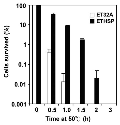
cells was 33%. The difference in survival rates at 50°C for 0.5 h was greater than 80 folds. Even after 1 h expo­sure to 50°C, the ETHSP cells still had a survival rate of about 10%, almost 1000-fold higher as compared with the ET32A cells, which had only 0.01% survival rate. Heating at 50°C for 1.5 h dropped the survival rate of the ETHSP cells to 1.8%, whereas no ET32A cells survived under the same condition. It was evident that 0.02% ETHSP cells could survive 2 h exposure to 50°C (Figure 4). It appeared that the transformed E. coli cells (ETHSP) producing the
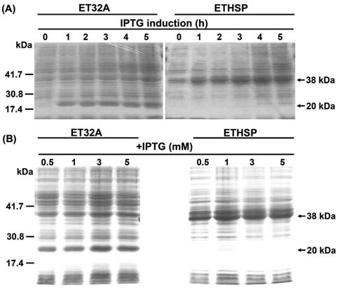
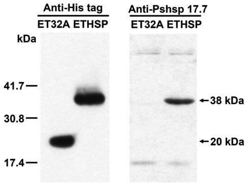
Figure 1. SDS-PAGE analysis of total proteins extracted from ET32A cells containing the pET32a expression vector and the ETHSP cells with the recombinant plasmid pETHSP overex-pressing recombinant Oshsp18.0-CII. A: Total proteins extracted from ET32A and ETHSP cells after 0, 1, 2, 3, 4, and 5 hour (h) 1 mM IPTG induction; B: Total proteins extracted from ET32A and ETHSP cells after 0.5, 1, 3, and 5 mM IPTG induction for 4 h. Ten [iL of protein samples were subjected to SDS-PAGE anal­ysis. The recombinant protein expressed in ET32A and ETHSP cells was ~20 kDa and ~38 kDa in size, respectively.
by 1 mM IPTG induction (Figure 1B). The western blot­ting analysis showed that the IPTG induced Oshsp18.0-CII fusion protein in the transformed ETHSP cells was histidine-tagged and recognized by the polyclonal antibod­ies against Pshsp17.7, the 17.7 kDa class II sHSP of pea (Figure 2).
Figure 2. Immunological gel blots of the recombinant proteins produced in ET32A and ETHSP cells. Monoclonal antibodies against histidine tag (Anti-His tag) and polyclonal antibodies against the 17.7 kDa class II HSP of pea (Anti-Pshsp 17.7) were used for blotting. The His-tagged recombinant Trx TagTM thi-oredoxin protein of ~20 kDa in ET32A and ETHSP cells and the His-tagged Oshsp18.0-CII fusion protein of ~38 kDa in ETHSP cells were detected after incubation 4 h with 1 mM IPTG.
Growth rates of the transformed E. coli cells
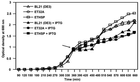
The ET32A and ETHSP cells had similar growth rates at 37°C when compared with the non-transformed E. coli BL21 (DE3) cells (Figure 3, open symbols), although slightly higher OD600 values were obtained in ETHSP cells after growth at 37°C for 480 min. After 1 mM IPTG in-duction, similar growth rates of the non-transformed BL21 (DE3), ET32A, and ETHSP cells were found (Figure 3, closed symbols). The growth rates of the above three cul-ture cells, however, were lower when subjected to 1 mM IPTG treatment than without IPTG. Overall, we observed no difference in the growth of these three E. coli cells at 37°C.
Thermotolerance of the E. coli cells containing the Oshsp18.0-CII fusion protein
Figure 3. Growth of wild-type and transformed E. coli BL21 (DE3) cells at 37°C with or without IPTG. Cultures of E. coli BL21 (DE3) cells (A and ▲), ET32A cells (O and •) carrying thepET32a vector, and ETHSP cells (口 and ) carrying the re­combinant pETHSP plasmid were grown at 37°C for 10 h. When OD600 reached to 0.8-1.0 (6 h after growth), half of the cultures were added with (closed symbols) or without (open symbols) IPTG to a final concentration of 1 mM, and subjected to further growth at 37°C for 4 h. Arrow indicates the addition of IPTG. Optical density at 600 nm was measured for cell growth.
To investigate the molecular chaperone function of Oshsp18.0-CII in vivo, the attenuation in lethality at 50°C was checked for the E. coli cells producing the re-combinant Oshsp18.0-CII protein. As shown in Figure 4, after exposing cells to 50°C for 0.5 h, the survival rate of ET32A cells dropped to 0.4%, whereas that of the ETHSP
CHANG et al. ― Protective function of a rice small HSP
295
Oshsp18.0-CII fusion protein was more thermotolerant than the cells harboring only the pET32a vector producing the recombinant Trx Tag™ thioredoxin protein.
Ultraviolet irradiation protection by rice Oshsp18.0-CII fusion protein in E. coli
Induction of the Oshsp18.0-CII fusion protein in the UV-sensitive E. coli strain BL21 (DE3) cells increased the survival of E. coli after UV irradiation (post-UV survival) (Figure 5). The data indicated that overproduction of Oshsp18.0-CII fusion protein protects ETHSP cells against UV irradiation at the dosages of 500 [J to 1700 [J. It is surprising that after IPTG induction, the post-UV survival of the ET32A strain harboring the control plasmid pET32a was higher than that of the plasmid-free non-transformed BL21 (DE3) cells. The increase in the post-UV survival of the ETHSP in comparison with other cells was observed when cells were subjected to 500 [J or higher UV irradia­tion. The significant increase in the post-UV survival of the ETHSP cells was from ~2.02-fold (at 500 [J UV) to ~7.29-fold (at 1700 [J) when compared with the ET32A cells. These results suggested that the Oshsp18.0-CII fu­sion protein may lead to UV-tolerance of E. coli and there­fore increased the post-UV survival.
Figure 5. Influence of Oshsp18.0-CII on cell viability of E. coli under UV irradiation. After 3 h of 1 mM IPTG induction, E. coli cells carrying pET32a vector (ET32A, spotted column), cells overproducing the Oshsp18.0-CII fusion protein (ETHSP, filled column), and non-transformed BL21 (DE3) cells (BL21 (DE3), open column) were diluted, plated on LA, and subjected to 0, 500, 900, 1300, and 1700 [J UV irradiation. Percentage of cells survived after UV irradiation was measured using the cell num­ber obtained at treatment of 0 [J as 100%. The percentage of the ET32A cells survived after 0, 500, 900, 1300, and 1700 [J UV irradiation was 100%, 33.5%, 10.4%, 4.6%, and 4.5%, respec­tively; that for BL21 (DE3) cells was 100%, 1.8%, 0.4%, 0.1%, and 0.1%, respectively; whereas that for ETHSP cells was 100%, 67.7%, 56.3%, 30.3%, and 32.8%, respectively. Values represent means SE (n = 3).
Thermo-stability of proteins prepared from the E. coli cells producing the Oshsp18.0-CII fusion protein
To understand if rice Oshsp18.0-CII could protect E. coli proteins from denaturation during heat stress, the pro­tein precipitation in the ET32A and ETHSP cell lysates were analyzed after heat treatments at 50, 60, 70, and 80°C for 15 min (Figure 6). The denatured proteins were precipitated by centrifugation after heat treatments. As the temperature was increased from 50°C to 80°C, an increase in the ratio of denatured proteins to soluble proteins in E. coli cell lysate was observed (Figure 6). The change of thermo-denatured proteins over the soluble proteins in ET32A cells after 15 min of 50°C, 60°C, 70°C, and 80°C treatments was 0.48, 1.45, 3.38, and 3.88-fold, respec-tively, whereas that in ETHSP cells was 0.4, 1.1, 2.17, and 2.73-fold, respectively. Nevertheless, in the lysate of ETH-SP cells, which produced Oshsp18.0-CII fusion protein, heat-denaturation of soluble proteins was less, especially at 70°C. At this temperature, some Oshsp18.0-CII fusion protein still remained in the soluble fraction without dena-turation, although at 80°C or higher temperature such as 100°C, most of the soluble proteins including Oshsp18.0-CII fusion protein were denatured as monitored by Anti-Pshsp 17.7 (data not shown).
Figure 4. Survival rates of the transformed E. coli ETHSP cells after heat treatments. After 4 h of 1 mM IPTG induction, the E. coli cells carrying pET32a expression vector (ET32A, open columns) and cells producing the Oshsp18.0-CII fusion pro­tein (ETHSP, filled columns) were heat-treated at 50°C for 0, 0.5, 1, 1.5, 2, and 3 h. Percentages of cells survived after 50°C treatment were measured using the cell numbers obtained at treatment of 0 h as 100%. The percentage of the ET32A cells survived after 0, 0.5, 1, 1.5, 2, and 3 h heat treatment was 100%, 0.4%, 0.01%, 0%, 0%, and 0%, respectively, while that of the ETHSP cells was 100%, 33%, 9.2%, 1.8%, 0.02%, and 0%, re­spectively. Values are means SE (n = 5).
DISCUSSION
For studying the function of HSP in vivo, null mutants are usually used and transformed with a specific HSP gene
296
Botanical Studies, Vol. 53, 2012
species for this protective function (Jinn et al., 1995). Transgenic expression of the rice sHSP17.7 (a class I sHSP) gene enhanced bacterial viability under heat stress, and recombinant sHSP17.7 protein prevented thermal ag­gregation of catalase in vitro (Murakami et al., 2004). It was demonstrated that the overexpression of a chloroplast small heat shock protein (Hsp21) in A. thaliana could enhance the tolerance against heat under high light condi­tions (Harndahl et al., 1999). Accumulation of sHSP in abundance could protect or repair plant cells from dam­ages caused by stresses (Krishna, 2003; Murakami et al., 2004). In this study, Oshsp18.0-CII was shown to provide thermotolerance and thermoprotection in E. coli. Whether Oshsp18.0-CII prevents thermal aggregation of other proteins or if it can form a protein complex with specific region important for binding to the substrate polypeptide warrant further investigation.
Reports have indicated that UV itself may induce the expression of specific HSP. HSP may provide an adaptive cellular response to tolerate the exposure of UV (Traut-inger et al., 1996). For two class II sHSP in rice, it was shown that Oshsp18.0-CII but not Oshsp19.0-CII was in-duced by UV (Sarkar et al., 2009). Therefore, the effect of Oshsp18.0-CII fusion protein on sensitivity to UV irradia-tion was observed in E. coli.
Figure 6. Thermo-denaturation of soluble proteins in cell lysate of transformed bacterial cells. The ET32A and ETHSP cells were subjected to 4 h of 1 mM IPTG induction for producing the cor­responding recombinant proteins. The cell lysate prepared from the IPTG-induced E. coli cells were heated for 15 min at 50, 60, 70, and 80°C. The denatured protein in pellet and in its corre­sponding supernatant was compared. Values represent means SE (n = 5).
Over-expression of a class I sHSP gene isolated from rice seedling, sHSP17.7, conferred heat tolerance in E. coli and both heat tolerance and UV-B resistance in rice plants (Murakami et al., 2004). Abundant accumulation of sHSP17.7 mRNA and protein also resulted in a significant increase in thermotolerance and UV-B resistance (302 nm, 3000 mJ cm-1) in transgenic rice plants (Murakami et al., 2004). Recently, Basha et al. (2010) described some mechanistic differences between class I and class II sHSP. Herein, the effect of a class II sHSP of rice, Oshsp18.0-CII, on decreasing sensitivity to UV irradiation in E. coli was demonstrated. It is also possible that the thioredoxin part of the fusion protein might play a role in the increase of tolerance against heat or UV. Thioredoxin is a small, ubiquitous protein which plays an important role in the antioxidant systems by repairing oxidative protein damage through the reversible oxidation of its active center dithiol to a disulfide (Seo and Lee, 2006). It was reported that thioredoxin is an essential protein in Bacillus subtilis. A variety of stresses, including heat, salt stress, and ethanol treatment, strongly enhanced the synthesis of thioredoxin in B. subtilis (Scharf et al., 1998). The increased levels of thioredoxin might help stressed B. subtilis cells to maintain the native and reduced state of cellular proteins (Scharf et al., 1998). Bone marrow cells from transgenic mice over-expressing human thioredoxin were more resistant to UV-C-induced cytocide compared with those from wild type C57BL/6 mice (Mitsui et al., 2002). In addition, LaVallie et al. (1993) reported that the thioredoxin tag in this fu-sion protein increased the intrinsic thermo-stability of the host cells. Therefore, by using another expression system without any polypeptide fused to the Oshsp18.0-CII may
to confirm the function of the transferred gene (Lee et al., 1994; Schirmer et al., 1994; Yeh et al., 1995; 1997). In order to study the function of the plant class II sHSP in vivo, which has not been studied before, a rice sHSP gene was transferred into E. coli that does not indigenously produce an equivalent HSP during heat stress, even though E. coli produces IbpA and IbpB (Vladimir et al. , 1995) which are not homologous to rice sHSP. In this study, the Oshsp18.0-CII cDNA, encoding an 18.0 kDa class II sHSP of rice, was introduced into the pET32a vector with 167 extra amino acids at the N-terminus. These 167 extra amino acids do not affect the function of the Oshsp18.0-CII protein, although the growth rate of E. coli after IPTG (1 mM) addition was slightly slower than the cells without IPTG. This may be due to the result of "metabolic bur-den" by channeling resources into the synthesis of a single foreign protein in E. coli (Dong et al., 1995; Hoffmann and Rinas, 2004). The function of plant class II sHSP in E. coli may be involved in providing thermotolerance by acting as chaperones similar to the class II sHSP from pea (HSP17.7; Lee et al., 1995) and TaHsp17.8C-II (Basha et al., 2004) in protecting E. coli proteins from heat denatur-ation.
Evidence is accumulating for the function of plant sHSP in heat-resistance. The rice class I sHSP (Oshsp16.9A) can protect E. coli proteins from heat denaturation and also in­crease thermotolerance of E. coli (Yeh et al., 1995; 1997), and the class I sHSP are exchangeable among different
CHANG et al. ― Protective function of a rice small HSP
297
provide further evidence for the role of Oshsp18.0-CII on UV tolerance and thermotolerance in E. coli. Neverthe­less, in the thioredoxin overproduced ET32A (vector only) cells, the UV protection was not as high as the ETHSP cells which overproduced thioredoxin-Oshsp18.0-CII fu­sion protein. This suggested that Oshsp18.0-CII did have some function on UV protection in E. coli. Actually, when thioredoxin gene in pETHSP was deleted from the ETHSP cells, the cells still showed higher UV tolerance than the control cells carrying the pET32a with thioredoxin gene deleted (Chang, unpublished results). Besides, when mea­suring the growth of the tested cell lines under heat condi­tion (50°C), the growth of ETHSP cells was much lower than that of the ET32A cells and the BL21 (DE3) control cells (Chang, unpublished results). In the future, transgenic plants overproducing the Oshsp18.0-CII may be needed for the in vivo study of its function. The correlation of sHSP and UV protection warrants further studies in both E. coli and plants.
and C.W. Wang. 2007. Induction of a cDNA clone from rice encoding a class II small heat shock protein by heat stress, mechanical injury, and salicylic acid. Plant Sci. 172: 64-75.
Dong, H., L. Nilsson, and C.G. Kurland. 1995. Gratuitous over-expression of genes in Escherichia coli leads to growth in­hibition and ribosome destruction. J. Bacteriol. 177: 1497­1504.
Ehrnsperger, M., S. Graber, M. Gaestel, and J. Buchner. 1997. Binding of non-native protein to HSP25 during heat shock creates a reservoir of folding intermediates for reactivation.
EMBO J. 16: 221-229.
Guan, J.C., T.L. Jinn, C.H. Yeh, S.P. Feng, Y.M. Chen, and C.Y Lin. 2004. Characterization of the genomic structures and selective expression profiles of nine class I small heat shock protein genes clustered on two chromosomes in rice (Oryza sativa L.). Plant Mol. Biol. 56: 795-809.
Harndahl, U., R.B. Hall, K.W. Osteryoung, E. Vierling, J.F. Bornman, and C. Sundby. 1999. The chloroplast small heat shock protein undergoes oxidation-dependent conforma-tional changes and may protect plants from oxidative stress. Cell Stress Chaperones 4: 129-138.
Heckathorn, S.A., J.K. Mueller, S. LaGuidice, B. Zhu, T. Barrett, B. Blair, and Y. Dong. 2004. Chloroplast small heat-shock proteins protect photosynthesis during heavy metal stress. Physiol. Biochem. 91: 1312-1318.
Helm, K.W., G.J. Lee, and E. Vierling. 1997. Expression and na­tive structure of cytosolic class II small heat-shock proteins. Plant Physiol. 114: 1477-1485.
Hoffmann, F. and U. Rinas. 2004. Stress induced by recombinant protein production in Escherichia coli. Adv. Biochem. Eng. Biotechnol. 89: 73-92.
Horwitz, J. 1992. a-Crystallin can function as a molecular chap-
erone. Proc. Natl. Acad. Sci. USA 89: 10449-10453. Hsieh, M.H., J.T. Chen, T.L. Jinn, Y.M. Chen, and C.Y. Lin.
1992. A class of soybean low molecular weight heat shock proteins: immunological study and quantitation. Plant Phy-siol. 99: 1279-1284.
Jakob, U., M. Gaestel, K. Engel, and J. Buchner. 1993. Small heat shock proteins are molecular chaperones. J. Biol. Chem. 268: 1517-1520.
Jinn, T.L., Y.M. Chen, and C.Y. Lin. 1995. Characterization and
physiological function of class I low-molecular-mass, heat shock protein complex in soybean. Plant Physiol. 108: 693­701.
Kotak, S., J. Larkindale, U. Lee, P. von Koskull-Doring, E. Vier-ling, and K.D. Scharf. 2007. Complexity of the heat stress response in plants. Curr. Opin. Plant Biol. 10: 310-316.
Krishna, P., R.F. Felsheim, J.C. Larkin, and A. Das. 1992. Struc­ture and light-induced expression of a small heat-shock pro­tein gene of Pharbitis nil. Plant Physiol. 100: 1772-1779.
Krishna, P. 2003. Plant responses to heat stress. Topics Curr.
Genet. 4: 73-101. LaVallie, E.R., E.A. DiBlasio, S. Kovacic, K.L. Grant, P.F.
Schendel, and J.M. McCoy. 1993. A thioredoxin gene fusion
Acknowledgements. We thank Dr. Chu-Yung Lin for his valuable suggestions and helpful discussion on the stress protection. We are grateful to Dr. Elizabeth Vierling for do­nating anti-PsHSP17.7, Drs. Peter P. Ueng and Chung-Jan Chang for English editing of the manuscripts, the technical help of Mr. Cheng-Wei Wang on the production of foreign protein in E. coli, and the assistance of Miss Ying-Ru Chen, Mrs. He-Wei Chen, Yi-Hung Lin, and Yung-Sheng Lin for preparation of this manuscript. We also thank the reviewers for their valuable suggestions to make this work to be presented in a much better way. This research was supported in part by the National Science Council, Taiwan, R.O.C. under grant numbers NSC89-2311-B-005-022 and NSC89-2313-B-005-159 to P.-F.L.C.; by the Ministry of Education, Taiwan, R.O.C. under the ATU plan; and also by National Chung Hsing University, Taiwan, R.O.C.
LITERATURE CITED
Banzet, N., C. Richaud, Y Deveaux, M. Kazmaier, J. Gagnon, and C. Triantaphylides. 1998. Accumulation of small heat shock proteins, including mitochondrial HSP22, induced by oxidative stress and adaptive response in tomato cells. Plant
J. 13: 519-527.
Basha, E., C. Jones, V Wysocki, and E. Vierling. 2010. Mecha­nistic differences between two conserved classes of small heat shock proteins found in the plant cytosol. J. Biol. Chem. 285: 11489-11497.
Basha, E., G.J. Lee, B. Demeler, and E. Vierling. 2004. Chap-erone activity of cytosolic small heat shock proteins from wheat. Eur. J. Biochem. 271: 1426-1436.
Chang, P.F.L. and C.Y. Lin. 2000. The discovery of the heat shock response in plants. In S.D. Kung and S.F. Yang (eds.), Discoveries in Plant Biology, Vol. 3. World Scientific Pub­lishing Co. Pte. Ltd., Singapore, pp. 347-370.
Chang, P.F.L., T.L. Jinnt, W.K. Huang, Y. Chen, H.M. Chang,
298
Botanical Studies, Vol. 53, 2012
expression system that circumvents inclusion body forma­tion in the E. coli cytoplasm. Nat. Biotechol. 11: 187-193.
Lee, G.J., A.M. Roseman, H.R. Saibil, and E. Vierling. 1997. A
small heat shock protein stably binds heat-denatured model substrates and can maintain a substrate in a folding compe­tent state. EMBO J. 16: 659-671.
Lee, G.J., N. Pokala, and E. Vierling. 1995. Structure and in vitro molecular chaperone activity of cytosolic small heat shock proteins from pea. J. Biol. Chem. 270: 10432-10438.
Lee, Y.R.J., R.T. Nagao, and J.L. Key. 1994. A soybean 101-kDa heat shock protein complements a yeast HSP104 deletion mutant in acquiring thermotolerance. Plant Cell 6: 1889­1897.
Lin, C.Y., C.H. Yeh, S.S. Tzeng, T.S. Tseng, F.C. Chang, Y.L.
Lee, T.L. Jinn, and P.F.L. Chang. 1997. Class I low-mo­lecular-weight heat-shock proteins in Oryza sativa. In J.C. Su (ed.), Proceedings of Fifth International Symposium on Rice Molecular Biology, Yi Hsien Publishing Co., Taipei, Taiwan, pp. 41-52.
Lin, W.C. and P.F.L. Chang. 2009. Approaches for acquired tol­erance to abiotic stress of economically important crops. In C.T. Hou and J.F. Shaw (eds.), Biocatalysis and Agricultural Biotechnology. CRC Press, Boca Raton, Florida, pp. 63-87.
Low, D., K. Brandle, L. Nover, and C. Forreiter. 2000. Cytosolic heat-stress proteins Hsp17.7 class I and Hsp17.3 class II of tomato act as molecular chaperones in vivo. Planta 211:575-582.
Miernyk, J.A. 1997. The 70 kDa stress-related proteins as mo­lecular chaperones. Trends Biochem. Sci. 2: 80-87.
Mitsui, A., J. Hamuro, H. Nakamura, N. Kondo, Y. Hirabayashi, S. Ishizaki-Koizumi, T. Hirakawa, T. Inoue, and J. Yodoi. 2002. Overexpression of human thioredoxin in transgenic mice controls oxidative stress and life span. Antioxid. Redox Signal. 4: 693-696.
Mogelsvang, S. and D.J. Simpson. 1998. Protein folding and transport from the endoplasmic reticulum to the Golgi ap­paratus in plants. J. Plant Physiol. 153: 1-15.
Murakami, T., S. Matsuba, H. Funatsuki, K. Kaeaguchi, H. Sar-uyama, M. Tanida, and Y. Sato. 2004. Over-expression of a small heat shock protein, sHSP17.7, confers both heat toler­ance and UV-B resistance to rice plants. Mol. Breed. 13:165-175.
Nieto-Sotelo, J., K.B. Kannan, L.M. Martinez, and C. Segal. 1999. Characterization of a maize heat-shock protein 101 gene, HSP101, encoding a ClpB/Hsp100 protein homo-
logue. Gene 230: 187-195.
Parsell, D.A. and S. Lindquist. 1993. The function of heat-shock proteins in stress tolerance: degradation and reactivation of damaged proteins. Annu. Rev. Genet. 27: 437-496.
Port, M., J. Tripp, D. Zielinski, C. Weber, D. Heerklotz, S. Winkelhaus, D. Bublak, and K.-D. Scharf. 2004. Role of
Hsp17.4-CII as coregulator and cytoplasmic retention fac­tor of tomato heat stress transcription factor HsfA2. Plant Physiol. 135: 1457-1470.
Sarkar, N.K., Y.K. Kim, and A. Grover. 2009. Rice sHsp genes: genomic organization and expression profiling under stress
and development. BMC Genomics 10: 393-410.
Scharf, C., S. Riethdorf, H. Ernst, S. Engelmann, U. Volker, and M. Hecker. 1998. Thioredoxin is an essential protein in­duced by multiple stresses in Bacillus subtilis. J. Bacteriol. 180: 1869-1877.
Scharf, K.D., M. Siddique, and E. Vierling. 2001. The expanding family of Arabidopsis thaliana small heat stress proteins and a new family of proteins containing a-crystallin do­mains (Acd proteins). Cell Stress Chaperones 6: 225-237.
Schirmer, E.C., J.R. Glover, M.A. Singer, and S. Lindquist. 1996. HSP100/Clp proteins: a common mechanism explains diverse functions. Trends Biochem. Sci. 21: 289-296.
Schirmer, E.C., S. Lindquist, and E. Vierling. 1994. An Arabi-dopsis heat shock protein complements a thermotolerace
defect in yeast. Plant Cell 6: 1899-1909.
Seo, H.J. and Y.N. Lee. 2006. Occurrence of thioredoxin re-ductase in Deinococcus species, the UV resistant bacteria. J. Microbiol. 44: 461-465.
Siddique, M., S. Gernhard, P. von Koskull-Doring, E. Vierling, and K.D. Scharf. 2008. The plant sHSP superfamily: five new members in Arabidopsis thaliana with unexpected properties. Cell Stress Chaperones 18: 183-197.
Sigler, P.B., Z. Xu, H.S. Rye, S.G. Burston, W.A. Fenton, and A.L. Horwich. 1998. Structure and function in GroEL-me-diated protein folding. Annu. Rev. Biochem. 67: 581-608.
Sun, W., C. Bernard, B. van de Cotte, M. van Montagu, and N. Verbruggen. 2001. At-HSP17.6A, encoding a small heat-shock protein in Arabidopsis, can enhance osmotolerance upon overexpression. Plant J. 27: 407-415.
Sun, W., M. van Montagu, and N. Verbruggen. 2002. Small heat shock proteins and stress tolerance in plants. Biochim. Bio-
phys. Acta. 1577: 1-9.
Sun, Y. and T.H. MacRae. 2005. Small heat shock proteins: mo­lecular structure and chaperone function. Cell. Mol. Life
Sci. 62: 2460-2476.
Trautinger, F., I. Kindas-Mugge, R.M. Knobler, and H. Honigs-mann. 1996. Stress proteins in the cellular response to ultra­violet radiation. J. Photochem. Photobiol. B. 35: 141-148.
Vierling, E. 1991. The roles of heat shock proteins in plants.
Annu. Rev. Plant Physiol. Plant Mol. Biol. 42: 579-620.
Vladimir, P.Z., A.Z. Galina, I.D. Alexander, G. Matthias, and K. Timo. 1995. Structural and functional homology between periplasmic bacterial molecular chaperones and small heat
shock proteins. FEMS Immunol. Med. Mic. 11: 265-272.
Wang, W., B. Vinocur, O. Shoseyov, and A. Altman. 2004. Role of plant heat-shock proteins and molecular chaperones in the abiotic stress response. Trends Plant Sci. 9: 244-252.
Wang, W., B. Vinocur, and A. Altman. 2003. Plant responses to drought, salinity and extreme temperatures: towards genetic engineering for stress tolerance. Planta 218: 1-14.
Waters, E.R., B.D. Aevermann, and Z. Sanders-Reed. 2008. Comparative analysis of the small heat shock proteins in
CHANG et al. ― Protective function of a rice small HSP
299
three angiosperm genomes identifies new subfamilies and reveals diverse evolutionary patterns. Cell Stress Chaper-
ones 13: 127-142.
Waters, E.R., G.J. Lee, and E. Vierling. 1996. Evolution, struc­ture and function of the small heat shock proteins in plants. J. Exp. Bot. 47: 325-338.
Yeh, C.H., K.W. Yeh, S.H. Wu, P.F.L. Chang, Y.M. Chen, and
C.Y. Lin. 1995. A recombinant rice 16.9-kDa heat shock
protein can provide thermoprotection in vitro. Plant Cell
Physiol. 36: 1341-1348. Yeh, C.H., P.F.L. Chang, K.W. Yeh, W.C. Lin, Y.M. Chen, and
C.Y. Lin. 1997. Expression of a gene encoding a 16.9-kDa heat-shock protein, Oshsp16.9, in Escherichia coli enhances
thermotolerance. Proc. Natl. Acad. Sci. USA 94: 10967­10972.
重組水稻第二族小分子量熱休克蛋白質Oshsp18.0-CII
大腸桿菌的保護功能
張碧芳1,2 黃文寬1 林盈宏1 陳裕星3 張道禾1
1國立中興大學植物病理學系
2國立中興大學農業生物科技發展中心
3行政院農委會台中區農業改良場
所有植物都會合成多種小分子量熱休克蛋白質。本研究在大腸桿菌(Escherichia coli)中大量表現
Oshsp18.0-CII 這個水稻18.0 kDa 第二族小分子量熱休克蛋白質的基因,以研究其功能。結果顯示除了
可因此提高大腸桿菌的耐熱性、且對大腸桿菌的水溶性蛋白質具有熱保護性之外,更可因此提高大腸桿
菌對紫外光的耐受性,其對50°C 一小時處理的耐熱性可比只轉入pET32a表現載體的對照組細胞增高
幾乎1000倍的存活率;而對1700 [J的紫外光照射的耐性約為只轉入pET32a表現載體之對照組細胞的
7.29倍。900 μJ的紫外光照射之後,未轉型的細胞幾乎無法存活,其存活率不高於0.4%
關鍵詞:熱休克;水稻;耐熱性;熱保護性;紫外光。