Bot. Bull. Acad. Sin. (1995) 36: 181-188

Hwang and Chen — Anatomical changes in Kandelia candel

Anatomical responses in Kandelia candel (L.) Druce seedlings growing in the presence of different concentrations of NaCl

Yuan-Hsun Hwang1 and Shuh-Chun Chen

Institute of Botany, Academia Sinica, Taipei 115, Taiwan, Republic of China

(Received January 24, 1995; Accepted June 7, 1995)

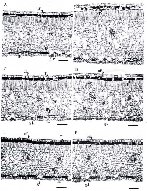
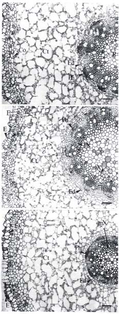
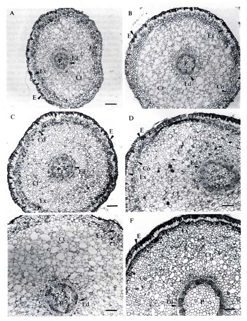
Abstract. We studied the histology of leaves and roots of Kandelia candel seedlings growing in various salinities (0_550 mM NaCl). The structural changes found were related mainly to growth status. The plants grew best in the presence of 50 and 100 mM NaCl, with greater leaf area and thickness, higher ratios of palisade to leaf thickness, and higher stele to root cross-section-area proportions. Lignification and/or suberization of the cell wall appeared farther from the root apex. Structural modifications associated with adaptation to a saline environment were not found in this study—physiological regulation may be more important.

Keywords: Anatomical changes; Kandelia candel; Mangrove; NaCl; Rhizophoraceae; Salinity.

Introduction

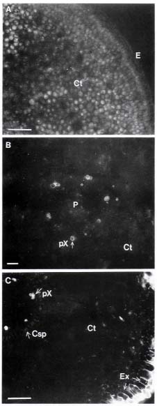
Plants that tolerate high salinity environments are termed halophytes. They always evolve structural modifications and physiological adaptations (Flowers et al., 1986). Some of the structural changes that occur in those halophytes that lack specialized salt-excretion organs such as salt glands or salt bladders include increased succulence, changed number and size of stomata, thickened cuticle, inhibited differentiation, and changed diameter and number of xylem vessels. There may also be extensive development of tyloses and earlier occurrence of Casparian strips and lignification (Flowers et al., 1986; Poljakoff-Mayber, 1975). Depending on the species, these changes may be either adaptations to salinity stress or signs of damage and disturbance of the normal life processes (Poljakoff-Mayber, 1975).

The mangrove Kandelia candel (L.) Druce grows in the intertidal zone along estuarian river banks, where the tidal salinity ranges between 10 and 36 parts per thousand (ppt) (Hwang, 1983). The optimal salinity for the growth of K. candel seedlings, however, is between 0 and 15 ppt (ca. 257 mM NaCl) (Hwang and Chen, 1995). The basic anatomical structures of this species have been studied by Chiang (1984). Structural changes were noticed when seedlings collected from saline environment in the field were grown in freshwater in a greenhouse for more than one year. These plants showed decreased thickness of leaves and cuticle layers, reduced tannin content in the tannic and palisade cell layers, increased intercellular spaces, and increased ratio of spongy cell layers to leaf (Chiang, 1984). The anatomical structure of K. candel changes in response to different salinity conditions, but whether these

changes are related to the development of salt tolerance was not clear. The present study investigates further the relationship between anatomical change and salt tolerance in K. candel.

Materials and Methods

Mature propagules of Kandelia candel (L.) Druce, collected from mature trees growing along the Tamshui River, Taipei, Taiwan, (121° 26' E, 25° 9' N), were cultivated on sand in pots, partially submerged in nutrient solutions. The culture solution was a modified Hoagland's solution, with NaCl added at the beginning to form 0, 50, 100, 250, 400 and 550 mM NaCl solutions. Details of the culture method were described by Hwang and Chen (1995). We used mature leaves and roots of four-month-old plants for our study, except for the autofluorescence study, which used root tissues of one-month-old plants.

Light Microscopy

Leaf area was measured with a leaf-area meter (model Li-3000A, LI-COR Inc., Lincoln, Nebraska, USA). Leaf discs (7 mm diameter) were cut with a paper punch along the mid-rib at the central part of mature leaves. Segments (0.5 cm long) of mature root were cut 2 cm behind the hypocotyl, and segments (0.5 cm long) of root tip were cut 0.5 cm behind the apex. The segments were fixed for 8 h in FAA (37% formalin, acetic acid, 50% alcohol = 5:5:90). The materials were then dehydrated through a TBA (tertiary butyl alcohol) series and embedded in paraplast (Oxford Labware, Maryland, USA). Cross sections (10 µm) were cut with a rotary microtome (Leitz, West Germany) and stained with 1% safranin O and 0.5% fast-green (Johansen, 1940).

1Corresponding author.


Botanical Bulletin of Academia Sinica, Vol. 36, 1995

We used autofluorescence to assay areas such as cell walls of the exodermis, endodermis, and protoxylem for the presence of lignin and/or suberin (Peterson et al., 1981). Twelve roots were randomly selected from three plants (four from each). Root length was measured, and unstained fresh sections (100 µm) were cut in series from the root apex with a vibratome (Lancer Vibratome 1000, Brunswick Co., Missouri, USA) and observed with a light microscope (Model BHS, Olympus Optical Co., Tokyo, Japan) equipped with an epifluorescence optics (exciting wavelengths 400_440 nm).

Scanning Electron Microscopy (SEM)

Stomata of leaves were observed by SEM. Leaf discs were fixed by vacuum infiltration with 5% glutaraldehyde in 0.1 M phosphate-buffered solution (pH 7.2), then post-fixed in 1% (v/v) OsO4 in 0.1 M phosphate-buffered solution. The fixed materials were then dehydrated by a 2,2-DMP and acetone fast-dehydration method (Lin et al., 1977). Dehydrated materials were critical-point dried with CO2, mounted, and coated with a gold ion coater (Eiko Engineering IB-2, Ibaraki, Japan). Electron-microscopy was carried out using a Zeiss DSM-950 scanning electron microscope operated at an accelerating voltage of 20 kV.

Thickness of leaf and of palisade and spongy parenchyma tissues, and diameter of root, cortex, and stele were measured with an image processor. Stomata number per leaf was estimated as the product of stomatal density and leaf area. Mean values of each parameter for all salinity treatments were compared using the SAS general linear model procedure (PROC GLM) one-way analysis of variance and Tukey's Studentized Range test (P Ł 0.05; SAS, 1989).

Results

Leaf Anatomy

Stomata were found only on the abaxial side of the leaves. The stomatal density was significantly higher on plants growing in the absence of NaCl (Table 1), but because the leaf area of plants grown in the absence of NaCl

was significantly smaller than that of those grown in low and medium salinity solutions, no differences in the estimated stomata number per leaf were found among plants grown in the presence of less than 250 mM NaCl (Table 1). Plants growing in 400 mM NaCl and above, however, had significantly lower leaf area and stomata number per leaf (Table 1). Apparently, the development of stomata and growth of leaf on plants growing in greater than 400 mM NaCl was inhibited.

The leaves of plants grown in low salinity were thicker than those of plants grown in high salinities (Table 2). The palisade-mesophyll ratio was highest for plants grown in 100 and 250 mM NaCl (Table 2, Figure 1). The spongy mesophyll portion, however, did not vary with salinity (Table 2). The increased leaf thickness seen in low salinity treatments seems to be a result of the increased thickness of the palisade mesophyll portion.

Root Anatomy

The mature-root cross section area did not show any trend associated with the salinity, although that of plants grown in the absence of NaCl was the largest (Table 3). The ratio of stele cross-section area to root section was higher in plants grown in 50_250 mM NaCl than in those

Table 2. Leaf thickness and ratio of spongy- and palisadal-parenchyma to leaf thickness (n=8, ± s.d.) in mature leaves of K. candel grown in different concentrations of NaCl.

Salinity Leaf thickness Sponge/Leaf Palisade/Leaf

(mM NaCl) (µm) (%) (%)

0 390 ± 40b 60.7 ± 5.2 21.1 ± 2.5 50 420 ± 40a 57.6 ± 3.5 22.5 ± 2.8 100 390 ± 30b 56.3 ± 3.2 26.2 ± 2.3 250 360 ± 20c 51.5 ± 3.5 27.1 ± 2.3 400 390 ± 40b 56.2 ± 4.0 24.5 ± 3.2 550 350 ± 20c 56.0 ± 4.0 22.9 ± 3.8

Different superscripts indicate that differences between salinity treatments are significant according to the Tukey's Studentized Range test (P Ł 0.05 ).

Table 1. Stomatal density, leaf area, and estimated number of stoma per mature leaf (n=4, ± s.d.) from K. candel grown in different concentrations of NaCl.

Salinity Stomatal density Leaf area Stomata/leaf

(mM NaCl) (no./cm2) (cm2) (no./leaf)

0 19905 ± 641a 13.51 ± 0.24c 268877 ± 3885a 50 15231 ± 1304b 16.13 ± 0.51b 245304 ± 14100a 100 14911 ± 265b 18.35 ± 0.79a 273512 ± 6956a 250 15194 ± 1492b 18.49 ± 0.70a 280810 ± 27035a 400 14133 ± 1277b 13.33 ± 0.83c 187642 ± 5690b 550 16721 ± 600b 10.88 ± 0.88d 181788 ± 14325b

Different superscripts indicate that differences between salinity treatments are significant according to the Tukey's Studentized Range test (P Ł 0.05 ).

Table 3. Mature root cross section area and the ratio of cortex and stele area to root cross section (n=6, ± s.d.) in K. candel grown in different concentrations of NaCl.

Salinity Root area Cortex/root Stele/Root

(mM NaCl) (mm2) (%) (%)

0 3.9 ± 0.5a 67.5 ± 5.5 10.9 ± 2.8

50 2.5 ± 0.6c 52.0 ± 1.7 14.6 ± 2.8

100 3.1 ± 0.4b 58.3 ± 2.8 14.4 ± 3.1

250 3.3 ± 0.6b 66.4 ± 3.6 10.7 ± 2.9

400 2.5 ± 0.2c 61.5 ± 3.3 9.3 ± 1.2

550 2.9 ± 0.2bc 62.0 ± 2.8 6.4 ± 0.8

Different superscripts indicate that differences between salinity treatments are significant according to the Tukey's Studentized Range test (P Ł 0.05 ).


Hwang and Chen — Anatomical changes in Kandelia candel

Figure 1. LM-micrographs showing cross sections of mature leaves from 4-month-old K. candel plants. Growth conditions were (mM NaCl): A, 0; B, 50; C, 100; D, 250; E, 400; and F, 550. Scale bar = 100 µm. lE, lower epidermis; uE, upper epidermis; lP, lower palisade parenchyma; uP, upper palisade parenchyma; S, stomata; Sp, spongy parenchyma; T, tannin layer; V, vascular bundle.


Botanical Bulletin of Academia Sinica, Vol. 36, 1995

grown in the absence of NaCl and in greater than 400 mM NaCl (Table 3, Figure 2). The cortex contained large lacunae, which are characteristic of hydrophytes, and the ratio of its area to that of the root cross section did not show any trend associated with the salinity (Table 3, Figure 2).

The cross-section area of root apical region (0.5 cm behind the tip) was larger in plants grown in greater than 250 mM NaCl. The root cross section area and the ratio of stele cross-section area to root section was largest in plants grown in 550 mM NaCl (Table 4, Figure 3).

The lignification and/or suberization of the cell walls of the exodermis, endodermis, and protoxylem within the root apical area was observed by UV autofluorescence. In all plants, autofluorescence was first observed in the protoxylem (Figure 4B) about 2 mm from the apex (Table 5), but the appearance of autofluorescence in exodermis and endodermis (Figure 4) was significantly closer to the apex in plants grown in high concentrations of NaCl (Table 5). High salinity, however, caused reduced root growth (Table 5), which could have affected the distance of the autofluorescence from the root tip.

Table 4. Apical root (0.5 cm behind the tip) cross section area and the ratio of cortex and stele area to root cross section (n=6, ± s.d.) in K. candel grown in different concentrations of NaCl.

Salinity Root area Cortex/root Stele/Root

(mM NaCl) (mm2) (%) (%)

0 0.9 ± 0.4c 60.6 ± 10.3 6.5 ± 1.4

50 0.7 ± 0.1c 60.7 ± 7.2 5.8 ± 1.1

100 1.1 ± 0.6c 60.4 ± 8.0 5.2 ± 1.7

250 2.3 ± 1.0b 61.7 ± 6.9 5.3 ± 1.2

400 2.2 ± 0.9b 58.4 ± 7.8 6.1 ± 1.6

550 3.0 ± 0.9a 61.2 ± 4.3 8.8 ± 2.4

Different superscripts indicate that differences between salinity treatments are significant according to the Tukey's Studentized Range test (P Ł 0.05 ).

Table 5. The length of root and distance (mm, n=12, ± s.d.) to the root tip when the first autofluorescence appeared in root tissues of one-month-old K. candel.

Salinity Root tissues

(mM NaCl) Length Exodermis Endodermis Protoxylem

0 49 ± 4b 2.6 ± 0.5ab 4.8 ± 0.5ab 2.1 ± 0.3

50 55 ± 5a 2.7 ± 0.5a 5.3 ± 0.6a 2.2 ± 0.4

100 45 ± 4b 2.3 ± 0.3ab 4.5 ± 0.5ab 1.9 ± 0.2

250 45 ± 5b 2.4 ± 0.6ab 4.4 ± 0.3b 2.1 ± 0.4

400 36 ± 5c 2.0 ± 0.3b 3.6 ± 0.4c 1.8 ± 0.3

550 31 ± 5c 2.0 ± 0.5b 3.6 ± 0.4c 1.9 ± 0.4

Different superscripts indicate that differences between salinity treatments are significant according to the Tukey's Studentized Range test (P Ł 0.05 ).


Figure 2. LM-micrographs showing cross sections of mature roots from 4-month-old K. candel plants. Growth conditions were (mM NaCl): A, 0; B, 100; C, 550. Scale bar = 100 µm. Co, cork layer; Ct, cortex; E, epidermis; Ed, endodermis; Ex, exodermis; mX, metaxylem; P, pith; Ph, phloem; pX, protoxylem.


Hwang and Chen — Anatomical changes in Kandelia candel

Figure 3. LM-micrographs showing cross sections of root tips from 4-month-old K. candel plants. Growth conditions were (mM NaCl): A, 0; B, 50; C, 100; D, 250; E, 400; and F, 550. Scale bar = 100 µm. Abbreviations are the same as for Figure 2.


Botanical Bulletin of Academia Sinica, Vol. 36, 1995

Discussion

The anatomical structure of K. candel was greatly affected by the NaCl concentration of the growth medium. In comparison to plants grown in the absence of NaCl, or in the presence of high concentrations of NaCl, those grown in low and medium concentrations of NaCl had greater leaf area (Table 1) and thickness (Table 2), higher palisade to leaf-thickness ratios (Table 2), and higher stele to root-section area ratios (Table 3). Lignification and/or suberization of the cell walls in the endodermis and exodermis appeared farther from the root tips (Table 5). Kandelia candel grows better in low salinity (about 86 mM NaCl) than in zero NaCl, and growth is greatly inhibited by salinities above 430 mM NaCl (Hwang and Chen, 1995). Similar growth responses were also found in other mangroves (Clough, 1984; Downton, 1982; Werner and Stelzer, 1990), although the beneficial role of sodium in the growth of C3 halophytes is still unclear (Brownell and Crossland, 1972; Murata et al., 1992).

The aspects of K. candel's leaf anatomy most obviously influenced by salinity were leaf area (Table 1) and leaf thickness (Table 2). High salinity caused lower stomatal number per leaf and reduced leaf thickness. These results, however, are not consistent with the findings of studies of other dicotyledonous halophytes, in which high salinity always caused increased leaf thickness and decreased stomatal density (Flowers et al., 1986). Popp et al. (1993) found a positive correlation between succulency and salinity in mangroves grown in the field, but not for those grown under glasshouse conditions. Whether the differences in leaf-response in K. candel are due to the differences in growth conditions needs further investigation.

The root tissues of other halophytes showed different growth responses to saline environments. Suaeda maritima grown in the presence of 340 mM NaCl had greater root stele diameter and cortical thickness than those grown in the absence of NaCl (Hajibagheri et al., 1985). Prosopis tamarugo showed smaller root diameter, lower cortex layer number, and smaller vascular-system size when grown in NaCl concentrations up to 400 mM (Serrato Valenti et al., 1991). In the present study, the larger ratio of stele to root cross section (Table 3, Figure 2) and greater root growth (Table 5) in plants grown in low and moderate salinities might indicate a beneficial influence of NaCl on the growth of K. candel. The trend was reversed when K. candel was cultured in high salinities (400 and 550 mM NaCl) (Table 2, Figure 1, Table 3, Figure 2). This could be the result of salt stress caused by the accumulation of Na+ and Cl- in the tissues (Hwang and Chen, 1995).

Early development of the Casparian strips, and their location close to the root apex was found in salt-grown Puccinelli peisonis (Stelzer and Lauchli, 1977) and in Suaeda maritima (Hajibagheri et al., 1985). Moreover, early lignification of secondary tissues, such as metaxylem, was believed to be a general characteristic of plants growing in habitats with a low water potential (Saadeddin and Doddema, 1986; Serrato Valenti et al., 1992). The in

Figure 4. LM-micrographs showing autofluorescence in cross sections of root tips from 1-month-old K. candel plants. Growth conditions and distance to the apex (mM NaCl, mm) were: A, no autofluorescence (250, 1.2); B, protoxylem showing autofluorescence (50, 3.8); C, protoxylem, endodermis and exodermis showing autofluorescence (50, 3.2). Scale bar = 100 µm. Csp = Casparian strip; other abbreviations are the same as for Figure 2.


Hwang and Chen — Anatomical changes in Kandelia candel

creased dimensions in the root apex region (Table 4, Figure 3) and the reduced distance of the Casparian strip on the endodermis to the root tip (Table 5, Figure 4C) found in K. candel grown in high salinities were consistent with those in S. maritima (Hajibagheri et al., 1985). Kandelia candel grown in high salinities, however, developed shorter roots than those grown in low salinities (Table 5), which is the reverse of findings with S. maritima (Hajibagheri et al., 1985). We suggest that the increased dimensions in the root apical region of K. candel is caused by an inhibition of cell division in the apical meristem.

Plants grown in high salinity environments face both ionic and water stresses. Kandelia candel does not possess salt glands, bladders, or other salt-secreting structures in its leaves or stem (Chiang, 1984)—absorbed salt accumulates in tissues and causes metabolic stress. In the absence of excretory organs, K. candel had to evolve other mechanisms of adaptation to survive in high ionic- and low osmotic-potential environments. We found no special structures in the roots and leaves of K. candel directly related to salt tolerance. We suggest that physiological regulation is more important than structural modifications in K. candel's adaptation to saline environments.

Acknowledgments. This research was supported by Academia Sinica, Taiwan, ROC. We thank Dr. R. Stelzer for his valuable criticism of the paper, Drs. Ho-Shii Chang, Wei-Chin Chang, Mei-Hwa Chen, and Tsung-Hsien Chen for kind permission to use their equipment for light microscopy, and Ms. Chyong-Ere Shieh for her assistance with scanning electron microscopy.

Literature Cited

Brownell, P. F. and C. J. Crossland. 1972. The requirement for sodium as a micronutrient by species having the C4 dicarboxylic photosynthetic pathway. Plant Physiol. 49: 794_797.

Chiang, S. H. T. 1984. A preliminary study on the histology of Kandelia candel (mangrove). Yushania 1: 7_21. (in Chinese)

Clough, B. F. 1984. Growth and salt balance of the mangroves Avicennia marina (Forsk.) Vierh. and Rhizophora stylosa Griff. in relation to salinity. Aust. J. Plant Physiol. 11: 419_430.

Downton, W. J. S. 1982. Growth and osmotic relations of the mangrove Avicennia marina, as influenced by salinity. Aust. J. Plant Physiol. 9: 519_528.

Flowers, T. J., M. A. Hajibagheri, and N. T. W. Clipson. 1986. Halophytes. Quart. Rev. Biol. 61: 313_337.

Hajibagheri, M. A., A. R. Yeo, and T. J. Flowers. 1985. Salt

tolerance in Suaeda maritima (L.) Dum.: Fine structure and ion concentrations in the apical region of roots. New Phytol. 99: 331_343.

Hwang, Y.-H. 1983. Dynamics of nutrient flow in Chuwei mangrove ecosystem. MS thesis, National Taiwan University, Taipei, Taiwan. (in Chinese)

Hwang, Y.-H. and S. C. Chen. 1995. Salt tolerance in seedlings of the mangrove Kandelia candel (L.) Druce, Rhizophoraceae. Bot. Bull. Acad. Sin. 36: 25_31.

Johansen, D. A. 1940. Plant Microtechnique. McGraw-Hill Book Company, New York and London, 523 pp.

Lin, C. H., R. H. Falk, and C. R. Stocking. 1977. Rapid chemical dehydration of plant material for light and electron microscopy with 2,2-dimethoxypropane and 2,2- diethoxypropane. Amer. J. Bot. 64: 602_605.

Murata, S., M. Kobayashi, T. Matoh, and J. Sekiya. 1992. Sodium stimulates regeneration of phosphoenolpyruvate in mesophyll chloroplast of Amaranthus tricolor. Plant Cell Physiol. 33: 1247_1250.

Peterson, C. A., M. E. Emanuel, and G. B. Humphreys. 1981. Pathway of movement of apoplastic fluorescent dye traces through the endodermis at the site of secondary root formation in corn (Zea mays) and broad bean (Vicia faba). Can. J. Bot. 59: 618_625.

Poljakoff-Mayber, A. 1975. Morphological and anatomical changes in plants as a response to salinity stress. In A. Poljakoff-Mayber and J. Gale (eds.), Plants in Saline Environments. Springer-Verlag, Berlin, pp. 97_117.

Popp, M., J. Polania, and M. Weiper. 1993. Physiological adaptations to different salinity levels in mangrove. In H. Lieth and A. Al Masoom (eds.), Towards the Rational Use of High Salinity Tolerant Plants. vol. 1. Kluwer Academic Publishers, Dordrecht, The Netherlands, pp. 217_224.

Saadeddin, R. and H. Doddema. 1986. Anatomy of the `extreme' halophyte Arthrocnemum fruticosum (L.) Moq. in relation to its physiology. Ann. Bot. 57: 531_544.

Serrato Valenti, G., M. Ferro, D. Ferraro, and F. Riveros. 1991. Anatomical changes in Prosopis tamarugo Phil. seedlings growing at different levels of NaCl salinity. Ann. Bot. 68: 47_53.

Serrato Valenti, G., L. Melone, O. Orsi, and F. Riveros. 1992. Anatomical changes in Prosopis cineraria (L.) Druce seedlings growing at different levels of NaCl salinity. Ann. Bot. 70: 399_404.

Stelzer, R. and A. Lauchli. 1977. Salt- and flooding tolerance of Puccinellia peisonis: I. The effect of NaCl- and KCl-Salinity on growth at varied oxygen supply to the root. Z. Pflanzenphysiol. 83: 35_42.

Werner, A. and R. Stelzer. 1990. Physiological responses of the mangrove Rhizophora mangle grown in the absence and presence of NaCl. Plant, Cell and Environ. 13: 243_255.


Botanical Bulletin of Academia Sinica, Vol. 36, 1995