Bot. Bull. Acad. Sin. (1996) 37: 51-59

Roberts et al. — Nitrate reductase during shoot morphogenesis

In vitro tobacco cultures provide a convenient system for

investigating nitrate reductase regulation during shoot

morphogenesis

M.A. Roberts1, M.P. Watt2,4, and B.I. Huckett3

1School of Biological and Medical Sciences, University of St Andrews, St Andrews, KY 16 9th FIFE, UK

2Biology Department, University of Natal, Private Bag X10, Dalbridge 4014, South Africa

3Biotechnology Department, SA Sugar Association Experiment Station, Private Bag X02, Mount Edgecombe 4300, South Africa

(Received June 13, 1995; Accepted October 26, 1995)

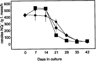
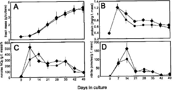
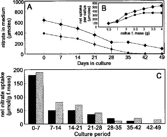
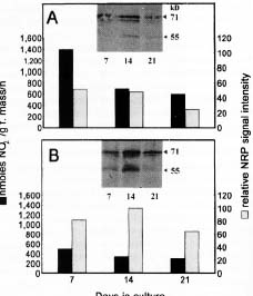
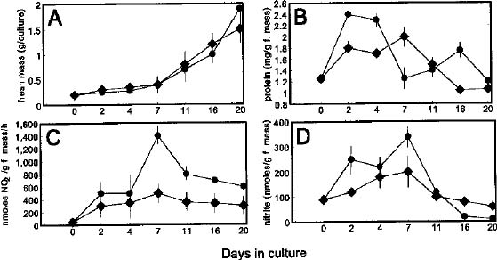
Abstract. Nicotiana tabacum callus culture was assessed in terms of its usefulness as a system for investigating the regulation of nitrate reductase (NR; EC 1.6.6.1) during shoot morphogenesis. Culture conditions for organogenesis and appropriate methodologies for extraction and assay of NR during callus growth and development were established. The most suitable medium comprised Murashige and Skoog salts and vitamins, sucrose, indoleacetic acid and kinetin. Provided that NO3_-N was present, callus growth and shoot morphogenesis occurred in the same culture step. Optimised in vivo, in situ, and in vitro NR assays yielded similar values and patterns during culture development, and the in vivo assay was selected for subsequent studies on the basis of ease and efficiency of use. With quantitative alteration in nitrogen supply (60 vs. 120 mM; 1:2 NH4+-N : NO3_-N) and variations in light regime (16 /8 h light/dark vs. continuous darkness), callus growth parameters were comparable in terms of nitrate uptake, increase in fresh mass and protein accumulation. However, a marked rise in the level of in vivo NR activity (NRA) was observed just prior to shoot primordia emergence in light/dark-grown but not in dark-grown calli. In protein blot analyses using a polyclonal antibody raised against spinach NR, major protein bands representing holo-NR cleavage products were identified at 71 and 55 kDa. Putative nitrate reductase protein (NRP) levels were compared with in vivo NRA during the final stages of callus differentiation under both light/dark and continuous dark conditions. NRP variations appeared to be much less marked than those of NRA, suggesting the possibility that post-translational modification is a significant feature of NR regulation. The potential value of the system for further studies of NR regulation at the molecular level is discussed.

Keywords: Callus; Differentiation; Morphogenesis; Nitrate reductase; Nitrate reductase assays; Tobacco.

Introduction

Nitrate reductase (NR; EC 1.6.6.1) is considered to be a limiting factor for higher plant growth, development and protein production, and much research has been undertaken involving the delineation of the regulatory properties of this enzyme (e.g. Solomonson and Barber, 1990) due to the potential for increasing nitrogen assimilation efficiency. Regulation of NR is achieved by several sensitive and complex control systems, with precise regulating mechanisms differing among higher plant species and responding variously to cellular conditions (Beevers and Hageman, 1969; Caboche et al., 1989; Campbell, 1989). Nitrate reductase activity (NRA) is known to change during organ and plant development (Kenis et al., 1992), as do the activities of other enzymes involved in the nitrate assimilation pathway (de la Haba et al., 1988). Nevertheless, much further work needs to be done on the mechanisms involved in developmental NR regulation in higher plants, including the possible roles of this enzyme in cell and organ differentiation processes.

Cellular differentiation and morphogenesis arise from progressive developmental interactions between cells and their environment, leading to the initiation of activity of specific genes, which may be regulatory or required for enhanced metabolism (Gyorgyey et al., 1991). The influence of certain endogenous factors (e.g. phytohormone levels) (Li et al., 1992) and environmental influences (e.g. light, temperature) (Thorpe, 1983; Tran Thanh Van and Trinh, 1990) on these processes is undisputed. Yet, as higher plant differentiation is difficult to decipher at the whole plant level due to numerous biological constraints (Tran Thanh Van and Trinh, 1990), research with whole plants has been restricted largely to descriptive analyses rather than the elucidation of causal mechanisms.

In vitro plant culture systems have been developed that facilitate morphological, physiological, cytological, biochemical and molecular work on differentiation (Tran Thanh Van and Trinh, 1990). Such cultures approximate the responses of their whole plant counterparts genetically and morphologically, and are highly suitable for growth and differentiation studies in terms of generation of material at the required stage of morphogenesis and manipulation of nutritional and environmental parameters (Bisbis

4Corresponding author.


Botanical Bulletin of Academia Sinica, Vol. 37, 1996

et al., 1993; Faure and Aarrouf, 1994). Some such studies have shown that in vitro growth and differentiation are affected by the composition of the nutrient medium, including the level and source of nitrogen (e.g. Thorpe, 1983; Grimes and Hodges, 1990; Hardy and Thorpe, 1990). Preliminary studies in our laboratory (Amory et al., 1989) have also indicated that the presence of nitrate in the culture medium is essential for shoot regeneration from callus cultures of tobacco.

The aim of this work was to investigate the merit of using in vitro cultures, in particular organogenic calli, to provide a system for studying regulation of NR during shoot morphogenesis. Towards this end, we established culture conditions for induction of high frequency shoot regeneration in tobacco, identified and optimised the most appropriate assay for the quantification of NRA in tobacco callus material, and made preliminary observations of the effects of nitrogen and light on nitrate assimilation during shoot morphogenesis.

Materials and Methods

Plant Material

Tobacco seeds (Nicotiana tabacum L. var. Samsun) were obtained from Karl Kunert (Laboratoire de Biologie Cellulaire, INRA, Versailles, France). Polyclonal antibody raised in rabbit against spinach NR was a gift from Ian Prosser (Long Ashton Research Station, Bristol, UK).

Establishment and Maintenance of in vitro Cultures

Tobacco plants were germinated from seeds under greenhouse conditions. Callus was established from surface-sterilised tobacco leaf segments, in darkness at 25°C, on 10 ml MS medium (Murashige and Skoog, 1962) supplemented with 20 g l-1 sucrose, 10 g l-1 agar, 2 mg l-1 indoleacetic acid (IAA) and 0.5 g l-1 kinetin (callus growth and differentiation medium). The generated calli were subcultured on to fresh medium (containing 40 mM NO3_-N and 20 mM NH4+-N as in MS formulation) and kept in the dark for 21 days. Thereafter, approximately 0.2 g fresh mass of callus was transferred to 10 ml callus growth and differentiation medium and used in the investigations. The experimental treatments involved culturing the calli on

different nitrogen regimes (Table 1), under constant darkness or 16/8 h light/dark (at 400 µE m-2 sec-1). In a few, preliminary, experiments calli were obtained by plating cell suspension cultures growing on 1 g l-1 2,4-ichlorophenoxy- acetic acid (2,4-D) on to callus growth and differentiation medium, at a volume of 1.0_1.5 ml per 10 ml solid medium.

NRA Assays

In vivo assay—Callus samples (0.2_0.35 g f. mass) were placed in glass centrifuge tubes containing 0.1 ml dH2O, sealed with Suba-seal bungs and wrapped in aluminum foil. The assay was carried out at 20°C with continuous N2 (gas) flushing, and terminated after 30 min by adding 3_5 ml boiling dH2O (Watt et al., 1986). Accumulated nitrite was quantitatively extracted from the callus by boiling for 30 min and, after centrifugation (5 min at 3,000 g), was assayed colourimetrically (Hageman and Reed, 1980).

In situ assay—Callus was vortexed for 3 min in 25 mM Hepes-KOH solubilisation buffer (pH 7.5) containing 250 mM Na2EDTA (1 ml buffer per 0.2 g of callus f. mass). An equal volume of incubation buffer (100 mM Hepes-KOH, pH 7.5, 20 mM KNO3, 4 mM methyl viologen) was added, and the reaction initiated with sodium dithionite (final concentration 5 mM). The assay was terminated after 0 or 10 min at 25°C by vortexing for 30 s to reduce excess dithionite (Larsson et al., 1985). One volume of formaldehyde (0.6% w/v) in Hepes-KOH (300 mM, pH 7.5) was added and the mixture vortexed for 3 min. Immediately after centrifugation (5 min at 3,000 g), the mixture was assayed for nitrite (Hageman and Reed, 1980).

In vitro assay—A crude enzyme extract was obtained by grinding callus with mortar and pestle (at 4°C) in the presence of acid-washed sand and potassium phosphate buffer (50 mM, pH 8.5) containing 2% (w/v) bovine serum albumin (2.5 ml buffer per 0.5 g f. mass callus). The homogenate was filtered through four layers of muslin cloth and the supernatant collected after centrifugation (3,000 g) for 10 min at 4°C. The enzyme extract was assayed for NRA in the presence of 9 volumes of potassium phosphate buffer (100 mM, pH 7.5) containing KNO3 (10 mM) and NADH (0.1 mM) for 30 min at 25°C. The

Table 1. Effect of different nitrogen regimes on in vitro tobacco callus growth and shoot morphogenesis, after 7 weeks in culture, under a light/dark photoperiod. Standard MS formulation contains 20 mM NH4+ -N + 40 mM NO3_-N.

Nitrogen regime Callus growth % Callus with shoots

0 N _ 0

60 mM L-glutamic acid _ 0

30 mM NH4+ -N _ 0

60 mM NH4+ -N _ 0

30 mM NO3_-N +++ 50

60 mM NO3_-N +++ 93

10 mM NH4+ -N + 50 mM NO3_-N +++ 95

20 mM NH4+ -N + 40 mM NO3_-N +++ 95

40 mM NH4+ -N + 80 mM NO3_-N +++ 95


Roberts et al. — Nitrate reductase during shoot morphogenesis

reaction was terminated by placing tubes in boiling water for 5 min, after which nitrite was determined colourimetrically (Hageman and Reed, 1980).

Determination of Nitrate Utilisation from Agar Growth Medium

Agar medium was frozen at -20°C for at least 24 h, thawed at room temperature and centrifuged at 40,000 g for 60 min (Lumsden et al., 1990) and the supernatant assayed for nitrate (Cataldo et al., 1975).

Protein Estimation

Protein was extracted from callus tissue with a Janke and Kunkel Ultra-turrax following the method of Wetter and Dyck (1983), except that octan-2-ol was used to prevent foaming. After precipitation of 100 µl crude protein extract in the presence of 6.06% (v/v) trichloroacetic acid and 0.016% (w/v) sodium deoxycholate, the latter to eliminate water-soluble interfering compounds (Bensadoun and Weinstein, 1976), protein was estimated using the Folin-Lowry assay, as described by Wetter (1984).

Protein Blot Analysis of NRP

Acetone powders were prepared from tobacco callus, using a modified Ibrahim and Cavia (1975) protocol as follows: callus (0.3_0.6 g f. mass) was ground to a powder in liquid N2 using a pestle and mortar, homogenised with cold 75% acetone, and then repeatedly washed under vacuum with very cold 100% acetone; after drying the residue with desiccated air, the powder was stored at -80°C. After resolubilisation of 50 µg samples in SDS treatment buffer [62.5 mM Tris-HCl, 2% (W/V) sodium dodecyl sulphate, 10% (V/V) glycerol, 5% (V/V) 2-mercaptoethanol, pH 6.8] for 1.5 h on ice with occasional mixing, the soluble fraction was boiled for 2 min, and a 1/10 volume of 0.1% bromophenol blue marker dye was added to each sample prior to separation in a 9% polyacrylamide gel. Protein molecular weight markers (Rainbow, Amersham) were run as standards. Protein bands were transferred by electroblotting to nitrocellulose (Towbin et al., 1979) at 45 mA for 14 h using the Hoefer TE 70 SemiPhor Semi-Dry Transfer Unit. Transfer efficiency was checked by staining the blotted gel with Coomassie Blue. Polyclonal antibody raised in rabbit against spinach NR was used as first antibody for NRP detection, with anti-rabbit IgG conjugated to alkaline phosphatase as secondary (indicator) antibody. Secondary antibody was detected by incubation in veronal acetate buffer, pH 9.5, containing 4 mM MgCl2, 0.2 mg ml-1 nitrobluetetrazolium and 0.1 mg ml-1 5-bromo-4-chloro-3-indolyl phosphate:toluidine salt (the last two reagents added dissolved in dimethylformamide).

Statistical Procedures

A minimum of three replicates was used. Regression analyses, T-tests for equality of means, ANOVAs (Analysis of Variance), and Student-Newman-Keuls (SNK) tests

(p<0.05) were performed using the Costat statistical package (Cohort Software, Berkeley, USA), while the Kolmogorov-Smirnov test was run from Statgraphics (Statistical Graphics Corporation, Rockville, USA). Outliers were eliminated when detected using Dixon's test statistic, as described by Sokal and Rohlf (1981).

Results and Discussion

Comparison of NR Assays

Nitrate reductase activity is most frequently estimated from leaf material using one of three assays, namely the in vivo (Watt et al., 1986), the in vitro (Hageman and Reed, 1980), and the in situ (Larsson et al., 1985) assays. Each has been subjected to valid criticisms (e.g. Brunswick and Cresswell, 1986; Heuer and Plaut, 1978; Huber et al., 1992a; Wray and Fido, 1990), and each requires optimisation according to the type of material under study. Consequently, such investigations were undertaken for tobacco callus in this investigation (results not shown).

In the initial part of this study, viz. comparison of assays for NR, cell suspension cultures were used to initiate callus to ensure cellular homogeneity. After 21 days in the dark, the calli were transferred to fresh medium and a 16/8 h light/day photoperiod (Figure 1, day 0) and assayed weekly for NRA using the optimised conditions for in vitro, in vivo, and in situ assays.

NRA measured by the three assays showed similar trends, with a decline after day 14 (Figure 1). Results were compared at each sampling point using a T-test for equality of means (p<0.05), which indicated that the only statistically significant differences occurred at days 21 and 28 when in vitro NRA was lower than in vivo and in situ NRA. The lesser values for in vitro NRA at these points could possibly have been caused by age related inhibitory substances exposed to NR during extraction (Hoarau et al., 1991).

Figure 1. In vivo, in situ and in vitro NRA profiles during tobacco callus growth and shoot morphogenesis. Callus was generated from cell suspension cultures on solid medium. Cultures were maintained in the dark for 21 days and then subcultured (day 0) on to fresh medium (containing 40 mM NO3_-N + 20 mM NH4+-N) and placed under a 16/8 h light/dark photoperiod regime for 42 days.


Botanical Bulletin of Academia Sinica, Vol. 37, 1996

The in vitro and in situ assays are assumed to measure NRA under optimal conditions, and hence provide a measure of the amount of potentially active enzyme (Padidam et al., 1991). For this reason, many studies have obtained in vitro NRA values several times higher than in vivo NRA (Duke and Duke, 1984). In contrast, other workers have found that in vivo activity was consistently higher than in vitro activity, attributing this to inactivators and organic compound inhibition during extraction in the latter (Jones et al., 1976). Here, the three assays generally gave similar results, indicating that physiological levels of reductant and substrate were probably non-limiting during most of the tobacco callus culture period. On the basis of these results, a single assay system was deemed sufficient to determine regulatory aspects of callus morphogenesis. The in vivo assay was selected for all subsequent studies, because it is easier to perform than the other assays and large numbers of samples can be handled simultaneously (Padidam et al., 1991).

The Effect of N Supply (60 mM and 120 mM N) on Tobacco Callus Growth, NO3_-N Assimilation and Shoot Morphogenesis

Higher plant species differ in ability to grow and differentiate on different sources of nitrogen, and a combination of nitrate- and ammonium-nitrogen has been found to be required for in vitro growth and differentiation in various higher plant species (Grimes and Hodges, 1990). In this study, the most effective media for callus growth and shoot morphogenesis were those containing nitrate, either singly (30_60 mM N) or combined with ammonium as in the MS formulation (Table 1).

Reduced callus growth and lack of differentiation with ammonium and glutamic acid (Table 1) implies that NO3_-N may be required for differentiation. Although inhibition of growth and morphogenesis in these media could have been due to alterations in pH (Jones et al., 1976), recent work in our laboratory on the effects of NH4+-N on tobacco callus has provided some evidence against this hypothesis (Sweby et al., 1994).

In preliminary experiments with tobacco callus initiated from cell suspension cultures, a decrease in enzyme activity in the callus was accompanied by substantial depletion in medium nitrate (results not shown). Therefore, NRA regulation and growth parameters during tobacco callus differentiation on 60 mM N (standard MS medium containing 40 mM NO3_-N and 20 mM NH4+-N) and 120 mM nitrogen (80 mM NO3_-N and 40 mM NH4+-N) were investigated. In these studies, subcultured tobacco callus was employed as inoculum, in preference to cell suspension cultures, because the former were easier to manipulate in vitro and shorter culture periods (approximately 20 to 25 days) were required to initiate shoot primordia.

The concentration of inorganic nitrogen used for in vitro tissue culture can affect growth and differentiation, with optimal levels being species dependent. Murashige and Skoog (1962) found 96 mM nitrogen to be slightly inhibitory for tobacco callus growth in comparison with 60 mM

nitrogen. For the tobacco variety and culture conditions in this study, callus fresh mass increases were equivalent for the two nitrogen treatments (Figure 2A). These growth curves are characteristic of in vitro culture development (Hardy and Thorpe, 1990). Soluble protein levels were significantly higher in the 120 mM nitrogen regime during the third to the fifth week in culture, but comparable at other sample points (Figure 2B), indicating that the supply of 120 mM nitrogen was not inhibitory to callus growth.

Following initial callus induction and growth, the first visible signs of differentiation were buds (shoot primordia), which developed into plantlets with shoots and roots. Shoot production was delayed in the higher nitrogen treatment, with 50% of the cultures showing visible bud formation after 30 days, compared with 23 days in the 60 mM nitrogen regime. Nevertheless, there was no significant difference (p<0.05) between the number of shoots produced per culture on the two nitrogen regimes after 8 weeks: 9.18 ± 6.14 for 60 mM nitrogen and 10.59 ± 5.77 for 120 mM nitrogen (mean ± standard error).

Differences in in vivo NRA (Figure 2C) between the two nitrogen treatments were not statistically significant (p<0.05), indicating that the developmental regulation of NR during tobacco callus differentiation was not affected by nitrogen level. Nitrate reductase activity reached maximal levels after day 7 for 120 mM and after day 14 for 60 mM nitrogen treatments. Single NRA peaks have been obtained for both differentiating and non-differentiating cultures (Evenson et al., 1988), but Hardy and Thorpe (1990) found differentiating tobacco callus cultures to have higher levels of NRA than non-differentiating cultures, suggesting an alteration in NR control. In this study, maximum NRA of callus was observed prior to the emergence of shoot buds in both nitrogen treatments (Figure 2C), when meristemoid formation would be occurring (Thorpe, 1983). As noted previously (Figure 1), after one to two

Figure 2. Effect of nitrogen supply on callus fresh mass (A), and levels of protein (B), NRA (C) and nitrite (D) during in vitro growth and shoot morphogenesis. Calli generated from leaf material were subcultured on to fresh medium (containing 60 mM nitrogen) and maintained in the dark for 21 days. At the end of the dark treatment, the calli were subcultured (day 0) on to 60 mM N (40 mM NO3_-N + 20 mM NH4+-N) or 120 mM N (80 mM NO3_-N + 40 mM NH4+-N) and placed under a 16/8 h light/dark photoperiod for 49 days.


Roberts et al. — Nitrate reductase during shoot morphogenesis

weeks in culture NRA decreased with time and this was the case even in calli supplied with 120 mM N (Figure 2C).

Hardy and Thorpe (1990) have shown that, under certain circumstances, nitrite reductase (NiR) may limit nitrogen assimilation during in vitro culture. Those workers reported that levels of nitrite in differentiating tobacco callus cultures were higher than in non-differentiating cultures, and nitrite levels increased in parallel with NRA. Similar results were obtained in the present study (Figure 2D), although the possibility that NiR might have been limiting nitrate assimilation was not investigated further.

Nitrate depletion from the culture medium resulted in similar trends for the two nitrogen regimes (Figure 3A), with net nitrate uptake per callus being generally higher for callus grown on 120 mM nitrogen (Figure 3B). Rapid net nitrate uptake during the initial stages of tobacco callus growth indicated that the reduced nitrogen source ammonium was not utilised preferentially, as has been observed for other cultures (Guerrero et al., 1981). Whereas maximal NRA coincided with highest net nitrate uptake values for 120 mM nitrogen (days 0 to 7), this was not observed for the lower nitrogen regime (Figures 2C, 3C). Thus it is not clear whether under the 60 mM N treatment the `excess' nitrate entering callus cells during the first week in culture was translocated into the storage (vacuolar) or metabolic (cytoplasmic) pool (Ferrari et al., 1973; Granstedt and Huffaker, 1982). Over the entire culture period, however, NRA was correlated broadly with net nitrate uptake, supporting the hypothesis that nitrate flux into cells regulates NRA (Heimer and Filner, 1971).

There was a direct relationship between tobacco callus fresh mass and net nitrate uptake (Figure 3B), as has been observed in other species (Lumsden et al., 1990). This proportional relationship between nitrate uptake and growth rate confirms that nitrate is an important nutrient for morphogenic cultures. In addition to its incorporation into organic nitrogen via nitrate assimilation, nitrate may have an osmoregulatory role during plant cell growth (Talouizite and Champigny, 1988). It was also observed that tobacco callus incorporated more nitrate per unit fresh mass when supplied with 120 mM nitrogen than with 60 mM nitrogen (Figures 3B and 3C).

In summary, most of the growth parameters examined here did not yield a significant difference between the 60 mM and 120 mM nitrogen regimes, and 120 mM nitrogen was found not to inhibit callus growth or protein accumulation. Although bud formation was delayed slightly in the higher nitrogen regime, differentiation into plantlets was not affected. In subsequent experiments, nitrogen was used at 120 mM nitrogen to ensure that nitrate was not a limiting factor for NR regulation.

The Effect of Light on Tobacco Callus Growth and NO3_-N Assimilation

In all the light studies, calli were cultured in either continuous darkness or a 16/8 h light/dark regime. Analyses were restricted to the first stages of differentiation (0_21

Figure 3. Nitrate utilisation during in vitro growth and shoot morphogenesis of calli grown on 60 mM or 120 mM N. Growth conditions as described in Figure 2.

days), where an increase of NRA had been observed previously under the light/dark regime (Figure 2C). Because of the known diurnal response of NR (Bowsher et al., 1991) and its response with a circadian rhythm in tobacco (Galangau et al., 1988), sampling for analysis was always conducted at the same time of day (6 hours after onset of light).

Tobacco callus growth was comparable in terms of fresh mass in the dark and dark/light treatments (Figure 4A). Protein levels were generally higher in calli grown under the photoperiod regime than under constant darkness for the initial culture period (Figure 4B), as was NRA (Figure 4C). Although shoot morphogenesis occurred in both treatments, in the light/dark regime callus differentiation was preceded by a marked increase in NRA not observed in the dark (Figure 4C). This is in agreement with previous findings that light does not affect meristemoid and bud formation on tobacco callus, but does affect the regulation of many genes, including NR (Deng et al., 1990; Pilgrim et al., 1993).

Callus nitrite accumulated to relatively high levels in both dark and light/dark regimes during the first seven days (Figure 4D). Callus nitrite was significantly higher in the dark treatment at day 16 and 20 (p<0.05), indicating that nitrite reductase activity may have been restricted in the absence of light. However, the fact that nitrite accumulated in the calli during early differentiation in both light/dark and dark treatments suggests that NiRA was regulated by factors other than the availability of light. Simultaneous regulation of NR and NiR induction by nitrate and light has been reported (Neininger et al., 1992)

Disappearance of nitrate from the culture medium followed similar trends for both treatments (results not shown) as observed previously (Figure 3). The similarity of net nitrate uptake kinetics in the dark and light/dark treatments implied that light did not significantly affect the nitrate uptake process.


Botanical Bulletin of Academia Sinica, Vol. 37, 1996

The Effect of Light on NR Regulation During Shoot Morphogenesis

The increase in NRA in response to photoperiod conditions (Figure 4C) is striking evidence for the importance of light in NR regulation during morphogenesis, and is compatible with previous observations that NR is light inducible (Deng et al., 1990), that NR fluctuates diurnally according to the pattern of light (Bowsher et al., 1991; Huber et al., 1992b) and that, overall, nitrate assimilation is coordinated with photosynthesis (Huber et al., 1994). Regulation of gene expression during differentiation has been described for several genes, for example the small subunit of ribulosebisphosphate carboxylase (Vernet et al., 1982) and heat shock genes (Gyorgyey et al., 1991), but no studies on developmental NR expression during in vitro differentiation have been reported. Consequently, it was of considerable interest to undertake a preliminary investigation into possible regulatory mechanisms operating in the tobacco callus differentiation system. This was done by characterising and assessing the levels of nitrate reductase protein (NRP) during and following the crucial period of callus development (day seven) in both the dark and light/dark regimes, and comparing NRP with NRA.

Protein blot analysis of tobacco callus NRP using a polyclonal antibody against spinach NR showed the presence of two major bands, with molecular mass of 55 and 71 kDa, respectively (Figure 5, insert). Holo-NR is a dimer with a native molecular mass of 200_230 kDa and a subunit size of 100_115 kDa; however, the enzyme is labile and the subunits extremely difficult to identify in intact form (Fido, 1987). Both band species obtained were regarded as cleaved subunits but, on the basis of several preliminary analyses, the 71 kDa polypeptide was interpreted as the most stable NR breakdown product, probably the Mo-containing domain, while the 55 kDa band was seen to be insufficiently stable to be a reliable index of NR. Hence relative NRP was quantified by densitometry

Figure 5. The effect of light regime on in vivo NRA and relative NRP levels of calli grown under a 16/8 h light/dark photoperiod (A) or constant darkness (B) during the initial stages of in vitro tobacco callus growth and shoot morphogenesis. Calli were produced as described in Figure 4. NRP levels were determined by protein blot immunodetection and densitometric measurement of the 71 kDa band (inserts); relative values were calculated by assigning 100% to the maximum signal. Total acetone-extracted protein load was 50 mg per lane.

measurement of the 71 kDa band (putative NR) at each sample point, the supposition being that the 71 kDa polypeptide was derived from holo-NR in a consistent ratio. Such an assumption is implicit in immunopurification techniques, employed routinely for NRP quantification, in which monoclonal antibodies specific for a single antigenic site may be used for the precipitation and detection of a specific NR subunit (Caboche et al., 1989; Huber et al., 1994).

The NRA and relative (putative) NRP levels were largely correlated under the light/dark but not in the dark regime (Figure 5). In the light/dark treated callus (Figure 5A), the peak of NRA at 7 days and its subsequent decline was generally reflected in the steady-state level of NRP. In the dark maintained callus (Figure 5B), NRA was relatively low at 7 days, subsequently decreasing further with time; however, NRP was usually high and did not match NRA decline, particularly at the 14 day sample point. Both NRA trends confirmed previous observations (Figure 4C). In mature leaves of spinach during a day/night cycle it has been observed that changes in NRP level are normally small and that the major changes in NRA that occur are the result of phosphorylation and dephosphorylation of the enzyme (Huber et al., 1992a). By con

Figure 4. Effect of light regime on callus fresh mass (A), and levels of protein (B), NRA (C) and nitrite (D) during in vitro growth and shoot morphogenesis. Calli generated from leaf material were subcultured on to fresh medium (containing 60 mM nitrogen) and maintained in the dark for 21 days. At the end of the dark treatment, the calli were subcultured (day 0) on to 120 mM nitrogen placed under constant darkness (D) or a 16/8 h light/dark photoperiod (L/D) for 49 days.


Roberts et al. — Nitrate reductase during shoot morphogenesis

trast, in maize leaves, it has been shown that light activation of NR involves responses in both steady-state levels of NRP and enzyme phosphorylation. The results presented in Figure 5 shows a trend similar to the former example: in contrast to NRA, putative NRP level is not changed substantially by the light/dark regime during shoot morphogenesis. It is tempting to speculate that during tobacco shoot morphogenesis NR regulation is accomplished largely by post-translational modification. Future studies undertaken using this tissue culture system to further clarify the operative mode of control should therefore include investigations into phosphorylation of NR in vivo.

Conclusions

For studies on nitrate assimilation during shoot morphogenesis in tobacco callus culture, a medium was required in which the hormones IAA and kinetin were provided and nitrate was the main source of N. A single NR assay system was adequate for determining NRA in developing callus, and the in vivo assay was the most suitable method for reasons of simplicity and efficiency. Growth parameters and patterns of NR expression were not significantly affected by nitrogen levels of 60 and 120 mM. During tobacco callus differentiation, light did not significantly affect tobacco callus growth in terms of nitrate uptake, increase in fresh mass and protein accumulation. However, light affected NRA significantly, indicating that NR is regulated in response to light, with light induced NRA peaking prior to visible bud emergence and declining gradually during shoot morphogenesis. NRP was much less responsive to light than NRA, suggesting the possibility that post-translational modification of NR is an important regulatory factor and that enzyme phosphorylation studies would be informative. The in vitro culture approach developed and described embodies a defined growth system which can be used for the controlled genesis of shoots. The system is effective and convenient, with the potential to govern various transitions from dark to light, and should prove useful in further investigations into developmental regulation, particularly at the molecular level.

Acknowledgements. This work was supported by the University of Natal Research Fund and the Foundation for Research Development.

Literature Cited

Amory, A., M.P. Watt, C.F. Cresswell, F. Blakeway, L. Ford, and B. Ripley. 1989. Effect of light and nitrogen nutrition on the metabolism of nitrogen by tobacco plant cell cultures. In C. Rigano (ed.), 2nd Advanced Course on Inorganic Nitrogen Metabolism, Universita Delgi Studi Napoli, Italy, pp. 131.

Beevers, L. and R.H. Hageman. 1969. Nitrate reduction in higher plants. Ann. Rev. Plant Physiol. 20: 495_522.

Bensadoun, A. and D. Weinstein. 1976. Assay of proteins in the presence of interfering materials. Anal. Biochem. 70:

241_250.

Bisbis, B., F. le Dily, C. Kevers, J-P. Billard, C. Huault, and T. Gaspar. 1993. Disturbed sugar metabolism in a fully habituated nonorganogenic callus of Beta vulgaris (L.). Plant Growth Regul. 13: 257_261.

Bowsher, C.G., D.M. Long, A. Oaks, and S.J. Rothstein. 1991. Effect of light/dark cycles on expression of nitrate assimilatory genes in maize shoots and roots. Plant Physiol. 95: 271_285.

Brunswick, P. and C.F. Cresswell. 1986. Limitations to the measurement of in vitro nitrate reductase assimilation by exogenous additives and endogenous interference factors in the leaves of Zea mays L. seedlings. Ann. Bot. 57: 859_868.

Caboche, M., I. Cherel, F. Galangau, M. Grandbastien, C. Meyer, T. Moureaux, F. Pelsy, P. Rouze, H. Vaucheret, F. Vedele, and M. Vincentz. 1989. Molecular genetics of nitrate reduction in Nicotiana. In J.L Wray and J.R. Kinghorn (eds.), Molecular and Genetic Aspects of Nitrate Assimilation, Oxford Science Publications, Oxford, pp. 186_196.

Campbell, W.H. 1989. Structure and regulation of nitrate reductase in higher plants. In J.L Wray and J.R Kinghorn (eds.), Molecular and Genetic Aspects of Nitrate Assimilation, Oxford Science Publications, Oxford, pp. 125_154.

Cataldo, D.A., M. Haroon, L.E. Schrader, and V.L. Youngs. 1975. Rapid colorimetric determination of nitrate in plant tissue by nitration of salicylic acid. Commun. Soil Sci. Plant Anal. 6: 71_80.

De La Haba, P., E. Aguerra, and J.M. Maldonado. 1988. Development of nitrogen-assimilating enzymes in sunflower cotyledons during germination as affected by the exogenous nitrogen source. Planta 173: 52_57.

Deng, M., T. Moureaux, M. Leydecker, and M. Caboche. 1990. Nitrate-reductase expression is under the control of a circadian rhythm and is light inducible in Nicotiana tabacum leaves. Planta 180: 257_261.

Duke, S.H. and S.O. Duke. 1984. Light control of extractable nitrate reductase activity in higher plants. Physiol. Plant 62: 485_493.

Evenson, K.J., D.S. Galitz, and D.G. Davis. 1988. The relationship of nitrogen source and in vivo nitrate reductase activity to root formation in Euphorbia esula cell suspension cultures. Plant Cell Rep. 7: 361_354.

Faure, O. and J. Aarrouf. 1994. Metabolism of reserve products during development of somatic embryos and germination of zygotic embryos in grapevine. Plant Sci. 96: 167_178.

Ferrari, T.E., O.C. Yoder, and P. Filner. 1973. Anaerobic nitrite production by plant cells and tissues: evidence for two nitrate pools. Plant Physiol. 51: 423_431.

Fido, R.J. 1987. Purification of nitrate reductase from spinach (Spinacea oleracea L.) by immunoaffinity chromatography using a monoclonal antibody. Plant Sci. 50: 111_115.

Galangau, F., F. Daniel-Vedele, T. Moureaux, M-F. Dorbe, M-T. Leydecker, and M. Caboche. 1988. Expression of leaf nitrate reductase genes from tomato and tobacco in relation to light-dark regimes and nitrate supply. Plant Physiol. 88: 383_388.

Granstedt, R.C. and R.C. Huffaker. 1982. Identification of the leaf vacuole as a major nitrate storage pool. Plant Physiol. 70: 410_413.

Grimes, H.D. and T.K. Hodges. 1990. The inorganic NO3- : NH4+ ratio influences plant regeneration and auxin sensitivity in


Botanical Bulletin of Academia Sinica, Vol. 37, 1996

primary callus derived from immature embryos of indica rice (Oryza sativa L.). J. Plant Physiol. 136: 362_367.

Guerrero, M.G., J.M. Vega, and M. Losada. 1981. The assimilatory nitrate-reducing system and its regulation. Ann. Rev. Plant Physiol. 32: 169_204.

Gyorgyey, J., A. Gartner, K. Nemeth, Z. Magyar, H. Hirt, E. Heberle-Bors, and D. Dudits. 1991. Alfalfa heat shock genes are differentially expressed during somatic embryogenesis. Plant Mol. Biol. 16: 999_1007.

Hageman, R.H. and A.J. Reed. 1980. Nitrate reductase from higher plants. Methods Enzymol. 69: 270_280.

Hardy, E.L. and T.A. Thorpe. 1990. Nitrate assimilation in shoot-forming tobacco callus cultures. In Vitro Cell. Dev. Biol. 26: 525_530.

Heimer, Y.M. and P. Filner. 1971. Regulation of the nitrate assimilation pathway in cultured tobacco cells. III. The nitrate uptake system. Biochim. Biophys. Acta 230: 362_372.

Heuer, B. and Z. Plaut. 1978. Reassessment of the in vivo assay for nitrate reductase in leaves. Physiol. Plant. 43: 306_312.

Hoarau, J., A. Nato, D. Lavergne, V. Flipo, and B. Hirel. 1991. Nitrate reductase activity changes during a culture cycle of tobacco cells: the participation of a membrane-bound form enzyme. Plant Sci. 79: 193_204.

Huber, J.L., S.C. Huber, W.H. Campbell, and M.G. Redinbaugh. 1992a. Reversible light/dark modulation of spinach leaf nitrate reductase activity involves protein phosphorylation. Arch. Biochem. Biophys. 296: 58_65.

Huber, J.L., M.G. Redinbaugh, S.C. Huber, and W.H. Campbell. 1994. Regulation of maize leaf nitrate reductase activity involves both gene expression and protein phosphorylation. Plant Physiol. 106: 1667_1674.

Huber, S.C., J.L. Huber, W.H. Campbell, and M.G. Redinbaugh. 1992b. Comparative studies of the light modulation of nitrate reductase and sucrose-phosphate synthase activities in spinach leaves. Plant Physiol. 100: 706_712.

Ibrahim, R.K. and E. Cavia. 1975. Acrylamide gel electrophoresis of proteins from intact and cultured plant tissues. Can. J. Bot. 53: 517_519.

Jones, R.W., A.J. Aabbot, E.J. Hewitt, D.M. James, and G.R. Best. 1976. Nitrate reductase activity and growth in Paul's Scarlet Rose cell suspension cultures in relation to nitrogen source and molybdenum. Planta 133: 27_34.

Kenis, J.D., S.T. Silvente, C.M. Luna, and W.H. Campbell. 1992. Induction of nitrate reductase in detached corn leaves: the effect of the age of the leaves. Physiol. Plant. 85: 49_56.

Larsson, M., C. Larsson, and M.G. Guerrero. 1985. Photosynthetic nitrogen metabolism in high and low CO2-adapted Scenedesmus. J. Exp. Bot. 36: 1373_1386.

Li, Y., G. Hagen, and T.J. Guilfoyle. 1992. Altered morphology in transgenic tobacco plants that overproduce cytokinins in specific tissues and organs. Dev. Biol. 153: 386_395.

Lumsden, P.J., S. Pryce, and C. Leifert. 1990. Effect of mineral nutrition on the growth and multiplication of in vitro cultured plants. In H.J.J. Nijkamp, L.H.W. Van der Plas, and J. Van Aartrijk (eds.), Progress in Plant Cellular and Molecular Biology, Kluwer, Dordrecht, pp. 108_113.

Murashige, T. and F. Skoog. 1962. A revised medium for rapid growth and bio assays with tobacco tissue cultures. Physiol.

Plant. 15: 472_497.

Neininger, A., J. Kronenberg, and H. Mohr. 1992. Coaction of light, nitrate and a plastidic factor in controlling nitrite-reductase gene expression in tobacco. Planta 187: 381_387.

Padidam, M., K. Venkateswarlu, and M.M. Johri. 1991. Ammonium represses NADPH-nitrate reductase in the moss Funaria hygrometrica. Plant Sci. 75: 185_194.

Pilgrim, M.L., T. Caspar, P.H. Quail, and C.R. McClung. 1993. Circadian and light-regulated expression of nitrate reductase in Arabidopsis. Plant Mol. Biol. 23: 349_364.

Santoro, L.G. and A.C.N. Magalhaes. 1983. Changes in nitrate reductase activity during development of soybean leaf. Z. Pflanzenphysiol. 112: 113_121.

Scholl, R.L., J.E. Harper, and R.H. Hageman. 1974. Improvements of the nitrite color development in assays of nitrate reductase by phenazine methosulfate and zinc acetate. Plant Physiol. 53: 825_828.

Sokal, R.R. and F.J. Rohlf. 1981. Biometry, W.H. Freeman & Co., San Francisco, pp. 412_414.

Solomonson, L.P. and M.J. Barber. 1990. Assimilatory nitrate reductase: functional properties and regulation. Ann. Rev. Plant Physiol. Plant Mol. Biol. 41: 225_253.

Sweby, D.L., B.I. Huckett, and M.P. Watt. 1994. Effects of nitrogen nutrition on salt-stressed Nicotiana tabacum var. Samsun in vitro plantlets. J. Exp. Bot. 45: 995_1008.

Talouizite, A. and M.L. Champigny. 1988. Response of wheat seedlings to short-term drought stress with particular respect to nitrate utilization. Plant Cell Environ. 11: 149_55.

Thorpe, T.A. 1983. Morphogenesis and regeneration in tissue culture. In L.D. Owens (ed.), Genetic Engineering: Applications to Agriculture, Rowman & Allanheld, London Toronto, pp. 285_303.

Towbin, H., T. Staehelin, and J. Gordon. 1979. Electrophoretic transfer of proteins from polyacrylamide gels to nitrocellulose sheets: procedure and some applications. Proc. Natl. Acad. Sci. USA 76: 4350_4354.

Tran Thanh Van, K. and T.H. Trinh. 1990. Organogenic differentiation. In S.S. Bhojwani (ed.), Plant Tissue Culture: Applications and Limitations, Elsevier, Amsterdam, pp. 34_53.

Vernet, T., J. Fleck, A. Durr, C. Fritsch, M. Pinck, and L. Hirth. 1982. Expression of the gene coding for the small subunit of ribulosebisphosphate carboxylase during differentiation of tobacco plant protoplasts. Eur. J. Biochem. 126: 489_494.

Watt, M.P., V.M. Gray, and C.F. Cresswell. 1986. The effect of oxygen on nitrate and nitrite assimilation in leaves of Zea mays L. under dark conditions. Planta 169: 339_344.

Wetter, L.R. 1984. Protein extraction and analysis. In I.K. Vasil (ed.), Cell Culture and Somatic Cell Genetics of Plants, vol. 1, Academic Press, London, pp. 651_658.

Wetter, L.R. and J. Dyck. 1983. Isoenzyme analysis of cultured cells and somatic hybrids. In D.A. Evans, W.R. Sharp, P.V. Ammirato, and Y. Yamada (eds.), Handbook of Plant Cell Culture, vol. 1, Macmillan, New York, pp. 607_628.

Wray, J.L. and R.J. Fido. 1990. Nitrate reductase and nitrite reductase. In P.J. Lea (ed.), Methods in Plant Biochemistry, vol. 3, Academic Press, London, pp. 241_256.


Roberts et al. — Nitrate reductase during shoot morphogenesis