Bot. Bull. Acad. Sin. (1996) 37: 113-119

Liu et al. — Effect of light on indole-3-acetic acid in soybean hypocotyls

Effect of light on endogenous indole-3-acetic acid, peroxidase and indole-3-acetic acid oxidase in soybean hypocotyls

Zin-Huang Liu1, Ho-Yih Liu and Hwei-Yi Wang

Department of Biology, Institute of Life Sciences, National Sun Yat-Sen University, Kaohsiung, Taiwan 804, Republic of China

(Received August 8, 1995; Accepted January 8, 1996)

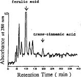
Abstract. Two days of light irradiance reduced the levels of endogenous indole-3-acetic acid (IAA) in soybean (Glycine max) hypocotyls. The key enzymes for IAA metabolic degradation are peroxidases and IAA oxidases. Peroxidase activity was enhanced about twofold by light. IAA oxidase activity also increased after irradiance. The lignin content and total phenolic compounds in soybean hypocotyls were enhanced 2_2.5 fold by light treatment. Light-induced inhibition of soybean hypocotyl growth might be due to the decline of endogenous IAA, which was degraded by IAA oxidases or peroxidases. The high levels of lignin were correlated with the increased peroxidase activity in light-treated tissues. Two phenolic compounds, ferulic acid and trans-cinnamic acid, which are precursors for lignin synthesis, greatly increased in light-treated soybean hypocotyls. This suggested that lignin synthesis catalyzed by peroxidase was enhanced during irradiance.

Keywords: Glycine max L.; IAA; Indole-3-acetic acid; Peroxidase.

Introduction

It is well known that light can inhibit the elongation of etiolated plant seedlings (Fletcher and Zalik, 1965; Jose and Vince-Prue, 1977; Ingram et al., 1983). Indole-3-acetic acid (IAA), a plant hormone, plays an important role in the promotion of hypocotyl elongation. Considerable attention has been paid to the possibility that light causes growth-inhibition by reducing the availability or effectiveness of IAA in the irradiated tissues (Iino, 1982; Jones et al., 1989; Jones et al., 1991). Free IAA can be modulated via several pathways, including IAA metabolism, synthesis and breakdown of conjugated IAA, and transport of IAA. One well known IAA metabolic pathway consists of oxidative decarboxylation of the side chain of IAA by IAA oxidase leading to the formation of either indole-3-methanol or 3-methylene oxindole (Grambow and Langenbeck-Sckwich, 1983; Beffa et al., 1990). Little is known concerning the correlation between levels of IAA and IAA oxidase activity during the processes of light-induced growth inhibition. One aim of this investigation was to examine the effect of light irradiance on soybean hypocotyl elongation through regulating the endogenous IAA levels which are modulated by IAA oxidase.

As the young seedlings in this study were exposed to light, their stems became more rigid. Hence the process of lignification in the cell wall may have occured. Numerous reports have indicated that particular peroxidases have the ability to catalyze polymerization of some aromatic alcohols to synthesized lignin (Bruce and West, 1989; Polle et al., 1994). In addition to examining the effect of light

on IAA levels, the effect of irradiance on lignification in soybean hypocotyl is also investigated.

Materials and Methods

Plant Materials

Soybean seeds (Glycine max) were obtained from the Asia Vegetable Research and Development Center, Taiwan. Dark-grown soybean seedlings were grown in vermiculite at 27 ± 0.5 °C for five days. Light-treated seedlings were grown in darkness for three days and then transferred to continuous light (16.7 W m-2) for two days. The apical region of each seedling's hypocotyl (2 cm) was excised after five days of growth for all experiments.

The Estimation of Indole-3-Acetic Acid

Extraction and partial purification—Five grams fresh weight of soybean hypocotyl segments were excised and immediately homogenized in 100 ml extracting solution with chilled motar and pestle (Guinnet et al., 1986; Dunlap and Guinn, 1989). The extracting solution containing 0.5 mg ml-1 butylated hydroxytoluene (BHT) in 80 ml of methanol and 0.5 mg ml-1 sodium ascorbate in 20 ml of distilled water prevented oxidation of IAA during extraction. The homogenates were filtered with suction through Whatman No.1 paper. Methanol was removed from the filtrate by rotary flash evaporation (RFE) at 35°C. The aqueous residue was adjusted to pH 8.0 with 0.2 N KOH and partitioned twice with an equal volume of ethyl acetate to remove phenolic compounds and other impurities. The ethyl acetate fraction was discarded and the residual ethyl acetate in aqueous phase was removed by RFE. The aque

1Corresponding author. Fax: 886-7-5319333.


Botanical Bulletin of Academia Sinica, Vol. 37, 1996

peroxidase and IAA oxidase activities. Protein concentration was determined by the method of Bradford (1976) using bovine serum albumin (BSA) as protein standard.

IAA oxidase assay—IAA oxidase activity was measured by the spectrophotometric method described by Beffa et al. (1990). Reaction mixtures contained 0.76 ml of 50 mM phosphate buffer (KH2PO4/Na2HPO4, pH 6.0), 0.01 ml of 5 mM MnCl2, 0.01 ml of 5 mM 2,4-dichlorophenol (DCP), 0.02 ml of 14.3 mM IAA and 0.2 ml of enzyme extract. The final volume of the reaction mixtures was 1 ml. Assays were conducted at 25.0 ± 0.5°C for 1 h. Salkowski reagent was used to determine IAA destruction at the wavelength of 535 nm after 1 h. One unit of IAA oxidase activity is equivalent to a DA535 of 1.0 for 1 mg of protein in 1 h. Each value represented the mean of three replicates.

Peroxidase assay—Peroxidase (EC 1.11.1.7) activity was determined spectrophotometrically by measuring the increase in absorbance at 470 nm after 30 min incubation at 30.0 ± 0.5 °C (Beffa et al., 1990; Liu and Lee, 1995). The reaction mixtures contained 0.1 ml of 50 mM H2O2, 0.02 ml of 250 mM quaiacol, 0.78 ml of 12.5 mM 3,3-dimethylglutaric acid (3,3-DGA)-NaOH and 0.1 ml of enzyme extract. The final volume of the reaction mixtures was 1 ml. One unit of peroxidase activity is equivalent to a DA470 of 1.0 for 1 mg of protein in 30 min. Each value represented the mean of three replicates.

Measurement of Lignin Content

The content of lignin was extracted and measured by the method of Bruce and West (1989). One gram fresh weight of soybean hypocotyls were ground in 25 ml of 80% ethanol with motar and pestle. The ground tissues were centrifuged at 20,000 g for 20 min at room temperature, and the residue was transferred to a glass petri dish for air drying. The resulting alcohol insoluble residue was used for lignin determination. Fifty mg of dried-residue in a glass screw-cap tube 5 ml of 2 N HCl and 0.5 ml of thioglycolic acid was added. The sealed tube was heated with a CSB-COD reactor (Model CR 1100, Bayern) at 100°C for 4 h. After cooling, the contents were centrifuged at 30,000 g for 20 min at 4°C. The supernatant was discarded and the pellet was washed once with 5 ml of water. The pellet was resuspended in 5 ml of 0.5 N NaOH and agitated gently at 25°C for 18 h to extract the lignin thioglycolate. The samples were centrifuged (30,000 g, 20 min) at 4°C and the supernatant was transferred to a test tube. One ml of HCl was added to the test tube and the lignin thioglycolic acid was allowed to precipitate at 4°C for 4 h. Following centrifugation at 30,000 g for 20 min, the pellets were dissolved in 10 ml of 0.5 N NaOH. The absorbance of the solution at 280 nm was measured. One unit of lignin content displayed corresponds to the lignin thioglycolic acid solution per gram of air dry hypocotyl tissues absorbed at the wavelength of 280 nm.

Analysis of Phenolic Compounds

Phenolic compounds were extracted by homogenizing five grams fresh weight of soybean hypocotyls in 100 ml

ous fraction was then adjusted to pH 2.5 with 0.2 N HCl and partitioned twice with an equal volume of ethyl acetate. The acidic ethyl acetate fraction was dried by RFE after removing the excess water with anhydrous sodium sulfate. The acidic residue was immediately dissolved in 1.0 ml of methanol.

Thin layer chromatography (TLC)—The acidic ethyl acetate residue in methanol was further purified by thin layer chromatography (TLC). TLC was performed on thin-layer plates (analytical Kieselgel 60, 20 × 20 cm, Schichtdicke, 0.2 mm, Merck). TLC plate was run with 100% methanol for cleaning and activated at 100°C for 1 h before loading. Up to 1.0 ml of extracts were loaded to the plate, and the plate was developed for about 1 h using benzene : acetone : acetic acid (10:4:1, v/v) as the solvent system. Identification of unknown was done by comparison with the Rfs of standard IAA running at the same time. After TLC development, the spots containing IAA were visualized by an UV lamp (Multiband UV-254/366 nm, Model UVGL-58, UVP). The IAA band on the TLC plate was scraped off and redissolved immediately in methanol. The samples were centrifuged (12,000 g, 20 min) and the supernatant was dried by RFE. The residue was dissolved in 1.0 ml of methanol and then estimated by HPLC.

High performance liquid chromatography (HPLC)—Samples partially purified by TLC were introduced to a reverse phase C-18 column (5 × 250 mm, TSK gel column, ODS-80 TM, Tosoh, Japan). The mobile phase was 35% methanol in 20 mM ammonium acetate buffer (pH 3.5) at a flow rate of 0.7 ml min-1. Column eluates were monitored with a fluorescence spectrophotometer (Model F-1050, Hitachi) using excitation and emission wavelengths of 280 nm and 360 nm respectively for detecting IAA. The retention time of eluate peaks and their areas were recorded by an integrator (Model D-2500, Hitachi). The estimation of endogenous IAA levels was done by HPLC-fluorescence methods developed by Crozier et al. (1980). The detection limit was 50 picograms.

Enzyme Preparation and Assay

Extraction and partial purification of enzymes—Five grams fresh weight of soybean apical hypocotyls were ground in 50 mM phosphate buffer (KH2PO4/Na2HPO4, pH 6.0) with chilled motar and pestle. The homogenate was centrifuged at 20,000 g for 20 min at 4°C, acetone was added to the supernatant to the final concentration of 50%, and the sample was centrifuged at 20,000 g for 20 min at 4°C to obtain the 50% acetone precipitated protein fraction. The precipitated protein fraction was dissolved in 2 ml of 50 mM phosphate buffer (pH 6.0). After centrifugation (20,000 g, 20 min) at 4°C, the supernatant was collected for analysis of enzyme activities.

For further enzyme fractionation, the 50% acetone precipitated protein fraction was applied to a column (1.0 × 90 cm) packed with Sephacryl S-100 gel (Pharmacia, Sweden) previously equilibrated with 50 mM phosphate buffer (pH 6.0) at a flow rate of 0.25 ml min-1. Fractions were collected (1.0 ml each fraction) for determination of


Liu et al. — Effect of light on indole-3-acetic acid in soybean hypocotyls

of 80% methanol with chilled motar and pestle. The sample was filtrated with suction through Whatman No.1 paper. Methanol in filtrate was removed by RFE at 35°C. The aqueous residue was adjusted to pH 7.6 with 0.2 N KOH and partitioned twice with an equal volume of diethyl ether. The organic fraction was collected and reduced to dryness by RFE. The residue was dissolved immediately in 1 ml of methanol. The quantitative assay of phenolic compounds was followed to Ettinger et al. (1951) using phenol as standard. The reaction mixtures contained 50 ml of 50 mg ml-1 NH4Cl (pH 10.0), 15 ml of 20 mg ml-1 aminoantipyrine, 15 ml of 80 mg ml-1 potassium ferricyanide, 2.5 ml of H2O and 100 ml of extract. The change in the absorbance at 510 nm was measured after 20 min incubation at 25.0 ± 0.5 °C. The concentration of phenolic compounds was expressed as mg l-1 of phenol per gram fresh weight of tissue.

Phenolic compounds were also separated and estimated by HPLC. Samples were injected into a reverse phase C-18 column (5 × 250 mm, TSK gel column, ODS-80TM, Tosoh, Japan) and separated with 40% methanol in 20 mM sodium acetate buffer (pH 5.0) at a flow rate of 0.7 ml min-1. Ferulic acid and trans-cinnamic acid were detected using an UV spectrophotometer (Model VL-613, Jasco) at 280 nm. The identity of these compounds in plant extracts was confirmed by co-chromatography with the authentic standards purchased from Sigma under the same HPLC conditions (Yalpani et al., 1993).

Results

Effects of Light on Hypocotyl Growth and IAA Levels

The preliminary test indicated that two days of continuous irradiance maximumly inhibited the elongation of soybean hypocotyl (data not shown). As shown in Table 1, two days of light irradiance reduced the length of soybean hypocotyl by 25%. The result confirmed the reports of previous studies using different plant species (Jose and Vince-Prue, 1977; Gaba and Black, 1979). Although the extracts of dark-grown and light-treated soybean hypocotyl after TLC purification were still far from being pure, the IAA peak was well separated from other impurities (Figure 1A and 1B). In order to make sure that the peak represented IAA, not the other similar compounds, another run of extract spiked with standard IAA showed that there was only one peak with the identical retention time (data not shown). The levels of IAA in light-treated soybean hypocotyl were reduced 28% (Table 1). The results confirmed the data reported by Fletcher and Zalik (1965) and Muir (1970). During the processes of IAA extraction, partition, and TLC purification, the recovery rate of IAA was 72%.

Enzymes Assay and Fractionation

IAA oxidase and peroxidase activities—As shown in Table 1 and 2, both IAA oxidase and peroxidase activities of light-treated soybean hypocotyls were higher than that of dark grown tissues. Peroxidase activity after irradiance

Figure 1. HPLC elution profile of soybean hypocotyl extracts of dark-grown (A) and light-grown seedlings (B) after TLC purification. Column: 5 × 250 mm, TSK gel; mobile phase: 35% methanol in 20 mM ammonium acetate buffer, pH 3.5; Flow rate: 0.7 ml min-1; Fluorometric detection with excitation and emission wavelengths of 280 nm and 360 nm, respectively.


Botanical Bulletin of Academia Sinica, Vol. 37, 1996

Table 1. The effect of light on length of soybean seedlings, IAA contents and IAA oxidase activity. One unit of IAA oxidase activity is equivalent to a DA535 of 1.0 for 1 mg of protein in 1 h.

Treatment Darkness Light

Lengtha (cm) 9.57 ± 0.34 7.24 ± 0.35

IAA contentb (ng/g FW) 9.06 ± 0.60 6.42 ± 0.53

IAA oxidase activityc 27.7 ± 6.40 33.5 ± 4.60

aValues are means ± SE of ten replications.

bData are means ± SE of five replications.

cValues are means ± SE of triplicate treatments.

lignin content of light-treated soybean hypocotyl was two times higher than that of dark grown tissues. Similarly, the total phenolic compounds of light-treated soybean hypocotyl were two and a half times higher than that of dark grown tissues. In addition, two phenolic compounds, ferulic acid and trans-cinnamic acid, which are precursors for lignin synthesis, were examined and estimated by HPLC. As shown in Figure 4, ferulic acid was eluted with the retention time at 9.3 min and trans-cinnamic acid was eluted at 20.7 min. The levels of ferulic acid and trans-cinnamic acid were greatly enhanced after irradiance (Table 2).

Discussion

The light-induced growth inhibition of young seedlings occurs in pea, bean, and corn. The regulation of pea stem

Table 2. The effect of light on levels of lignin, ferulic acid, trans-cinnamic acid and peroxidase activity in soybean hypocotyls. One unit of peroxidase activity is equivalent to a DA470 of 1.0 for 1 mg of protein in 30 min. One unit of lignin content displayed correspond to the absorbance at 280 nm, of the lignin thioglycolic acid solution per gram of air dry hypocotyl tissues. The concentration of total phenolic compounds were expressed as mg l-1 of phenol per gram fresh weight of tissue.

Treatment Darkness Light

Lignina 4.92 ± 0.66 10.76 ± 1.41

Total phenolic compounda 10.62 ± 1.74 24.49 ± 3.85

Ferulic acidb (mg/g FW) 3.29 ± 0.56 8.51 ± 0.42

trans-cinnamic acidb (mg/g FW) 50.90 ± 13.20 224.9 ± 11.2

Peroxidase activityb 158.60 ± 3.10 280.70 ± 8.10

aValues are means ± SE of four replications.

bData are means ± SE of three replications.

was enhanced about twofold. IAA oxidase activity was increased by 17%. The 50% acetone precipitation of soybean hypocotyl extract obtained from dark grown and light-treated soybean seedlings were applied to a Sephacryl S-100 column (1.0 × 90 cm, Bio-Rad). The crude extract of dark grown soybean hypocotyls eluted from the Sephacryl S-100 column was given in Figure 2. There were three peaks of peroxidase activity with molecular weights estimated to be 90 kDa, 23 kDa, and 18 kDa, respectively. Two peaks of IAA oxidase activity with molecular weights estimated to be 23 kDa and 18 kDa were observed in dark grown soybean hypocotyl extracts. In light-treated tissues, a similar elution profile was obtained (Figure 3). Among these peroxidase isozymes, the activity of peroxidase (90 kDa) in light-treated tissues was about three times higher than that of dark grown tissues. In IAA oxidase isozymes, there was no significant difference between light-treated and dark-grown tissues in terms of IAA oxidase activity. The elution profiles of soybean hypocotyl extracts (Figure 2 and Figure 3) confirmed the data in published papers that dual catalysis was present in IAA oxidase and peroxidase isozymes (Grambow and Langenbeck-Sckwich, 1983; Pressey, 1990; Quesada et al., 1992).

Effect of Light on Levels of Lignin and Phenolic Compounds

To investigate how light affects the lignification of soybean hypocotyl, the levels of lignin and phenolic compounds have to be measured. As shown in Table 2, the

Figure 2. Elution profile of 50% acetone precipitated protein fraction of dark-grown soybean seedlings on a Sephacryl S-100 column. Protein concentration is given in mg ml-1. One unit of IAA oxidase activity is equivalent to a DA535 of 1.0 for 1 mg of protein in 1 h. One unit of peroxidase activity is equivalent to a DA470 of 1.0 for 1 mg of protein in 30 min. The molecular weight corresponding to each peak fraction was expressed in kDa.

Figure 3. Elution profile of 50% acetone precipitated protein fraction of light-grown soybean seedlings on a Sephacryl S-100 column. Protein concentration, IAA oxidase and peroxidase activities are defined as in Figure 2.


Liu et al. — Effect of light on indole-3-acetic acid in soybean hypocotyls

gation, we obtained three peaks of peroxidase activity obtained with molecular weights estimated to be 90 kDa, 23 kDa, and 18 kDa, respectively. As far as the relationship between IAA oxidase and peroxidase is concerned, our data (Figure 2 and 3) confirmed the dual catalysis of these two groups of isozymes reported by many investigators (Grambow and Langenbeck-Schwich, 1983; Pressey, 1990; Quesada et al., 1992). In this study, IAA oxidase could be considered as the IAA oxidation activity by peroxidases (Nakajima and Yamazaki, 1979; Pressey, 1990). The 90 kDa peroxidase activity in light-treated soybean hypocotyl enhanced about threefold by light are probably associated with the degradation of IAA. In addition, the levels of two phenolic compounds, ferulic acid and trans-cinnamic acid, which are precursors for lignin synthesis (Nimz, 1974), also significantly increased (Table 2). Light-induced inhibition of soybean hypocotyl growth might be due to the decline of endogenous IAA which was degraded by IAA oxidase. The inhibition of soybean hypocotyl growth by light was accompanied by the increase of levels of both lignin precorsors (ferulic acid and trans-cinnamic acid) and lignin. This suggested that lignin synthesis catalyzed by peroxidase was enhanced during irradiance. Peroxidases involved in lignification processes are known to be localized in the cell wall (Sato et al., 1993), yet, some investigators have reported that the isoperoxidases involved in polymerization of lignin monomer were localized in the free space of the wall. The phosphate buffer used in this study for extracting the cytoplasmic enzymes could then solubilize these isoperoxidases (Mäder et al., 1975; Imberty et al., 1985). Numerous reports have indicated that either anionic (acidic) or cationic (alkaline) peroxidase isozymes are involved in lignification (Sato et al., 1993; Polle et al., 1994). The activity of peroxidase, especially the 90 kDa peak, might be correlated with the lignin synthesis previously reported by these investigators. A large amount of enzyme extract for the estimation of molecular weight as well as pI determination of peroxidase isozymes is needed to elucidate the detailed relationship between the lignification of cell wall and the action of peroxidases.

Acknowledgements. The authors wish to thank Dr. T.-Y. Feng, Research Fellow of the Institute of Botany, Academia Sinica (ROC), for his discussion and suggestions.

Literature Cited

Beffa, R., H. V. Martin, and P.-E. Pilet. 1990. In vitro oxidation of indoleacetic acid by soluble auxin-oxidases and peroxidases from maize roots. Plant Physiol. 94: 485_491.

Bradford, M. M. 1976. A rapid and sensitive method for the quantitation of microgram quantities of protein utilizing the principle of protein-dye binding. Anal. Boichem. 72: 248_254.

Bruce, R. J. and C. A. West. 1989. Elicitation of lignin biosynthesis and isoperoxidase activity by pectic fragments in suspension cultures of castor bean. Plant Physiol. 91: 889_897.

Campell, B. R. and B. A. Bonner. 1986. Evidence for phyto

Figure 4. HPLC spectrum of phenolic compounds in soybean hypocotyl. Column: 5 × 250 mm, TSK gel; mobile phase: 40% methanol in 20 mM sodium actate buffer, pH 5.0; Flow rate: 0.7 ml min-1; phenolic compounds were detected with UV spectrophotometer at 280 nm.

elongation by the levels of endogenous gibberellin (GA1), another plant hormone, has been reported by many investigators (Ross et al., 1989; Weller et al., 1994). They found that GA1 was either absent, or present in underdetectable levels in the dwarf pea grown in light. Campell and Bonner (1986) suggested that light can prevent the accumulation of biologically active GA1. In our studies, two days of irradiance reduced the length of soybean hypocotyls by about 25%, which was accompanied by a 28% decline of IAA content in light-treated hypocotyls (Table 1 ). In this study, the enhancement of IAA oxidase activity by light suggests that this may be the result of the decrease of IAA levels in light-treated hypocotyls. Some investigators have reported that the decline of IAA levels in light-treated tissues was due to the decrease of IAA transport or IAA synthesis (Sherwin and Furuya, 1973; Jones et al., 1991). However, our data indicate that the decline in IAA content may be due to the increased degradation of IAA, which is catalyzed by IAA oxidase. IAA oxidase might degrade IAA through the oxidation of the indolenucleus (Tsurumi and Wada, 1980) or through the oxidative decarboxylation of the side chain (Beffa et al., 1990). After gel filtration chromatography on a Sephacryl S-100 column, two IAA oxidases (23 kDa and 18 kDa) were obtained in our study (Figure 2 and 3). Further purification of these two isozymes will help us elucidate the detailed IAA metabolism in soybean hypocotyls. In light-mediated processes of photomorphogenesis, phytohormone regulates plant growth probably through the regulation of free IAA levels or auxin receptors (Jones et al., 1989).

The lignin content and total phenolic compounds of light-treated soybean hypocotyl were remarkably enhanced by irradiance. The high levels of lignin were correlated with the increased peroxidase activity in light-treated tissues (Table 2). Peroxidases are isozymes. In our investi


Botanical Bulletin of Academia Sinica, Vol. 37, 1996

chrome regulation of gibberellin A20 3b-hydroxylation in shoots of dwarf (lele) Pisum sativum L. Plant Physiol. 82: 909_915.

Crozier, A., K. Loferski, J. B. Zaeer, and R. O. Morris. 1980. Analysis of picogram quantities of indole-3-acetic acid by high performance liquid chromatography-fluorescence procedures. Planta 150: 366_370.

Dunlap, J. R. and G. Guinn. 1989. A simple purification of indole-3-acetic acid and abscisic acid for GC-SIM-MS analysis by micro-filtration of aqueous sample through nylon. Plant Physiol. 90: 197_201.

Ettinger, M. B., C. C. Ruchhoft, and R. J. Lishka. 1951. Sensitive 4-aminoantipyrine method for phenolic compounds. Anal. Chem. 23: 1783.

Fletcher, R. A. and S. Zalik. 1965. Effect of light quality on growth and free IAA content in Phaseolus vulgaris. Plant Physiol. 39: 328_331.

Gaba, V. and M. Black. 1979. Two separate photoreceptors control hypocotyl elongation in green seedlings. Nature 278: 51_54.

Grambow, H. J. and B. Langenbeck-Schwich. 1983. The relationship between oxidase activity, peroxidase activity, hydrogen peroxide, and phenolic compounds in the degradation of indole-3-acetic acid in vitro. Planta 157: 131_137.

Guinnet, G., D. L. Brummett, and R. C. Beier. 1986. Purification and measurement of abscisic acid and indoleacetic acid by high performance liquid chromatography. Plant Physiol. 81: 997_1002.

Iino, M. 1982. Inhibitory action of red light on the growth of the maize mesocotyl: evaluation of the auxin hypothesis. Planta 156: 388_395.

Imberty, A., R. Goldberg, and A.-M. Catesson. 1985. Isolation and characterization of Populus isoperoxidases involved in the last step of lignin formation. Planta 164: 221_226.

Ingram, T. J., J.B. Reid, W. C. Potts, and I. C. Murfet. 1983. Internode length in Pisum. IV. The effect of the Le gene on gibberellin metabolism. Physiol. Plant. 59: 607_616.

Jose, A. M. and D. Vince-Prue. 1977. Action spectra for the inhibition of growth in radish hypocotyls. Planta 136: 131_134.

Jones, A. M., P. M. Lamerson, and M. A. Venis. 1989. Comparison of site I auxin binding and a 22-kilodalton auxin-binding protein in maize. Planta 179: 409_413.

Jones, A. M., D. S. Cochran, P. M. Lamerson, M. L. Evans, and J. D. Cohen. 1991. Red light-regulated growth. 1. Changes in the abundance of indoleacetic acid and a 22-kilodalton

auxin-binding protein in the maize mesocotyl. Plant Physiol. 97: 352_358.

Liu, Z.-H. and B.-H. Lee. 1995. Changes of endogenous indole-3-acetic acid, peroxidases, and auxin oxidases during pollen germination in maize. Bot. Bull. Acad. Sin. 36: 53_58.

Mäder, M., Y. Meyer, and M. Bopp. 1975. Lokalisation der Peroxidase Isoenzyme in Protoplasten und Zellwänder von Nicotiana tabacum L. Planta 122: 250_268.

Muir, R. M. 1970. The control of growth by the synthesis of IAA and its conjugation. In D. J. Carr (ed.), Plant Growth Substances. Springer-Verlag, Berlin, Heidlberg, New York, pp. 96_101.

Nakajima, R. and I. Yamazaki. 1979. The mechanism of Indole-3-acetic Acid Oxidation by Horsrradish peroxidases. J. Biol. Chem. 254: 872_878.

Nimz, H. 1974. Beech lignin-proposal of a consititutional scheme. Angew. Chem. Int. Ed. Engl. 74: 313_321.

Polle, A., T. Otter, and F. Seifert. 1994. Apoplastic peroxidases and lignification in needles of Norway spruce (Picea abies L.). Plant Physiol. 106: 53_60.

Pressey, R. 1990. Anions activate the oxidation of indoleacetic acid by peroxidases from tomato and other sources. Plant Physiol. 93: 798_804.

Quesada, M. A., C. Sánchez-Roldán, A. Heredia, V. Valpuesta, and M. J. Bukovac. 1992. Peroxidase and IAA oxidase activities and peroxidase isoenzymes in the pericarp of seeded and seedless "Redhaven" peach fruit. J. Plant Growth Regul. 11: 1_6.

Ross, J. J., J. B. Reid, P. Gaskin, and J. MacMillan. 1989. Internode length in Pisum. Estimation of GA1 levels in genotypes Le, le, and led. Physiol. Plant. 76: 173_176.

Sato, Y., M. Sugiyama, R. J. Górecki, H. Fukuda, and A. Komamine. 1993. Interrelationship between lignin deposition and the activities of peroxidase isoenzymes in differentiating tracheary elements of Zinnia. Planta 189: 584_589.

Sherwin, J. E. and M. Furuya. 1973. A red-far red reversible effect on uptake of exogenous indole-acetic acid in etiolated rice coleoptiles. Plant Physiol. 51: 295_298.

Tsurumi, S. and S. Wada. 1980. Metabolism of indole-3-acetic acid and natural occurrence of dioxindole-3-acetic acid derivatives in Vicia roots. Plant Cell Physiol. 21: 1515_1525.

Weller, J. L., J. J. Ross, and J. B. Reid. 1994. Gibberellins and phytochrome regulation of stem elongation in pea. Planta 192: 489_496.

Yalpani, N., J. León, M. A. Lawton, and I. Raskin. 1993. Pathway of salicylic acid biosynthesis in healthy and virus-inoculated tobacco. Plant Physiol. 103: 315_321.


Liu et al. — Effect of light on indole-3-acetic acid in soybean hypocotyls