Bot. Bull. Acad. Sin. (1996) 37: 121-126

Yang et al. — Pigment and surfactant

Pigment solubilization of the chloroplast thylakoid membranes by a surfactant

Chi-Ming Yang1, Jen-Chieh Hsu, Yih-Kuang Lu, and Ming-Horng Yin

Institute of Botany, Academia Sinica, Nankang, Taipei, Taiwan 115, Republic of China

(Received February 13, 1995; Accepted January 29, 1996)

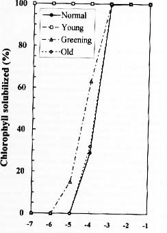
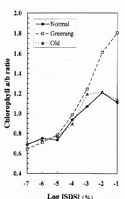
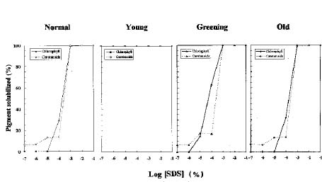
Abstract. We examined the influence of various concentrations of sodium dodecyl sulfate (SDS) on the pigment solubilization of the chloroplast thylakoid membranes isolated from leaves at different developing stages of normal and Golden-leaves figs (Ficus microcarpa). When the SDS concentration was lower than 10-5 %, more carotenoids, especially neoxanthin, were released than chlorophyll (Chl), and more Chl b was released than Chl a. When SDS concentration was between 10-5 % and 10-3 %, more Chl molecules were released than carotenoids. As the SDS concentration was increased above 10-3 %, all Chl molecules and carotenoids were solubilized. Among the solubilized carotenoids, the release sequence from thylakoid membrane may be neoxanthin, antheraxanthin, (lutein, violaxanthin, taraxanthin, b-carotene), a-carotene, and zeaxanthin. We conclude that in the pigment-protein complexes, carotenoids (especially neoxanthin) are more susceptible to SDS than Chl molecules, that Chl b is more susceptible to SDS than Chl a, and that neoxanthin may be directly exposed to the stroma face of the thylakoid membrane.

Keywords: Carotenoid; Chlorophyll; Neoxanthin; Pigment-protein complex; Release sequence; Solubilization; Surfactant; Thylakoid membrane.

Introduction

Surfactants have been widely used to study the organization and constituents of biological membranes (Helenius and Simons, 1975; Rosen, 1978). Much of our current knowledge about the structure and molecular organization of the higher plant photosynthetic apparatus is derived from studies using surfactants. The isolation of pigment-protein complexes from higher plant chloroplasts was performed by the aid of surfactants (Markwell et al., 1978; Markwell, 1986). Many surfactant systems developed to fractionate photosynthetic pigment-protein complexes, however, may not fully solubilize the complexes prior to the electrophoretic step (Allgood et al., 1991). The lateral distribution of thylakoid membrane components was obtained by differential solubilization of the chloroplast thylakoid membranes with surfactants (see refs. of Markwell and Thornber, 1982; Bartzatt et al., 1983). Many photochemical activities of photosynthetic apparatus are dramatically altered after surfactant treatment (Apostolova and Ivanov, 1995). Although it has been reported that the photosynthetic apparatus of one organism Dunaliella teriolecta appears to be unusually sensitive to surfactant Triton X-100 (Sukenik et al., 1989), the surfactant concentrations usually used in the litera

ture have produced similar results with most species. Chloroplast thylakoid protein phosphatase was monitored by Triton X-100 to be membrane surface-associated (Sun et al., 1989). Studies at sub-solubilizing concentrations have indicated that the primary interaction between the surfactant and the thylakoid membrane may involve adsorption at the membrane/solution interface rather than insertion of surfactant molecules into the membrane, and that surfactants may specifically interact with exposed PSI components on the surface of the thylakoid membrane (Bartzatt et al., 1983).

The perturbation of Chl molecules within pigment-protein complexes is a sensitive indicator of change in the local environment of thylakoid membranes. Little information is available about the selective effect of surfactants on the thylakoid membrane components. There have been no reports at all, however, on the selective or differential solubilization of Chl a and b and carotenoids from the thylakoid membrane. The aim of this research is to investigate the selective effect of surfactants on the pigment solubilization of the chloroplast thylakoid membrane in higher plants.

Materials and Methods

Plants

About one-year-old normal and Golden-leaves figs (Ficus microcarpa cv. Golden-leaves) about 50 cm in height were purchased from a local nursery and grown for 6 weeks in a soil-vermiculite mixture in a greenhouse under natural light in the summer.

1Corresponding author. Fax: 886-2-7827954.


Botanical Bulletin of Academia Sinica, Vol. 37, 1996

Pigment Determination

Leaves were harvested and thylakoid membranes were isolated as previously described (Markwell, 1986). The concentrations of Chl and carotenoid were determined according to the spectrophotometric methods of Porra et al (1989) and Kirk and Allen (1965), respectively, following the extraction of leaf or thylakoid membranes with 80% acetone. Absorbance was measured with a Hitachi U2000 UV-visible spectrophotometer.

Pigment Solubilization

To study the interaction of surfactant with thylakoid membranes, 1 ml of fresh SDS solution in 50 mM Tris-HCl (pH 8.0) was added to an equal volume of fresh thylakoid membranes containing 15 µg ml-1, both being twice the desired final concentration (Bartzatt et al., 1983; Lu et al., 1995). The mixture was incubated at 25°C for 10 min prior to measuring the solubilization of pigments. The mixture was centrifuged at 3,000 g for 10 min at room temperature. The concentrations of Chl and carotenoid in the supernatant were determined as described above by mixing 0.2 ml supernatant liquid and 0.8 ml acetone. The centrifugal force used to analyze the samples following SDS treatment was lower than that used by other authors (Bartzatt et al., 1983). The low centrifugal force was used to avoid mechanically disturbing the possible weak interaction between the thylakoid membranes and the SDS concentration, which was much lower than that used in the literature. The results were reproducible. No significant difference between the low centrifugal force used in the present research and the high centrifugal force used in the literature (Bartzatt et al., 1983) was observed.

High Performance Liquid Chromatography (HPLC)

The above supernatant was combined with 1/5 the volume of 0.5 M KCl, stored at 4°C for 1 h, and then centrifuged at 3,000 g for 10 min at 4°C. The solubilized pigments in the supernatant were extracted with an equal volume of ether according to the method of Braumann and Grimme (1981). After being dried in nitrogen, the solubilized pigments were dissolved in 1 ml of ether. The sample was stored in the dark at -80°C until analyzed. The sample was filtrated with Nylon acrodisk (13 mm HPLC certified 0.45 µm) just before injection. HPLC was performed on a Vercopak inertsil 10 ODS 25 × 4.6 mm C18 reverse phases column (10 µm particle size). The sample was injected into the column by a Waters U6K injector, and two mobile phases were set up as previously described (Lu et al., 1995). Mobile phases were pumped by a Waters M510 high pressure pump and Waters M680 automatic gradient controller at the flow rate of 2.5 ml min-1. Peaks were detected at 445 nm by a Waters Lamba-Max M481 detector and were identified by standard methods as previously described (Val et al., 1986).

Results and Discussion

Bartzatt et al. (1983) demonstrated that the presence of thylakoid membranes at a concentration of 30 µg Chl ml-1 slightly increased the amount of SDS needed to reach the critical micelle concentration. In this report, half of the above concentration of thylakoid membrane isolated from the leaf of normal fig and the young, greening, and old leaves of Golden-leaves fig was used. In addition, the concentration of SDS used in this report was about 1,000-fold lower than that used in a previous study (Bartzatt et al., 1983).

As the concentration of SDS added to the thylakoid membrane solution (15 mg g-1 of Chl) increased, the amount of Chl released from thylakoid membranes also increased (Figure 1). The thylakoid membranes isolated from the young leaf of Golden-leaves fig were completely solubilized when SDS concentration was as low as 10-7 % (equivalent to 10-6 mg ml-1). Chl solubilization of the

Figure 1. Effect of SDS concentration on the chlorophyll solubilization of the thylakoid membrane isolated from normal fig and the young, greening and old leaves of Golden-leaves fig. Relative amount of total chlorophyll remaining in the supernatant fraction following centrifugation at 3,000 g for 10 min is shown versus SDS concentration. Thylakoid membrane concentration was 15 µg ml-1 of chlorophyll.


Yang et al. — Pigment and surfactant

greening leaf of Golden-leaves fig begins when SDS concentration attains a level of 10-6 %, and that of the leaf of normal fig and the old leaf of Golden-leaves fig at a level of 10-5 %. At the level of 10-5 %, about 65% of Chl was released from the greening leaf of Golden-leaves fig, and about 30% from the leaf of normal and the old leaf of Golden-leaves fig. All Chl molecules were totally solubilized from the thylakoid membranes when the SDS concentration reached 10-3 %. The results were consistent with the transmission electron microscopic data, which showed that the young leaf of Golden-leaves fig contains no more than a single layer of thylakoid membrane and a very limited amount of stacked grana, and that the greening leaf of Golden-leaves fig contains several layers of thylakoid membrane and less stacked grana than that of the leaf of normal fig and the old leaf of Golden-leaves. The latter two should have similar chloroplasts (data not shown). In other words, the younger the leaf, the more accessible

to SDS the thylakoid membranes are due to the lack of stacked grana.

The a/b ratios of Chl remaining in the supernatant fractions following centrifugation were determined to compare the release of Chl a and b from the thylakoid membranes (Figure 2). When SDS concentration was lower than 10-5 %, the Chl a/b ratio was approximately 0.7 in all kinds of thylakoid membranes except the young leaf of Golden-leaves fig, indicating that relatively more Chl b was released than Chl a at low level of SDS. In contrast, as the SDS concentration was increased above 10-3 %, the Chl a/b ratio was approximately 1.2 in the leaf of normal fig and the old leaf of Golden-leaves fig and was greater than 1.2 in the greening leaf of Golden-leaves fig, indicating that relatively more Chl a was released than Chl b at a high level of SDS. When the SDS concentration is between 10-4 and 10-3 %, almost equal

Figure 3. Effect of SDS concentration on the carotenoid solubilization of the thylakoid membrane isolated from normal fig and the young, greening and old leaves of Golden-leaves fig. Relative amount of total carotenoid remaining in the supernatant fraction following centrifugation at 3,000 g for 10 min is shown versus SDS concentration. Thylakoid membrane concentration was 15 µg ml-1 of chlorophyll.

Figure 2. Effect of SDS concentration on the a/b ratios of chlorophyll solubilized from the thylakoid membrane isolated from normal fig and the greening and old leaves of Golden-leaves fig.


Botanical Bulletin of Academia Sinica, Vol. 37, 1996

carotene were abundantly released, occupying 11.3, 4.8, 3.5, and 1.0%, respectively. About 16% of solubilized pigments were xanthophyll cycle components . Therefore, it seems that the release sequence of pigments in the thylakoid membrane is: (i) neoxanthin is the first release carotenoid; (ii) antheraxanthin is the second; (iii) lutein, b-carotene, violaxanthin, and taraxanthin are the third release; (iv) a-carotene is the fourth release; and (v) zeaxanthin is the most difficult release.

As surfactant concentration increases, three types of interaction between the membrane and the surfactants take place (Bartzatt et al., 1983). At a low level of surfactants there is a high-affinity interaction between monomeric surfactant molecules and the membrane, occurring largely at the membrane/solution interface. Stroma-exposed pigment-protein complexes in the thylakoid membrane and surface-exposed pigments in the pigment-protein complexes appear to be more susceptible to surfactants. As the surfactant concentration increases further, significant amounts of surfactant are incorporated into the membrane, disrupting intercomplex interaction and releasing the stroma- and/or surface-exposed pigments. After still further increases in surfactant, leading to a concentration that exceeds the critical micelle concentration, solubilization of thylakoid membrane occurs. At this level, intracomplex interactions between pigments and polypeptides are totally disrupted, causing the release of all pigments.

It has been established that core complex (CCI) of photosystem I (PSI) contains 1 b-carotene and 40 chlorophyll a; light-harvesting complex I (LHCI) of PSI contains lutein, violaxanthin and neoxanthin; CCII of PSII contains 50 chlorophylls and 7 b-carotene; and LHCII of PSII

amounts of Chl a and b may be released. All Chl and carotenoid molecules are associated with several polypeptides to form pigment-protein complexes (Markwell et al., 1979; Staehelin, 1986). Photosynthetic pigment-protein complexes contain various ratios of Chl a and b and those pigments are located in different sites of the complexes (Lam et al., 1984a and 1984b; Jansson, 1994; Kuhlbrandt et al., 1994). Therefore, it is reasonable to conclude that Chl molecules leased than carotenoids. While the leaf of normal fig and the old leaf of Golden-leaves fig began the solubilization of Chl at a level of 10-5 %, the greening leaf of Golden-leaves fig began at 10-6 %. Regardless of leaf type, all pigments were totally solubilized when SDS concentration was higher than 10-3 %. That is, a high level of SDS can solubilize the normal thylakoid membrane containing grana and release all pigments.

To determine the release sequence of carotenoids when thylakoid membrane was solubilized with various concentrations of SDS, the pigments in the solubilized fraction were extracted, concentrated, and analyzed by HPLC (Table 1). When the SDS concentration was equal to or less than 10-3 %, around 90% of solubilized carotenoids were neoxanthin, only around 6% or less were chlorophyll and around 3% were xanthophyll cycle components (violaxanthin + antheraxanthin + zeaxanthin). When the SDS concentration was between 10-3 % and 10-1 %, besides neoxanthin, lutein, chlorophyll a and b were abundantly released, occupying approximately 11, 11.5, and 11.3%, respectively. About 5% were xanthophyll cycle components and 3% were b-carotene. When the SDS concentration was higher than 10-1%, besides the above pigments, violaxanthin, taraxanthin, zeaxanthin, and a-

Figure 4. Effect of SDS concentration and age of leaf on the pigment solubilization of the thylakoid membrane. Relative amount of total chlorophyll and carotenoid remaining in the supernatant fraction following centrifugation at 3,000 g for 10 min is shown versus SDS concentration. Thylakoid membrane concentration is 15 µg ml-1 of chlorophyll.


Yang et al. — Pigment and surfactant

Table 1. Percentage of carotenoids in the SDS-solubilized fraction. The result was the mean of three determinations. The standard deviation was less than 10%.

Pigments SDS concentration (%)

0 10-5 10-4 10-3 10-2 10-1 1 10

Neoxanthin (N) 2.8 87.5 91.0 88.5 59.0 54.0 21.9 8.0

Violaxanthin (V) 6.0 0.3 0.4 0.3 1.8 1.9 11.3 11.4

Taraxanthin (T) 5.2 0.3 0.3 0.3 1.0 0.8 4.2 5.5

Antheraxanthin (A) 1.2 0.2 0.4 0.7 1.3 1.0 0.8 1.3

Lutein (L) 19.4 2.1 2.0 1.7 10.5 12.0 17.7 23.3

Zeaxanthin (Z) 1.8 2.4 1.9 2.3 1.9 1.9 3.7 3.2

Chlorophyll b (b) 17.1 2.1 0.9 2.2 11.5 11.6 16.9 21.2

Chlorophyll a (a) 38.5 3.9 2.3 2.2 9.8 12.9 18.2 18.9

a-carotene (a) 0.7 0.2 0.2 0.1 0.2 0.7 1.1 0.9

b-carotene (b) 7.3 1.1 0.7 1.7 3.1 3.2 4.5 6.2

V+A+Z 9.0 2.9 2.7 3.1 4.8 5.0 15.8 15.9

Total 100 100 100 100 100 100 100 100

contains 4 chlorophyll a, 3 chlorophyll b, and 2 luteins (Lam et al., 1984a and 1984b; Jansson, 1994; Kuhlbrandt et al., 1994). The two luteins form an internal cross-brace in the center of the LHCII, providing a direct, strong link between the peptide loops at both surfaces (Kuhlbrandt et al., 1994). Therefore, it seems reasonable to propose that: (i) neoxanthin of LHCI is located at the hydrophilic surface of this pigment-protein complex or is directly exposed to the stroma and is the most accessible component of PSI to a low concentration of SDS, taking place prior to the release of intact pigment-protein complex from thylakoid membranes or the disruption of intercomplexes such as the interaction between LHCI and CCI, or the interaction between LHCII and CCII; (ii) some of the pigments which may be located at the contact faces of pigment-protein complexes are solubilized at the same moment that the intercomplex interaction is disrupted as SDS concentration increases; (iii) when the SDS concentration closes to critical micelle concentration, the intracomplex interaction between pigments and peptides begins to be disrupted, leading to the pigments located at the most inner site, such as lutein in LHCI or LHCII, being released from the complexes; and (iv) when SDS concentration increases further, all components in the thylakoid membrane are totally solubilized.

In this report, we have characterized the pigment solubilization of the thylakoid membranes at low concentration of SDS and have shown: (a) the maturity of thylakoid membranes affects the solubilization of pigments; (b) at a low level of SDS, more carotenoids were solubilized than Chl molecules and more Chl b was solubilized than Chl a; (c) at a medium level of SDS, more Chl molecules were solubilized than carotenoid; and (d) at a high level of SDS, all pigments were completely solubilized. It appears that carotenoids are more susceptible to SDS than Chl b, which is more susceptible than Chl a. Among the solubilized carotenoids, neoxanthin is the most easily accessible to low concentrations of surfactant, indicating it is directly exposed to the stroma face of the thylakoid membrane.

Literature Cited

Allgood, L., R. D. Curtright, and J. P. Markwell. 1991. Solubilization of photosynthetic pigment-protein complexes. Photochem. Photobiol. 54: 459_463.

Apostolova, E. L. and A. G. Ivanov. 1995. Influence of Triton X-100 on the structure and functions of pea thylakoid membranes. J. Plant Physiol. 145: 239_244.

Bartzatt, R. A., C. M. Yang, and J. P. Markwell. 1983. The interaction of surfactants with the chloroplast thylakoid membrane at sub-solubilizing concentration. Biochim. Biophys. Acta 725: 341_348.

Braumann, T. and L. H. Grimme. 1981. Reversed-phase high performance liquid chromatography of chlorophylls and carotenoids. Biochim. Biophys. Acta 637: 8_17.

Helenius, A. and K. Simons. 1975. Solubilization of membranes by detergents. Biochim. Biophys. Acta 425: 29_79.

Jansson, S. 1994. The light-harvesting chlorophyll a/b-binding proteins. Biochim. Biophys. Acta 1184: 1_19.

Kirk, J. T. and R. L. Allen. 1965. Dependence of chloroplast pigment synthesis on protein synthesis. Biochem. Biophys. Res. Comm. 21: 523_530.

Kuhlbrandt, W., D. N. Wang, and Y. Fujiyoshi. 1994. Atomic model of plant light-harvesting complex. Nature 367: 614_621.

Lam, E., W. Irtiz, and R. Malkin. 1984a. Chlorophyll a/b proteins of photosystem I. FEBS Lett. 168: 10_14.

Lam, E., W. Ortiz, S. Mayfield, and R. Malkin. 1984b. Isolation and characterization of a light-harvesting chlorophyll a/b protein complex associated with photosystem I. Plant Physiol. 74: 650_655.

Lu, Y. K., C. M. Yang, and Y. R. Chen. 1995. Characterization of the thylakoid membrane in a chlorophyll-deficient ch5 mutant of Arabidopsis thaliana. Bot. Bull. Acad. Sin. 36: 33_40.

Markwell, J. P. 1986. Electrophoretic analysis of photosynthetic pigment-protein complexes. In M. F. Hipkins and N. R. Baker (eds.), Photosynthesis Energy Transduction: A Practical Approach, IRL press, Oxford, England, pp. 27_49.

Markwell, J. P., S. Reinman, and J. P. Thornber. 1978. Chlorophyll-protein complexes from higher plants: a procedure for improved stability and fractionation. Arch. Biochem.


Botanical Bulletin of Academia Sinica, Vol. 37, 1996

Biophys. 190: 136_141.

Markwell, J. P. and J. P. Thornber. 1982. Treatment of the thylakoid membrane with surfactants. Assessment of effectiveness using the chlorophyll a absortion spectrum. Plant Physiol. 70: 633_636.

Markwell, J. P., J. P. Thornber, and R. T. Boggs. 1979. Higher plants chloroplasts: evidence that all the chlorophyll exists as chlorophyll-protein complexes. Proc. Natl. Acad. Sci. USA 76: 1233_1235.

Porra, R. J., W. A. Thompson, and P. E. Kriedelmann. 1989. Determination of accurate extractions and simultaneous equations for assaying chlorophylls a and b extracted with four different solvents: verification of the concentrations of chlorophyll standards by atomic absorption spectroscopy. Biochim. Biophys. Acta 975: 384_394.

Rosen, M. J. 1978. Surfactants and Interfacial Phenomena. Wiley, New York, pp. 26_122.

Staehelin, L. A. 1986. chloroplast structure and supramolecular organization of photosynthetic membranes. Encycl. Plant Physiol. 19: 1_84.

Sukenik, A., P. G. Falkowski, and J. Bennet. 1989. Energy transfer in the light-harvesting complex II of Dunaliella teriolecta is unusually sensitive to Triton X-100. Photo. Res. 21: 37_44.

Sun, G., D. Bailey, M. W. Jones, and J. P. Markwell. 1989. Chloroplast thylakoid protein phosphatase is a membrane surface-associated activity. Plant Physiol. 89: 238_243.

Val, J., J. Abadia, L. Heras, and E. Monge. 1986. Higher plant photosynthetic pigments analysis. Determination of carotenoids and chlorophylls by HPLC. J. Micronutr. Anal. 2: 305_312.