Bot. Bull. Acad. Sin. (1996) 37: 127-131

Wei et al. — Structure of extracellular polysaccharide

Structural studies of extracellular polysaccharide produced by Rhizobium fredii Tu6, a polysaccharide with nonasaccharide repeating units

Guor-Jien Wei1, Shuh-Chyung Song2, Liang-Ping Lin2, Guor-Rong Her1,3

1Department of Chemistry, 2Department of Agricultural Chemistry, National Taiwan University, Taipei, Taiwan, Republic of China

(Received October 7, 1995; Accepted February 14, 1996)

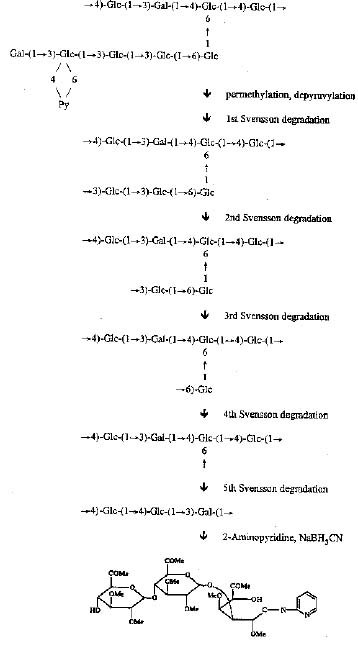
Abstract. Polysaccharide secreted by Rhizobium fredii Tu6 was investigated with composition analysis, linkage analysis, Svensson degradations, 1H-NMR, and desorption chemical ionization/mass spectrometry. The polysaccharide is composed of a nonasaccharide repeating unit (seven glucose, two galactose) with five sugar residues in the side chain and four sugar residues in the back bone. The terminal residue of the side chain is a galactose residue. The second residue from the terminal end is a 3-linked glucose substituted by a pyruvate group at the 4, 6-positions. Monosaccharide sequence was determined by Svensson degradations followed by linkage analysis. After removal of a pyruvate group from the methylated polysaccharide, the sugar residues in the side chain were removed sequentially by Svensson degradations. The reaction product of each degradation was then analyzed by methylation analysis. The polysaccharide secreted at pH 5 medium has a structure very similar to the polysaccharide secreted in the medium of pH 7 except that about 30% of the terminal galactose residues are also substituted at the 4, 6-positions by a second pyruvate group.

Keywords: Desorption chemical ionization/mass spectrometry (DCI/MS); Extracellular polysaccharide; Rhizobium fredii Tu6; Svensson degradation.

Introduction

Rhizobium species can infect leguminous plants and form nodules. The exopolysaccharides (EPS) excreted by the strains of Rhizobium play an important role in the infection of legumes and the Rhizobium / legume symbiosis (Phillip-Hollingsworth et al., 1989). Rhizobium fredii Tu6, a fast growing bacterium under neutral (pH 7) and acidic (pH 5) conditions, was isolated at Ping-Tong, Taiwan (Cheng and Lin, 1984). We have found a difference in morphological exhibition between neutral and acidic culture environments (unpublished data). We assume that EPS plays not only an important role in infection processes, but also a protective role in environmental adaptation. We now describe an investigation of the structure of the polysaccharide excreted by this bacterium. It is hoped that the investigation of the structure of EPS can provide some clues to the mechanism of infection and/or protection.

Materials and Methods

Isolation of the Polysaccharide

One and half day old (for pH 7) and two day old (for pH 5) cultures of R. fredii Tu6 were grown in B III medium (Bishop et al., 1976) and centrifuged. The polysac

charide was precipitated with cetyltrimethylammonium bromide. The precipitate was washed with ethanol, dialyzed against water, and lyophilized. The resulting crude exopolysaccharide was purified by a DEAE-sepharose column. Fractions containing exopolysaccharide were identified with a phenol-sulfuric acid test.

Proton NMR Spectroscopy

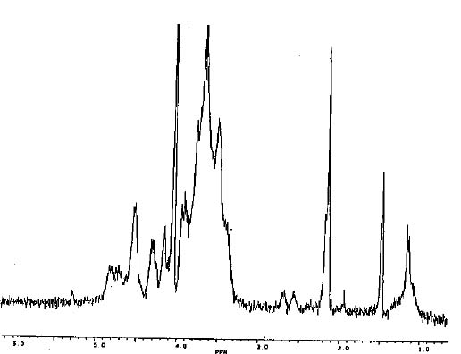
Samples for n.m.r. spectroscopy were lyophilized several times with D2O, and dissolved in D2O at a concentration of 1_3 mg/ml. Spectra were obtained with a Bruker 300-MHz instrument, with the probe heated to 60°C. The signal at d 1.47 was assigned to the methyl protons of pyruvic acid residues; the signal at d 2.13 was assigned to the methyl protons of O-acetyl groups; and the signals at d 2.57 and d 2.68 were assigned to the methylene protons of O-succinyl groups (Figure 1). The signal at d 1.17 remains to be assigned.

Desorption Chemical Ionization Mass Spectrometry

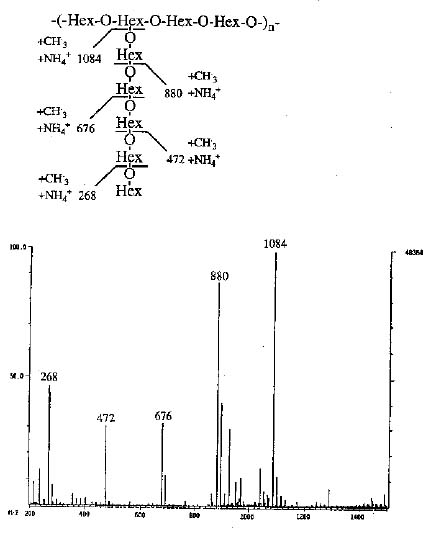
DCI/MS experiments were performed on a Finnigan TSQ-46C triplequadrupole mass spectrometer (Finnigan MAT, CA). Ammonia (from San-Fu Co., Hsinchu, Taiwan) was used as the reagent gas. The sample was air-dried on a platinum or rhenium emitter; the emitter was heated by a separated power supply at a heating rate of 50 mA/sec until the maximum current of 1.3 A was reached.

3Corresponding author. Fax: (02) 3638058.


Botanical Bulletin of Academia Sinica, Vol. 37, 1996

Results and Discussion

The results of the composition analysis suggested repeating units of nine sugars consisting of seven glucose and two galactose. Proton NMR obtained at 60°C showed that the EPS was substituted with pyruvate, acetyl, succinyl, and one unidentified group (Figure 1).

Desorption Chemical Ionization (DCI) mass spectrometry has proved to be a powerful technique for the determination of polysaccharide sequences (Her et al., 1990). Although no molecular ion can be detected, useful structural information may be obtained by DCI/MS with the detection of pyrolytic fragments. The formation of fragments along the backbone requires the cleavage of two bonds whereas a single bond cleavage is needed for the formation of a fragment in the side chain. Consequently, fragment ions from the side chain should be more abundant than backbone ions. The ammonia DCI mass spectrum obtained with permethylated and depyruvylated EPS (Figure 2) suggested that the side chain could consist of five sugar residues because the intensities of the signals decreased sharply after the fifth sugar residue.

Glycosidic linkages of the polysaccharide were determined by GC and GC/MS after permethylation, hydrolysis, reduction, and peracetylation (Harris and Henery, 1984). A total of nine sugar residues, including one non-reducing terminal galactose, two 3-substituted glucoses, two 4-substituted glucoses, one 3-substituted galactose, one 6-substituted glucose, one 4,6-disubstituted glucose,

Figure 1. The NMR spectrum of the extracellular polysaccharide secreted by Rhizobium fredii Tu6.

Composition Analysis

The glycosyl composition of the purified polysaccharide was determined by hydrolysis, reduction and acetylation followed by gas-liquid chromatography analysis of the alditol acetates (Biermann and McGinnis, 1989).

Linkage Analysis

Partially methylated alditol acetates were prepared (Biermann and McGinnis, 1989) and analyzed with a Hewlett Packard 5890 series 2 GC (Hewlett-Packard, PA, USA) and a VG-MD800 GC/MS instrument (Fisons, Manchester, UK). For GC separation, a DB-5 capillary column (30-m) was used. The column was temperature programmed by holding for 1 min at 75°C, increasing to 150°C at a rate of 20°C/min, and finally to 300°C at a rate of 2°C/min. Identification of alditol acetates was achieved by comparison of GC relative retention times and electron impact mass spectra of standard compounds.

Svensson Degradation (Jansson et al., 1977)

The EPS was permethylated, reduced by LiAlH4, depyruvylated (with 50% acetic acid at 100°C for 2 h) and then oxidized by DMSO activated by either oxalyl chloride (Mancuso et al., 1978) or P2O5 (Taber et al., 1987). After the oxidation, triethylamine (1 ml) was then added and stirred for 30 min. The solution was warmed to room temperature, dialyzed, and concentrated to dryness under vacuum. The sample was redissolved in 1 ml methylene chloride, treated with sodium ethoxide in ethanol (1 M, 1 ml), stored at room temperature for 1.5 h, neutralized with diluted hydrochloric acid, mixed with 1.5 ml 50% acetic acid, and heated to 100°C for 3 h. The solution was dried under vacuum and an aliquot of the sample was used for linkage analysis.

PA (2-Aminopyridine) Labeled Trisaccharide

The reductive amination procedure developed by Hase et al. (1979) was adopted for the preparation of PA labeled trisaccharide.

Figure 2. The ammonia DCI mass spectrum obtained with permethylated and depyruvylated EPS.


Wei et al. — Structure of extracellular polysaccharide

and one 3,4,6-trisubstituted glucose were detected. Pyruvic acid was linked to the 4,6 positions of the 3,4,6-trisubstituted glucose because linkage analysis of the depyruvylated polysaccharide showed the disappearance of the signal corresponding to 3,4,6-trisubstituted glucose and an increase in the intensity of the 3-substituted glucose. The branching point of the polysaccharide was indicated by the 4,6-disubstituted glucosyl residue.

Monosaccharide sequence was determined mainly by the Svensson degradation method (Figure 3). This method requires selective exposure of the hydroxyl group followed by oxidation of the exposed hydroxyl group, base-catalyzed b-elimination, and mild acid hydrolysis. The residue with the free hydroxyl group is degraded, and the sugars linked to this residue are released as a nonreducing and a reducing moieties. The free hydroxyl group in the reducing moiety provides a point of oxidation for a second Svensson degradation.

The pyruvate group in EPS Tu6 was reduced and hydrolyzed (Taylor and Conrad, 1972). After removal of the pyruvate group, the sugar residues in the side chain were removed sequentially by Svensson degradations. Linkage analysis of the first four degradation products are shown in Table 1. Seven sugar residues were detected for the first Svensson degradation indicating the substitution of pyruvate group on the glucose residue next to the terminal galactose (Gal1-3Glc(py)-). The third sugar residue was assigned as a 3 substituted glucose because linkage analysis of the repermethylated first degradation product showed the appearance of a terminal glucose and a decrease of the signal corresponding to 3 substituted glucose (Gal1-3Glc(py)1-3Glc-). This assignment is consistent with the linkage analysis of the second degradation product (see Table 1). Based on this approach, the fourth and fifth sugar residues in the side chain were designated a 3 linked glucose and a 6 linked glucose, respectively (Gal1-3Glc(py)1-3Glc1-3Glc1-6Glc). The side chain was believed to consist of five sugar residues because no signal corresponding to terminal sugar residue was observed in the linkage analysis of the repermethylated fourth degradation product. These data are in agreement with the results obtained in the DCI mass spectrum.

Table 1. Glycosyl linkage composition of the native and Svensson degradation products.

EPS 1st 1st* 2nd 2nd* 3rd* 4th*

Ter-Glc 1.1 1.1 0.9

Ter-Gal 1

3-Glc 1.7 1.7 0.9 0.9

4-Glc 2.3 2.3 1.9 2.1 2.3 2.4 3.4

3-Gal 1.1 1.0 1.0 1.0 0.9 1 1

6-Glc 1.1 1.0 1.0 1.0 0.7

4,6-Glc 1.1 0.8 1.0 1.1 0.7 0.9

3,4,6-Glc 1.1

*Repermethylated the Svensson degradation products.

Because the side chain consists of five sugar residues (Gal1-3Glc(py)1-3Glc1-3Glc1-6Glc), the repeating unit of the backbone should consist of four sugar residues. These four sugars consist of three 4 substituted glucose (including the 4,6 linked branching sugar) and one 3 substituted galactose. The side chain was cleaved with the first four degradation reactions. The fifth oxidation reaction should oxidize the branching sugar, and a trisaccharide was therefore left after the reaction. It is known that the branching sugar is a 4, 6 disubstituted glucose, so there are three possible arrangements for the three remaining sugars (4Glc1-4Glc1-3Gal-, 4Glc1-3Gal1-4Glc-, 3Gal1-4Glc1-4Glc-). Linkage analysis of the repermethylated trisaccharide could determine the terminal sugar but not the order of the other two sugars. In order to determine the sequence of the trisaccharide, the reducing sugar was labeled with 2-aminopyridine by reductive amination (Hase et al., 1979) before the permethylation and linkage analysis. The result showed that the terminal sugar was a 3 substituted galactose. This data suggested a sequence of 4Glc (branching sugar)-4Glc-4Glc-3Gal- for the backbone.

Figure 3. Sequence analysis of the extracellular polysaccharide secreted by Rhizobium fredii Tu6.


Botanical Bulletin of Academia Sinica, Vol. 37, 1996

EPS isolated from pH 5 medium and pH 7 medium have a very similar structure, except that the terminal galactose of the EPS obtained under pH 7 was partially substituted (about 30%) with pyruvic acid. As has been shown in R. trifolii (Abe et al., 1984), the non-carbohydrate substitutions of the R. fredii EPS change during culture age.

Me COOH

\ /

C

/ \

4 6

\ /

Glc-(6¬1)-Glc-(3¬1)-Glc-(3¬1)-Glc-(3 ¬1)-Gal

1

¯

6

-[-Glc-(1®4)-Glc-(1®4)-Glc-(1 ®3)-Gal-(1®4)-]n-

Acknowledgments. This work was supported by The National Research Council of the Republic of China.

Literature Cited

Abe, M., J. Sherwood, R. Hollingsworth, and F. Dazzo. 1984. Stimulation of clover root hair infection by lectin-binding oligosaccharides from the capsular and extracellular polysaccharides of Rhizobium trifolii. J. Bacteriol. 160: 517_520.

Biermann, C. J. and G. D. McGinnis. 1989. Analysis of Carbohydrates by GLC and MS. CRC Press, Inc., Boca Raton, Florida.

Bishop, P. E., J. G. Guevara, J. A. Engelke, and H. J. Evans. 1976. Relation between glutamine synthetase and nitrogenase activities in the symbiotic association between

Rhizobium japonicum and Glycine max. Plant Physiol. 57: 542_546.

Cheng, R. H. and L. P. Lin. 1984. Effects of soil acidity on growth of Rhizobium japonicum and symbiotic dinitrogen fixation. Mem. Coll. Agric. Natl. Taiwan Univ. 24: 63_73.

Harris, P. J. and R. J. Henery. 1984. An improved procedure for the methylation analysis of oligosaccharides and polysaccharides. Carbohydr. Res. 127: 59_73.

Hase, S., S. Hara, and Y. Matsushima. 1979. Tagging of sugars with a fluorescent compound, 2-aminopyridine. J. Biochem. 85: 217_220.

Her, G. R., J. Glazebrook, G. C. Walker, and V. N. Reinhold. 1990. Structural studies of a novel exopolysaccharide produced by a mutant of Rhizobium meliloti strain Rm1021. Carbohydr. Res. 198: 305_312.

Jansson, P. E., L. Kenne, B. Lindberg, H. Ljunggren, J. Lonngren, U. Ruden, and S. Svensson. 1977. Demonstration of an octasaccharide repeating unit in the extracellular polysaccharide of Rhizobium meliloti. J. Am. Chem. Soc. 99: 3812_3815.

Mancuso, A. J., S. L. Huang, and D. Swern. 1978. Oxidation of long-chain and related alcohols to carbonyls by dimethyl sulfoxide "Activated" by oxalyl chloride. J. Org. Chem. 43: 2480_2482.

Phillip-Hollingsworth, S., R. I. Hollingsworth, and F. B. Dazzo. 1989. Host-range related structural features of the acidic extracellular polysaccharides of Rhizobium trifolii and Rhizobium leguminosarum. J. Biol. Chem. 264: 1461_1466.

Taber, D. F., J. C. Amedio, Jr. and K. Y. Jung. 1987. P2O5/DMSO /Triethylamine (PDT): A convenient procedure for oxidation of alcohols to ketones and aldehydes. J. Org. Chem. 52: 5621_5622.

Taylor, R. L. and H. E. Conrad. 1972. Stoichiometric depolymerization of polyuronides and glycosamino-glycuronans to mono-saccharides following reduction of their carbodiimide-activated carboxyl groups. Biochemistry 11: 1383_1388.


Wei et al. — Structure of extracellular polysaccharide