Bot. Bull. Acad. Sin. (1996) 37: 181-190

Yeh and Chu — PWV coat protein transgenic tobacco

Production and evaluation of transgenic tobacco plants expressing the coat protein gene of passionfruit woodiness virus

Shyi-Dong Yeh1 and Fang-Hua Chu

Department of Plant Pathology, National Chung Hsing University, Taichung, Taiwan, Republic of China

(Received January 17, 1996; Accepted May 1, 1996)

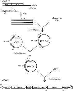
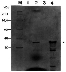
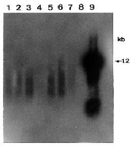
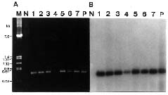
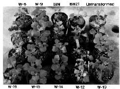
Abstract. Passionfruit woodiness virus (PWV), a member of the plant potyvirus group, is the major limiting factor for growing passionfruit in Taiwan. The coding sequence of the coat protein (CP) gene of PWV was modified to have an initiation codon and a leader sequence for translation. Analyses by in vitro transcription, in vitro translation, and immunoprecipitation verified that the modified PWV CP gene correctly encoded and expressed a protein of 36 kDa reacting to PWV CP antiserum. The CP gene was further inserted in a Ti binary vector and transferred to tobacco Nicotiana benthamiana by Agrobacterium-mediated transformation. Analyses of the putative transgenic plants by PCR amplification coupled with Southern blotting revealed an expected DNA fragment of 0.7 kb reacting with the probe specific to the PWV CP gene. Western blotting and Northern blotting were also used to further detect the expression of the PWV CP gene. The transgene-derived PWV CP was not detected but the degraded forms of the CP transcript were present. The putative transgenic tobacco lines of w-6, 9, 10, 12, 14, 15, and 16 were subjected to inoculation with different concentrations of PWV prepared from PWV-infected tobacco leaves. All plants of the four lines w-9, 10, 14, and 15 remained symptomless up to six weeks after inoculation. Some plants of two lines (w-6 and 9) showed a delay of 3 to 10 days in symptom development. All plants of line w-12 showed symptoms at the same time as the controls. Inoculated plants of lines w-9, 10, 14, and 15 were further tested by ELISA and bioassays on the local lesion host Chenopodium quinoa and the systemic host N. benthamiana. The negative results indicated that the complete resistance was provided by the apparent inhibition of PWV replication.

Keywords: Passionfruit woodiness virus; Transgenic tobacco plants; Coat protein gene; Potyvirus.

Introduction

Passionfruit woodiness virus (PWV), first described by McKnight (1953), is a potyvirus species, with flexuous particles of 750 × 12 nm and a genome consisting of a single stranded RNA of positive polarity (Taylor and Greber, 1973; Hollings and Brunt, 1981). PWV has been reported to infect plants of 44 species in 21 genera of 6 families (Edwardson and Christie, 1991). The virus is transmitted by aphids in a nonpersistent manner (Siaw, 1971) and mechanically by grafting or by contaminated cutting tools (Nattrass, 1944; Wang, 1983). The passionfruit woodiness disease caused by PWV is characterized by foliar symptoms of mosaic, distortion and rugosity, and the production of woody and severely malformed fruits (Taylor and Greber, 1973; Wang, 1983). Another feature of PWV infection is that the virus induces characteristic pinwheel and tubular inclusion bodies in the cytoplasm of infected cells (Moghal and Francki, 1981; Pares and McGechan, 1975; Lin et al., 1987; Jan and Yeh, 1995).

An isolate of PWV was first recorded in Taiwan in 1981 (Chang et al., 1981) and since then it has widely spread in the passionfruit-growing areas across the island as a major limiting factor on the production of the passionfruit

cultivar Tainung No. 1 (TN-1; a hybrid of Passiflora edulis Sims × P. edulis f. flavicarpa Degener). The virus has a single type coat protein (CP) of 36 kDa and produces a cylindrical inclusion protein (CIP) of 66 kDa and an amorphous inclusion protein (AIP) of 51 kDa in the cytoplasm of infected cells (Jan and Yeh, 1995). Antisera produced against the CP or virions of PWV have been widely used for detection of the virus by serological means (Lin et al., 1988; Chang, 1992). Since passionfruit in Taiwan is commercially propagated by grafting, indexing the presence of PWV in scions is important for the production of virus-free seedlings.

Plant viruses cause significant losses to crops worldwide and the basic approaches for controlling them have not been overly successful. The new concept of nonconventional resistance, which involves transforming plants with nucleic acid sequences that interfere with the viral infection cycle, is a promising new approach. Since the initial demonstration that expression of the CP gene of tobacco mosaic virus (TMV) in transgenic tobacco could provide effective resistance against TMV infection (Powell-Abel et al., 1986), genetically engineered CP-mediated protection has been used with success against several virus groups (for reviews, see Beachy et al., 1990; Hull and Davies, 1992; Fitchen and Beachy, 1993). In each case, expression of a viral CP gene conferred protection against different strains of the same virus and the resistance was nuclearly inherited.

1Corresponding author. Fax: 04-2877585; E-mail: sdyeh@ nchu.edu.tw.


Botanical Bulletin of Academia Sinica, Vol. 37, 1996

As an alternative to traditional plant breeding for disease control, CP-mediated protection has been performed for the potyvirus group. Examples include successful resistance to infection caused by potato virus Y (PVY) (Lawson et al., 1990; Van Der Vlugt et al., 1992; Farinelli et al., 1992), papaya ringspot virus (PRSV) (Ling et al., 1991), lettuce mosaic virus (LMV) (Dinant et al., 1993), soybean mosaic virus (SMV) (Stark and Beachy, 1989), tobacco etch virus (TEV) (Lindbo and Dougherty, 1992), watermelon mosaic virus (WMV) (Namba et al., 1992), and zucchini yellow mosaic virus (ZYMV) (Namba et al., 1992). The transgenic plants have been found to be effective in protecting tobacco, potato, and papaya against virus infection, showing significant delay in symptom development, attenuated symptoms, or complete resistance (Fitchen and Beachy, 1993). PWV causes a great loss in passionfruit production in Taiwan. Lack of resistant material makes the problem difficult to solve. Thus, CP-mediated protection becomes an attractive approach for control of PWV. The 3'-terminal region of a Taiwan PWV isolate has recently been cloned and sequenced. A cDNA clone containing 1623 nucleotide residues, reflecting part of the NIb gene, the entire CP gene, and the complete 3'-noncoding region, was obtained (Chen, 1992). In this investigation, the CP coding sequence of PWV was modified to have an initiation codon and a leader sequence for translation. The modified PWV CP gene was analyzed by in vitro transcription, in vitro translation, and immunoprecipitation to verify the construction. The CP gene was further inserted in a Ti binary vector and transferred to tobacco mediated by Agrobacterium. The putative transgenic plants were analyzed by PCR amplification coupled with Southern blotting, and by Western and Northern blotting to detect the presence and the expression of the PWV CP gene. The putative transgenic tobacco lines were subjected to PWV inoculation to evaluate their resistance to PWV infection under greenhouse conditions.

Materials and Methods

Virus Source

The virus isolate PWV-TW was originally isolated from diseased plants of golden passionfruit grown at Fengshan Tropical Horticultural Experiment Station in Kaohsiung (Wang, 1983; Chang, 1992). The virus was propagated in Nicotiana benthamiana Domin. Virus purification followed the procedure described by Gonsalves and Ishii (1980).

PCR Cloning and Construction of the PWV CP Gene

A genomic library was previously constructed from PWV RNA and a clone pPWV17 corresponding to the 3' terminal region of PWV RNA was selected, which covered part of the NIb gene, the entire CP gene, and the complete 3'-untranslated region (Chen, 1992). Since potyviral proteins are processed from polyproteins (Dougherty and Carrington, 1988), polymerase chain reaction (PCR) was

performed to introduce an ATG translation initiation codon within the context of a NcoI restriction site at the N terminus of the CP reading frame. The plasmid pPWV17 was used as a template to introduce a NcoI site at the 5' end of the CP coding sequence by the upstream sense primer 5'-ACCATGGGCACCAAATCAGAAGACGATA-3'. The downstream antisense primer at the 3' end of the untranslated region was oligo(dT)15 with a XbaI site. PCR amplification was performed using 50 ng of each primer and 40 ng of pPWV17 template in a total reaction volume of 100 ml as described by Ling et al. (1991). The PCR product was recovered from the agarose gel, digested with XbaI, and cloned into the SmaI/XbaI sites of the pBluescript KS(-) vector (Stratagene Co.) to generate pPWVCP. The NcoI/SacI fragment of pPWVCP was subcloned in the plasmid pGCP (Cheng, 1990) to replace the PRSV (papaya ringspot virus) CP gene, resulting in a clone pGWCP, which contained the PWV CP coding sequence with a GUS leader sequence and an initiation codon for efficient translation (Figure 1). The sequence of the modified PWV CP gene in pPWVCP and pGWCP was verified by the dideoxynucleotide chain-termination method (Sanger et al., 1977), using ssDNA generated by VCSM13 (Stragene) and dsDNA prepared from the selected clones (Holmes and Quigley, 1981).

Figure 1. Construction of the CP gene of PWV for expression in plants. The PWV CP gene was modified from the sequence in pPWV17 (Chen, 1992) by addition of an in frame ATG codon at its 5' end for translation initiation in a NcoI context CCATGG and introduced in pBI121 to replace GUS gene to generate pBWCP, under the control of a cauliflower mosaic virus (CaMV) 35S promoter and a nopaline synthase (NOS) terminator. The detailed procedure is described in Materials and Methods.


Yeh and Chu — PWV coat protein transgenic tobacco

In vitro Expression of the PWV CP Gene

The clones pPWVCP and pGWCP containing the entire PWV CP gene were analyzed by in vitro transcription and translation. RNA transcripts were synthesized from the plasmid by run-off transcription using T3 or T7 RNA polymerase according to manufacturer's directions (Stratagene). After the synthesis of RNA transcripts, the DNA templates were removed by addition of 10 units RNase-free DNase, incubated at 37°C for 5 min. The RNA transcripts were dissolved in 25 ml of RNase-free TE (10 mM Tris-HCl, pH 8.0, 1 mM EDTA) after phenol/ chloroform extraction and ethanol precipitation, and then directly used as templates for in vitro translation. The molecular size and quantity of RNA transcripts were estimated by 1% agarose gel electrophoresis.

In vitro translation was performed using the rabbit reticulocyte lysate translation system (Stratagene). The translation mixture containing the in vitro transcript and [35S] methionine (>1000 Ci/mmole, Amersham) was incubated at 30°C for 60 min. Protein products were subsequently analyzed by immunoprecipitation with the antiserum against PWV CP (Jan and Yeh, 1995), following the procedure described by Dougherty and Hiebert (1980). The reacted proteins were layered on 12% SDS-polyacrylamide gel and analyzed by electrophoresis and autoradiography.

Construction of PWV CP Gene in Ti Vector

The constructed PWV CP gene of 1.2 kb was excised from the plasmid pGWCP as an SmaI-SacI fragment and subcloned in the binary vector pBI121 (Clontech Co.), which contained the CaMV (cauliflower mosaic virus) 35S promoter, GUS (b-glucuronidase) gene from Escherichia coli, T-DNA border fragments (BR and BL), the promoter and the terminator of nopaline synthase (NOS), and the kanamycin selection marker NPT II (neomycin phosphotransferase II). The excised PWV CP fragment was ligated into the SmaI/SacI site of pBI121 to replace the GUS reading frame, and thus ensured the orientation of the insert in between the CaMV 35S promoter and the NOS terminator. The resulting clone pBWCP was confirmed by restriction enzyme digestion and mobilized to the Agrobacterium tumefaciens disarmed strain LBA 4404 by triparental mating as described by Rogers et al. (1986). The transformed Agrobacterium containing pBWCP was used for plant transformation.

Plant Transformation

The leaf-disk transformation method (Horsch et al., 1985) was used to introduce the T-DNA region of pBWCP into plant cells. The leaf disks were also transformed with the control Agrobacterium LBA4404 without pBWCP, LBA4404 with pBI121, or LBA4404 with pBIN which is a plasmid without GUS gene derived from pBI121. Agrobacterium tumefaciens was cultured in LB containing 50 mg/l kanamycin and 100 mg/l streptomycin at 28°C overnight. Tobacco plants of N. benthamiana were grown in greenhouse and used for transformation. Leaf pieces with developing callus were transferred once every three

weeks to the fresh selection medium until shoots developed. Shoots were excised and cultured on the hormone-free medium for rooting (MS medium containing 200 mg/l kanamycin). Shoots that rooted in the presence of kanamycin were transplanted in vermiculite soil for further analyses.

PCR Amplification Coupled with Southern Hybridization Analysis

The presence of PWV CP gene in the putative transgenic tobacco plants was detected by PCR using two primers PWV-P2 (5'-167AAGACAGGGATGTCGATGC TGGC189-3') and PWV-P3 (5'-875CGGTTTACCCAACC TTCACTGCG897-3') specific to the PWV CP gene. The total DNA was prepared following the procedure described by Mettler (1987). One mg of total DNA was used for PCR under the same program described above. PCR products in agarose gel were further analyzed by Southern blotting on nylon membrane. The specific probe to the PWV CP gene was prepared from the SmaI/SacI fragment of pGWCP, which was labeled with 32P-dCTP by nick translation according to Maniatis et al. (1982). The hybridization conditions followed the manufacturer's protocol (Amersham). Autoradiography was carried out at room temperature.

Analysis of CP Expression by Western Blotting

Tissue samples of 16 tobacco independent lines regenerated were ground in 3 vol. (v/w) of dissociation buffer (0.1M Tris-HCl pH 7.2, 2% SDS, 2% 2-mercaptoethanol, 10% sucrose and 0.005% bromophenol blue and 0.01 M EDTA) and boiled for 3 min. The extracts were centrifuged at 10,000 rpm (8000 g) for 5 min and the dissociated proteins remained in the supernatant were separated in 12% polyacrylamide gel containing SDS (Laemmli, 1970). The gel was electro-blotted onto a polyvinylidene difluoride (PVDF) membrane (Millipore), using a Bio-Rad transblot apparatus. The membrane was preincubated with TSW buffer (10 mM Tris-HCl, pH 7.4, 0.9% NaCl, 0.25% gelatin, 0.1% triton X-100 and 0.02% SDS) for 1 h, repeated washing for 30 min, and then incubated with 1:1000 dilution of polyclonal antiserum to PWV CP (Jan and Yeh, 1995) for 1 h. After rinsing 3 times with TSW solution the membrane was incubated with 1:2000 dilution of alkaline phosphatase-conjugated anti-rabbit IgG for 1 h in TSW solution. After rinsing 2 times with TSW buffer the membrane was incubated with the colorization buffer (100 mM Tris-Cl, pH 9.5, containing 100 mM NaCl and 5 mM MgCl2). Subsequently, color developing solution was prepared by adding 33 ml NBT (para-nitro blue tetrazolium chloride) and 16.5 ml BCIP (5-bromo-4-chloro-3-indoyl phosphate paratoluidine salt) to 5 ml of the buffer in the last step. The reactions were stopped by placing the membrane in distilled water.

Total RNA Extraction and Northern Hybridization

Total RNA was isolated from leaves of untransformed and transgenic tobacco plants by the method of Verwoerd et al. (1989) and Shrizadegan et al. (1991). The total RNAs


Botanical Bulletin of Academia Sinica, Vol. 37, 1996

extracted from plants were separated in a 1% agarose gel, transferred to Hybond-N nylon membranes (Amersham) with a MilliBlotTM-V Transfer System (Millipore), and reacted with the radioactive probe specific to the CP gene of PWV following the instructions in the manufacturer's manual (Amersham). Autoradiography was done at room temperature.

Evaluation of CP-Mediated Resistance in Transgenic Tobacco Plants

The putative transgenic tobacco plants expressing the PWV CP gene and control plants at the stage of 7_9 leaves were dusted with carborundum and the three youngest fully expanded leaves were rubbed with 1:10, 1:20, 1:50, and 1:100 dilutions of inocula prepared from PWV-infected tobacco leaves in 0.01 M potassium phosphate buffer, pH 7.0. Corresponding dilutions of inocula were also applied onto leaves of Chenopodium quinoa, the local lesion host, to check for the virus concentration. Symptom development was recorded daily after inoculation.

Indirect ELISA described by Yeh and Gonsalves (1984) was used to monitor PWV in the inoculated plants, using PWV CP antiserum (Jan and Yeh, 1995) diluted at 1:1000. Levels of the PWV antigen were determined by the OD405 and recorded by a Bio-Tek microplate reader.

Plants that did not show symptoms were retained for further observation up to six weeks. The sap extracted from leaves of symptomless plants was applied to the local lesion host C. quinoa and the systemic host N. benthamiana to check for the presence of virus.

Results

Cloning and Construction of the CP Gene

Two primers were designed to amplify the DNA fragment from pPWV17 by PCR, which reflects the complete CP coding sequence and the entire 3' untranslated region. When the PCR product was analyzed by agarose gels, a dsDNA of 1.2 kb was obtained. The PCR DNA product was then cloned into SmaI/XbaI sites of the pBluescript KS(-) vector. The selected clone pPWVCP contained ATG in frame with the CP coding sequence. Since the initiation codon ATG was designed in the context of NcoI restriction site, it was confirmed by NcoI digestion. DNA sequencing for the 5' ends of the insert also verified the correctives of the construction (data not shown). To further facilitate expression, the CP coding sequence with ATG was transferred to pGCP to obtain pGWCP which contained the translational GUS leader upstream from the start codon (Figure 1).

In vitro Expression of the CP Gene

Cloning in the transcriptional vector facilitated the synthesis of an in vitro transcript of the modified PWV CP gene. The clones pPWVCP and pGWCP were linearized by SacI digestion and run-off transcription was carried out by T3 and T7 RNA polymerase, respectively. Compari

son of the transcripts synthesized from pPWVCP and pGWCP with the marker RNA revealed that the two transcripts had the correct sizes. The transcripts of the clone were 1.2 kb similar to the viral cDNA in length (data not shown).

The pGWCP transcript was translated in rabbit reticulocyte lysate and the translation products were analyzed by polyacrylamide gels. The results are shown in Figure 2. Translation of this transcript in vitro resulted in a protein product of 36 kDa (Figure 2, lane 2), similar to the authentic viral CP. The protein reacted with the antibody to the PWV CP (Figure 2, lane 4), but not with the normal pre-immune serum (Figure 2, lane 3). This result clearly indicated that the pGWCP clone encoded and expressed the PWV coat protein correctly.

The engineered PWV CP gene was removed from pGWCP by SmaI/SacI digestion and cloned in a Ti binary vector pBI121 to replace the GUS gene. The final construct of pBWCP is shown in Figure 1. The plasmid in E. coli was transferred to the avirulent Agrobacterium strain LBA4404 by triparental mating. The resulting Agrobacterium (LBA4404/pBWCP) was used to transform tobacco.

Figure 2. Identification and immunoprecipitation analysis of the products of in vitro translation in rabbit reticulocyte lysate system using in vitro transcripts generated from the clone pGWCP. The proteins were analyzed in a 12% polyacrylamide gel. Lane M, [14C]-labeled protein markers. Lane 1, translation without exogenous RNA. Lane 2, translation products from the transcript generated from pGWCP. Lanes 3 and 4, translation products were immunoprecipitated with the pre-immune serum and the antiserum to PWV CP, respectively. The arrow indicates the position of a major protein of 36 kDa which reacted with the antiserum to PWV CP.


Yeh and Chu — PWV coat protein transgenic tobacco

Analyses of Putative Transgenic Tobacco Plants

PCR detection coupled with Southern hybridization — Leaf pieces of tobacco N. benthamiana were used for transformation. Eight weeks after cocultivation, the transformed callus was regenerated into shoots. Two to three weeks after rooting on MS medium containing 100 mg/ml kanamycin, plantlets were transferred to vermiculite soil and placed in the temperature-controlled (25_30°C) greenhouse. Plants of 16 kanamycin-resistant independent tobacco lines were obtained. PCR amplification was performed to analyze the presence of the PWV CP gene in tobacco plants, using plant DNA extracted from different transgenic tobacco lines and primers specific to the PWV CP gene. A dsDNA of 0.7 kb was amplified from the transgenic lines w-6, 9, 10, 12, 14, 15, and 16 (Figure 3A). The size of 0.7 kb was similar to the expected size of the span between the two PWV CP specific primers. PCR products were further analyzed by Southern blot using a specific probe to the PWV CP gene. The results of hybridization indicated that seven transformed lines—w-6, 9, 10, 12, 14, 15, and 16—carried CP sequences (Figure 3B), but line w-12 showed a weak signal. The similar PWV-CP specific fragment was also detected in the other 8 putative transgenic lines (data not shown).

Expression of the CP in transgenic plants — The PCR positive lines were further tested for the expression of the CP gene by Western blot, indirect ELISA, and northern blot. When proteins extracted from the putative transgenic tobacco were applied for Western blot analysis and ELISA, PWV CP was not detected in these plants (data not shown), even though the detection levels were sensitive up to 5-20 ng as shown in the positive controls. When transcripts in the PCR positive lines were analyzed by Northern blot, the positive signals were distributed from 0.7 to 1.2 kb (Figure 4), below the positive control of the transcript from

Figure 4. Northern blot analysis of putative PWV CP-transgenic lines of N. benthamiana. After blotting, total RNA extracted from plants was hybridized to PWV CP probe. Lane 1 to 7, putative transgenic lines w-6, 9, 10, 12, 14, 15, and 16, respectively. Lane 8, an untransformed tobacco. Lane 9, In vitro transcript generated from pGWCP.

the pGWCP, possibly due to degradation of the PWV CP transcript. The intact transcript of 1.2 kb was in trace amount or hardly detectable (Figure 4).

Evaluation of Resistance to PWV Infection in Transgenic Tobacco

Symptom development on CP-transgenic tobacco — Symptom development of the putative transgenic lines after inoculation with PWV CP were summarized in Table 1. All of the PWV-inoculated untransformed plants showed severe symptoms 4 days after inoculation, however, the four lines of W-9, 10, 14, and 15 were symptomless 20 days after inoculation (Figure 5). Infected leaves exhibited severe mosaic symptom, and the growth of infected plants was greatly reduced (Figure 6). Symptom development in control plants transformed with vector only was similar to the untransformed plants in time of appearance and severity of symptoms. All plants of four lines of w-9, 10, 14, and 15 remained symptomless during the period of the experiments, which lasted up to six weeks after inoculation (Figure 5). Some plants of two lines (w-6 and 9) showed delays of 3 to 10 days in symptom expression. All plants of line w-12 showed symptoms at the same time as the controls (Table 1). In all experiments, inocula were prepared by grounding the PWV-infected leaves of tobacco in inoculation buffer with dilution ratios of 1:10, 1:20, 1:50, and 1:100 (w/v); and

Figure 3. PCR amplification coupled with Southern hybridization analysis on putative PWV CP-transgenic lines of N. benthamiana. A, an ethidium bromide-stained gel showing a 0.7 kb fragment amplified by PCR with primers specific to PWV CP gene. B, products were blotted and hybridized with the specific probe to PWV CP gene. Lane M, DNA markers. Lane N, product from an untransformed plant. Lane 1_7, products from transgenic lines w-6, w-9, w-10, w-12, w-14, w-15, and w-16, respectively. Lane P, product from a pGWCP clone. The PWV CP probe was a SmaI/SacI DNA fragment from pGWCP.


Botanical Bulletin of Academia Sinica, Vol. 37, 1996

Figure 5. Symptoms on PWV transformants 20 days after inoculation with 1:10 dilution of inoculum prepared from PWV-infected leaves of N. benthamiana. Plants are those described in Table 1. Four plants in the upper two rows or the lower two rows represent a specific transgenic line as marked. A plant of C. quinoa with local lesions at the right upper corner indicates the concentration of the virus applied.

Figure 6. A PWV CP-transgenic plant of N. benthamiana showing complete resistance to PWV infection 20 days after inoculation after inoculation with PWV (1:10 dilution). Right, untransformed control plant showing typical severe symptoms of mosaic; left, plant of the CP-transgenic line w-10 which remained symptomless and in which PWV was not detectable either by ELISA or bioassays.

Table 1. Reactions of transgenic lines of N. benthamiana following inoculation with PWV.

Transgenic Dilution of ELISAb Numberc of plants with PWV symptoms

lines inoculaa at different time interval (days) after challenge inoculation

4 8 12 40

Normal

BI121

BIN

W-12

1:10 1.093-2.700 16 16 16 16

1:20 1.081-2.700 16 16 16 16

1:50 1.101-2.700 16 16 16 16

1:100 1.111-2.700 16 16 16 16

W-6

1:10 2.042 0 1 4 4

1:20 0.221 0 0 0 0 1:50 1.420 0 1 1 1

1:100 1.454 0 1 1 1

W-16

1:10 1.428 0 4 4 4

1:20 1.355 0 4 4 4

1:50 1.365 0 1 4 4

1:100 1.344 0 1 1 1

W-9

W-10

W-14

W-15

1:10 0.000-0.168 0 0 0 0 1:20 0.009-0.226 0 0 0 0

1:50 0.000-0.156 0 0 0 0

1:100 0.000-0.131 0 0 0 0

aIn all experiments, inocula were prepared by grounding the PWV-infected leaves of tobacco in 0.01 potassium phosphate buffer (pH 7.0) with dilution ratios of 1:10, 1:20, 1:50, and 1:100, which yielded 192, 132, 72, and 56 local lesions (averages of 8 leaves) per leaf on the host C. quinoa, respectively.

bLeaf samples were taken for ELISA test 20 days after inoculation with PWV. Prior to inoculation, the accumulation level of CP were not significantly different between PWV CP transgenic lines and control plants. The numbers refer to A405 readings recorded 30 min following the addition of substrate.

cIn each experiment, four plants from individual lines were inoculated.


Yeh and Chu — PWV coat protein transgenic tobacco

the inocula yielded an average of 192, 132, 72, and 56 local lesions per leaf (8 leaves for each dilution) on the host C. quinoa, respectively.

ELISA detection of virus concentration—Levels of PWV antigens were determined in plants of all lines. The concentration of PWV antigens was determined by ELISA 20 days after inoculation. The ELISA data were closely correlated with the observation of symptom appearance (Table 1). Four lines of w-9, 10, 14 and 15 were symptomless 20 days after inoculation, and the results of ELISA were also negative.

Detection of virus by bioassay—Inoculated plants of line w-9, 10, 14, and 15 did not have any indication of infection on the mechanically inoculated leaves nor on leaves that subsequently developed throughout the experiment. Tests to recover PWV from the resistant w-9, 10, 14, and 15 plants by using leaf extracts to inoculate the local lesion host C. quinoa and the systemic host N. benthamiana were all negative. This indicates an apparent inhibition of PWV replication.

Discussion

PWV is a member of the plant potyvirus group. The potyvirus CP is processed from the C terminal region of the viral polyprotein. To study the feasibility of using genetically engineered CP-mediated protection against PWV infection, the PWV CP gene was constructed and transferred to tobacco plants. Results of sequence analyses, in vitro transcription, in vitro translation and immunoprecipitation confirmed that the PWV CP construct can be expressed as a protein. Although the addition of the ATG in NcoI context CCATGG created two additional amino acid residues methionime and glycine, at the N-terminus of the PWV CP coding sequence, the modified CP gene was expressed correctly in reticulocyte lysate system, and a major protein of 36 kDa was produced. This result was confirmed by immunoreaction with PWV CP antiserum.

The 35S-PWV CP chimaeric gene was introduced into N. benthamiana to assess the possibility of using CP-mediated protection against PWV infection in transgenic plants. Among the kanamycin-resistant tobacco lines of w-6, 9, 10, 12, 14, 15, and 16, the existence of PWV CP reading frame was confirmed by PCR analysis, but the expressed PWV CP was not detected in any transgenic line. A possible explanation suggested by Farinelli et al. (1992) is that the product of the transgene is unstable in plant cells, possibly by absence of the protection at the N-terminal extreme of the constructed CP. The addition of the two amino acids may hamper the blocking of the modified N-terminus of the CP, which is a general trend for most of potyvirus, and thus makes the CP unstable. It is also possible that the transgenic CP is synthesized but rapidly degraded and thus hardly detectable by ELISA or Western blot analysis. Another possibility is that the unassembled CP is degraded during extraction.

To further check the transcription of the CP gene in transgenic plants, total RNA were analyzed by Northern

blot. Most of the PWV CP transcripts were in degraded forms, and intact transcript of 1.2 kb was in trace amount or hardly detectable. It is possible that the unfavorable process during extraction or blotting made the CP mRNA degraded. Another explanation is that the transgene transcript was degraded in plant cells and what we observed is similar to the RNA-mediated resistance reported for potyvirus (Lindbo and Dougherty, 1992).

Since the initial demonstration that expression of the CP gene of TMV in transgenic plants could provide effective protection against this virus (Powell-Abel et al., 1986), CP-mediated protection has been used with success against several virus groups (Hull and Davies, 1992; Fitchen and Beachy, 1993). Although all of the transgenic plants demonstrated the resistance conferred by expression of genes based upon CP sequences, they differ in important ways. In TMV CP transgenic plants the level of resistance was proportional to the amount of TMV CP accumulated in the cells, and plants that accumulated CP gene transcripts but not the CP itself were not resistant to infection (Wisniewski et al., 1990). From a mechanistic standpoint, the most important difference is the likelihood that the accumulation of RNA, rather than of protein, is responsible for resistance observed in some of the examples. In several cases, resistant plants have been generated by transformation with CP gene constructs designed to be transcribed into translationally incompetent mRNA. The mRNA resistance was in some cases as effective as that conferred by translationally competent constructs, suggesting that RNA may be the active entity, even when protein does accumulate. The examples of resistance to potato leafroll virus (Kawchuk et al., 1991) and to tomato spotted wilt virus (De Haan et al., 1992) appear to be of this type. In resistance to potyviruses, the role played by RNA is less certain. The examples of resistance to potato virus Y (Lawson et al., 1990; van der Vlugt et al., 1992; Farinelli and Malno, 1993) and tobacco etch virus (TEV) (Lindbo and Dougherty, 1992) have been considered as RNA-mediated protection.

PVY is the type member of the potyvirus group. Although highly effective protection was observed in PVY CP-resistant potato plants, no transgene-derived CP could be detected (Farinelli et al., 1992; van der Vlugt et al., 1992). This protein could be detected only in those plants that became infected with another PVY strain and only by detection with strain-specific monoclonal antibodies. This is possibly due to the instability of the unassembled CP. To further demonstrate a lack of a positive correlation between coat protein levels and protection, a gene lacking an initiating methionine codon was constructed. Transgenic plants carrying this gene were as resistant to PVY infection as the plants carrying the construct with the initiation signals added (Van Der Vlugt et al., 1992).

TEV is another member of the potyvirus group. The transgenic plants for a mutated frameshift construct that did not accumulate the CP were partially protected from the virus. Transgenic plant lines expressing truncated genes appeared to have more effective resistance than lines


Botanical Bulletin of Academia Sinica, Vol. 37, 1996

PWV resistant transgenic plant lines are readily immune by persistent inoculations on the same plant; 3) to test whether they are field resistant to PWV infection by exposing the transgenic plants to PWV under the continuous challenge by aphids; 4) to find out whether these plants are protected once the virus invades the vascular system, by grafting the apical parts of untransformed plants onto transformed tobacco stocks previously inoculated with PWV; 5) to study viral replication in inoculated leaves or protoplasts of the resistant lines.

PWV infection is a global problem and has become a major limiting factor in the production of passionfruit. To date, there are no reports of natural resistance to this virus among passionfruits. Thus, the successful production of the PWV CP-transgenic tobacco resistant to PWV infection provides a model system for the construction of PWV CP-transgenic passionfruit plants for control of the passionfruit woodiness disease.

Literature Cited

Beachy, R. N., S. Loesch-Fries, and N. E Tumer. 1990. Coat protein-mediated resistance against virus infection. Annu. Rev. Phytopathol. 28: 451_474.

Chang, C. A., H. L. Wang, Y. L. Chen, and T. Y. Chou. 1981. The investigation and identification of virus diseases of passionfruit in Taiwan. Plant Prot. Bull. (Taiwan) 23: 267. (Abstract)

Chang, C. A. 1992. Characterization and comparison of passionfruit mottle virus, a newly recognized potyvirus with passionfruit woodiness virus. Phytopathology 82: 1358_1363.

Chen, M. C. 1992. The nucleotide sequence of the 3'-terminal region of passionfruit woodiness virus. Master Thesis. Department of Plant Pathology, National Chung-Hsing University Taichung, Taiwan, ROC, 39 pp.

Cheng, Y. H. 1990. Expression of foreign genes in papaya cells by Ti binary vector. Master Thesis. Department of Plant Pathology, National Chung-Hsing University Taichung, Taiwan, ROC, 63 pp.

De Haan, P. J. J. L. Gielen, M. Prins, I. G. Wijkamp, and A. Van Schepen. 1992. Characterization of RNA-mediated resistance to tomato spotted wilt virus in transgenic tobacco plants. Bio/Technology 10: 1133_1137.

Dinant, S., F. Blaise, C. Kusiak, S. Astier-manifacier, and J. Albouy. 1993. Heterologous resistance to potato virus Y in transgenic tobacco plants expressing the coat protein gene of lettuce mosaic potyvirus. Phytopathology 83: 818_824.

Dougherty, W. G. and J. C. Carrington. 1988. Expression and function of potyviral gene products. Annu. Rev. Phytopathol. 26: 123_143.

Dougherty, W. G. and E. Hiebert. 1980. Translation of potyvirus RNA in a rabbit reticulocyte lysate: reaction condition and identification of coat protein as one of the products of in vitro translation of tobacco etch and pepper mottle viral RNAs. Virology 101: 466_474.

Edwardson, J. R. and R. G. Christie. 1991. The potyvirus group. Volume II, University of Florida, Florida Agricultural Experiment Station Agronomy Department, Plant Virus Laboratory Building 164, Gainesville, Florida 32611, 696 pp.

expressing the entire gene, although only a few lines were analyzed and the variation between lines may be great. Many individual plants of a line expressing the gene construct without the initiating methionine codon remained entirely asymptomatic and did not accumulate virus (Lindbo and Dougherty, 1992).

Virus-resistant transgenic plants have also been produced with CP sequences of other potyviruses such as soybean mosaic virus (SMV) (Stark and Beachy, 1989), papaya ringspot virus (PRSV) (Ling et al., 1991; Fitch et al., 1992), watermelon mosaic virus 2 (WMV 2) (Namba et al., 1992), zucchini yellow mosaic virus (ZYMV) (Namba et al., 1992; Fang and Grumet, 1993), plum pox virus (PPV) (Regner et al., 1992), and lettuce mosaic virus (LMV) (Dinant et al., 1993). In general, most plants which accumulate a low level CP show no symptoms or developing symptoms that were delayed in appearance, and show a significant decrease in viral accumulation. Several reports indicate that in the cases of CP-mediated protection with potyviruses, the most resistant lines are not necessarily those that accumulate the highest levels of CP. Some possibilities were considered: 1) the CP mRNA is sufficient to provide resistance to the virus, perhaps by hybridization to the minus-strand, or because the 3' untranslated region can be used to initiate abortive minus-strand synthesis (Farinelli and Malno, 1993); 2) the CP mRNA might interfere with replication, or compete directly for specific viral or host proteins essential for replication; 3) prior existent CP mRNA might trigger the host defensive system to overcome the infection by the challenge virus; 4) because the potyvirus CP is encoded as part of a polyprotein, the constructed CP mRNA might result in dysfunctional CP that interferes with the virus' ability to infect.

In this study, despite the fact that all of the transgenic plants lines expressed PWV CP at hardly detectable levels, four PWV CP transgenic lines showed complete resistance to PWV when challenged with different concentrations of PWV. Tests to recover PWV from the inoculated symptomless plants on a local lesion host and on a systemic host were all negative, indicating that the complete resistance was mediated by the apparent inhibition of PWV replication. The resistance of the four lines was not overcome even when challenged with a high dosage of the virus, such as virus concentration of 192 local lesions per leaf. Thus, in this case, it is likely that the resistance to PWV is not mediated by CP accumulation but mediated by the RNA transcripts of the CP gene. Thus, our results are similar to previous reports of RNA-mediated resistance in other potyviruses like TEV (Lindbo and Dougherty, 1992) and PVY (Lawson et al., 1990; Van Der Vlugt et al., 1992; Farinelli and Malnoë, 1993).

For further evaluation of resistance to PWV infection in transgenic tobacco lines, several approaches can be used: 1) to select R1 and R2 generations of self-fertilized progenies for determining the inheritance pattern of the CP gene and to study the differences in resistance between the heterozygote and homozygote plants; 2) to test whether


Yeh and Chu — PWV coat protein transgenic tobacco

Fang, G. and R. Grumet. 1993. Genetic engineering of potyvirus resistance using constructs derived from the zucchini yellow mosaic virus coat protein gene. Mol. Plant-Microbe Interact. 6: 358_367.

Farinelli, L. and P. Malnoë. 1993. Coat protein gene-mediated resistance to potato virus Y in tobacco examination of the resistance mechanisms—Is the transgenic coat protein required for protection? Mol. Plant-Microbe Interact. 6: 284_292.

Farinelli, L., P. Malnoë, and G. F. Collet. 1992. Heterologous encapsidation of potato virus Y strain O (PVY0) with the transgenic coat protein of PVY strain N (PVYN) in Solanum tuburosum cv. Bintje. Bio/Technology 10: 1020_1025.

Fitch, M. M. M., R. M. Manshardt, D. Gonsalves, J. L. Slightom, and J. C. Sanford. 1992. Virus resistant papaya plants derived from tissues bombarded with the coat protein gene of papaya ringspot virus. Bio/Technology 10: 1466_1472.

Fitchen, J. H. and R. Beachy. 1993. Genetically engineered protection against viruses in transgenic plants. Ann. Rev. Microbiol. 47: 739_763.

Gonsalves, D. and M. Ishii. 1980. Purification and serology of papaya ringspot virus. Phytopathology 70: 1027_1032

Hollings, M. and A. A. Brunt. 1981. Potyvirus Group. CMI/AAB Description of Plant Viruses. No. 245.

Holmes, D. S. and M. Quigley. 1981. A rapid boiling method for the preparation of bacterial plasmids. Anal. Biochem. 114: 193_197.

Horsch, R. B., J. E. Fry, N. L. Hottmann, D. Eichholtz, S. G. Rogers, and R. T. Fraley. 1985. A simple and general method for transferring genes to plants. Science 227: 1229_1231.

Hull, R. and J. W. Davies. 1992. Approaches to nonconventional control of plant virus disease. Crit. Rev. Plant Sci. 11: 17_33.

Jan, F. J. and S. D. Yeh. 1995. Purification, in situ localization, and comparative serological properties of passionfruit woodiness virus-encoded amorphous inclusion protein and two other virus proteins. Phytopathology 85: 64_71.

Kawchuk, L. M., R. R. Martin, and J. McPherson. 1991. Sense and antisense RNA-mediated resistance to potato leafroll virus in Russet Burbank potato plants. Mol. Plant-Microbe Interact. 4: 227_253.

Laemmli, U. K. 1970. Cleavage of structural proteins during the assembly of the head of bacteriophage T4. Nature (London) 227: 680_685.

Lawson, C., W. Kaniewski, L. Haley, R. Rozman, C. Newell, P. Sanders, and N. E. Tumer. 1990. Engineering resistance to mixed virus infection in a commerical potato cultivar: resistance to potato virus X and potato virus Y in transgenic Russet Burbank. Bio/Technology 8: 127_134.

Lin, N. S., Y. H. Hsu, N. Wang, and Y. K. Chen. 1987. Immunochemical detection and localization of passionfruit woodiness virus protein in infected cells. Plant Prot. Bull. 29: 439. (Abstract)

Lin, N. S., N. Wang, and Y. H. Hsu. 1988. Sequential appearance of capsid protein and cylindrical inclusion protein in root-tip cells following infection with passionfruit woodiness virus. J. Ultrastructure Res. 100: 201_211.

Lindbo, J. A. and W. G. Dougherty. 1992. Pathogen-derived resistance to a potyvirus: immune and resistant phenotypes

in transgenic tobacco expressing altered forms of a potyvirus coat protein nucleotide sequence. Mol. Plant-Microbe Interact. 5: 144_153.

Ling, K., S. Namba, C. Gonsalves, J. L. Slightom, and D. Gonsalves. 1991. Protection against detrimental effect of potyvirus infection in transgenic tobacco plants expressing the papaya ringspot virus coat protein gene. Bio/Technology. 9: 752_758.

Maniatis, T., E. F. Fritsch, and J. Sambrook. 1982. Molecular Cloning A Laboratory Manual, Cold Spring Harbor, New York, 545 pp.

Mettler, I. J., W. R. Center, and S. C. Co. 1987. A simple and rapid method for minipreparation of DNA from tissue cultured plant cells. Plant Mol. Biol. Rep. 5: 346_349.

McKnight, T. 1953. The woodiness virus of the passion vine (Passiflora edulis Sims) Qd. J. Agric. 10: 4_35.

Moghal, S. M. and R. I. B. Francki. 1981. Toward a system for the identification and classification of potyviruses II. Virus particle length, symptomatology, and cytopathology of six distinct viruses. Virology 11: 210_216.

Namba, S., K. Ling, C. Gonsalves, J. L. Slightom, and D. Gonsalves. 1992. Protection of transgenic plants expressing the coat protein gene of watermelon mosaic virus 2 or zucchini yellow mosaic virus against six potyviruses. Phytopathology 82: 940_946.

Nattrass, R. M. 1944. The transmission of the virus of the "woodiness" disease of passionfruit (Passiflora edulis) by single leaf grafts. Ann. Appl. Biol. 31: 310_311.

Pares, R. D. and J. K. McGechan. 1975. The distribution and ultrastructural effects of passionfruit woodiness virus in bean leaves. Aust. J. Bot. 23: 905_914.

Powell-Abel, P., R. S. Nelson, B. De, N. Hoffmann, S. G. Rogers, R. T. Fraley, and R. N. Beachy. 1986. Delay of disease development in transgenic plants that express the tobacco mosaic virus coat protein gene. Science 232: 738_742.

Regner, F., A. C. Machado, M. L. C. Machado, H. Steinkellner, D. Mattanovich, V. Hanzer, H. Weiss, and H. Katinger. 1992. Coat protein mediated resistance to plum pox virus in Nicotiana clevelandii and Nicotiana benthamiana. Plant Cell Rep. 11: 30_33.

Rogers, S. G., R. B. Horsch, and R. T. Fraley. 1986. Gene transfer in plants : Production of transformed plants using Ti plasmid vectors. Methods Enzymol. 118: 627_640.

Sanger, F., S. Nicklen, and A. R. Coulson. 1977. DNA sequencing with chain-terminating inhibitors. Proc. Natl. Acad. Sci. USA. 74: 5463_5467.

Shrizadegan, M., P. Christie, and J. R. Seemann. 1991. An efficient method for RNA from tissue cultured plant cells. Nucleic Acids Res. 19: 6055.

Siaw, A.B. 1971. Transmission of papaya leaf reduction virus by Myzus persicae. Plant Dis. 55: 526_529.

Stark, D. M. and R. N. Beachy. 1989. Protection against potyvirus infection in transgenic plants: evidence for broad spectrum resistance. Bio/Technology 7: 1257_1262.

Taylor, R. H. and R. S. Greber. 1973. Passionfruit Woodiness Virus. CMI/AAB Description of Plant Viruses No. 122.

Van Der Vlugt, R. A. A., R. K. Ruiter, and R. Goldbach. 1992. Evidence for sense RNA-mediated protection to PVYN in tobacco plants transformed with the viral coat protein cistron. Plant Mol. Biol. 20: 631_639.


Botanical Bulletin of Academia Sinica, Vol. 37, 1996

Verwoerd, T. C., B. M. M. Dekker, and A. Hoekema. 1989. A small-scale procedure for the rapid isolation of plant RNAs. Nucleic Acids Res. 17: 2362.

Wang, H. L. 1983. Studies on passionfruit woodiness virus in Taiwan. Master Thesis, Department of Plant Pathology, National Chung-Hsing University, Taichung, Taiwan, 84 pp.

Wisniewski, L. A., P. A. Powell, R. S. Nelson, and R. N. Beachy. 1990. Local and systemic spread of tobacco mosaic virus in transgenic tobacco. Plant Cell 2: 559_567.

Yeh, S. D. and D. Gonsalves. 1984. Evaluation of induced mutants of papaya ringspot virus for control by cross protection. Phytopathology 74: 1086_1091.