Bot. Bull. Acad. Sin. (1996) 37: 213-218

Hannweg et al. — Micropropagation of Bowiea volubilis

A simple method for the micropropagation of Bowiea volubilis from inflorescence explants

K. Hannweg, M.P. Watt1, and P. Berjak

Centre for Indigenous Plant Use Research, Biology Department, University of Natal, Private Bag X10, Dalbridge 4014, Republic of South Africa

(Received February 26, 1996; Accepted June 17, 1996)

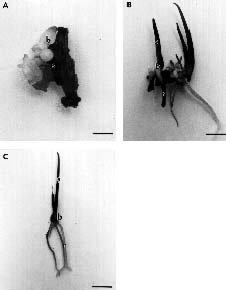
Abstract. The bulb of Bowiea volubilis (Liliaceae) is important in traditional medicine in South Africa. Although this species can be macro- and micropropagated, present methods relying on the use of bulb-scale pieces can result in the destruction of the parent plant. In this study, a protocol for the organogenic plant regeneration from inflorescence pieces was established. It involves the culture of 10 mm long pieces of inflorescence stem on MS basal nutrient medium, 30 g l-1 sucrose, 10 g l-1 agar, 1 mg l-1 2,4-D and 1 mg l-1 BAP, in the dark for 6_8 weeks. The explants are then transferred to fresh medium without growth regulators, where bulblet development, shoot elongation and rooting occur within 4_5 weeks. Successful hardening-off is achieved using conditions of relatively low humidty. As the inflorescence is very long (up to 20_30 m in total) and the protocol results in approximately 4.6 plantlets per explant, thousands of plantlets can potentially be produced in this manner from a single inflorescence.

Keywords: Conservation; Ethnobotany; Medicinal plant; Propagation.

Introduction

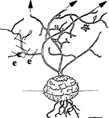
Bowiea volubilis (Zulu name = igibisila), a member of the Liliaceae, is a geophyte endemic to the central and eastern regions of South Africa (Dyer, 1964). The bulb is globose, up to 150 mm in diameter and may be subterranean or exposed. The leaves are lanceolate, canaliculate, up to 350 mm long and 5 mm in diameter. The stem producing the annual inflorescence can be up to 3 m long and appears during the summer months.

Although the bulb of B. volubilis has long been known to be poisonous (Watt and Breyer-Brandwijk, 1962), it forms an important and very popular component of traditional medicine in South Africa. It has been reported to contain three active cardiac glucosides, one of which appears to act as a heart stimulant (Watt and Breyer-Brandwijk, 1962). It is alleged to be of medicinal value and preparations of the bulb alone or mixed with parts of other species are used widely as a purgative, a remedy for dropsy and female infertility, and as components of skin and eye lotions. The bulb is also purported to have magical properties. It is sprinkled on impis' to ward off enemies and is used as an aphrodisiac (Watt and Breyer-Brandwijk, 1962; Mkhize, pers. comm.).

At present, the demand for Boweia volubilis is satisfied primarily from remaining wild populations. However, its popularity as an herbal medicine coupled with the rapid human population expansion and dramatic decrease in the areas of indigenous vegetation have led to the

overexploitation and depletion of wild stocks and consequent rapid increases in prices. In 1990, the sale value of Bowiea volubilis was $5.10 per dozen plants, and over two tons of bulb material were sold each year (Buckas, pers. comm.). It is not suprising, therefore, that even eight years ago B. volubilis was one of the top five medicinal species nominated by herb traders as having become scarce (Cunningham, 1988).

Very little is known about the natural propagation of B. volubilis. However, it has been reported that few seeds are set (Dyer, 1964) and hence progeny numbers are low. Traditional methods of vegetative propagation of bulbs such as daughter-bulb formation have been attempted in nurseries, but these techniques provide an extremely slow rate of multiplication. Thus, there has been a distinct need to investigate the potential of in vitro propagation systems for this plant, as has been successfully achieved with other bulbous species of commercial value (e.g. Allium spp.: Havraneck and Novak, 1976; Amaranthus spp.: Flores et al., 1981).

Bowiea volubilis has been previously propagated using tissue culture techniques. Jha and Sen (1986) used a shake culture system and Cook et al. (1988) successfully produced B. volubilis in solid culture using bulb scale material as explants. However, the removal of the inner bulb scales (which are the youngest, least contaminated, and most responsive to culture) results in the destruction of the parent plant, a great disadvantage in terms of conservation of an endangered species. In this study, we investigated the feasibility of establishing a protocol for the micropropagation of B. volubilis from inflorescence explants.

1 Corresponding author. Fax: 27-31-2602029/1195; Email: watt@biology.und.ac.za.


Botanical Bulletin of Academia Sinica, Vol. 37, 1996

Materials and Methods

Explant Type and Surface Sterilisation

Pieces (500 mm long) of the main inflorescence stem from a mature plant (bulb radius 75 mm) were washed and surface-sterilised. This was carried out by dipping the plant material into 70% [v/v] ethanol for 2_3 minutes, whereupon it was transferred to a solution of 1% sodium hypochlorite containing 5 drops of Tween-20 for 20 minutes. Subsequently, 10 mm-long explants were cut from the inflorescence branches, as depicted in Figure 1, and placed in culture.

Culture Conditions

Several nutrient media (Dollfus and Nicolas-Prat, 1969; Dunstan and Short, 1977; Margara, 1969; Murashige and Skoog, 1962; Ziv et al., 1970) supplemented with various types, concentrations, and combinations of plant growth regulators were tested for induction of organogenesis. The Murashige and Skoog (1962) basal nutrient formulation (macro- and micronutrients only, no vitamins) (MS - vitamins) was tested also (Table 1). After 6_8 weeks on induction media, explants were transferred to MS basal nutrient medium containing no plant growth regulators for 4_5 weeks. All regenerated plantlets were then excised from the original explant and subcultured onto similar fresh medium in bigger bottles for plantlet growth. All culture media were supplemented with 30 g l-1 sucrose and 10 g l-1 agar, pH 5.6. Explants were maintained at 25°C in the dark for the induction stage and subsequently in a daily 16 hour light/8 hour dark photoperiod, at 200 µEm-2 s-1 light intensity.

Hardening-Off and Acclimatisation of Plantlets

Regenerated plantlets were transplanted into a mixture (1:1) of autoclaved potting soil and washed coarse river sand. Three different methods were used in order to determine the most appropriate hardening-off procedure: 1)

Plants were tightly covered with polythene bags (to ensure high humidity) for 7 days after which the humidity of the microclimate of each plantlet was reduced gradually by opening the bags daily for increasingly longer periods over 2_3 weeks; 2) Plants were loosely covered with polythene bags (to achieve medium relative humidity) and the plantlets maintained at this humidity level for 2_3 weeks; 3) Plants were not covered. They were mist-sprayed twice daily with tap water (low humidity), ensuring that the soil/sand mixture was kept fairly moist while relative humidity remained low.

Figure 1. Diagrammatic representation of Boweia volubilis and sampling of inflorescence stem pieces used as explants (e). Bar = 6 cm

Table 1. The effect of composition of induction media on subsequent plantlet regeneration.

Induction medium % Explants exhibiting Number of

Nutrient formulation Plant growth regulators (mg 1-1) organogenesis plantlets per explant

Murashige and Skoog (1) 2,4-D + (1) BAP 77.9h (P) 4.6

(1965) basal nutrients (0) NAA + (10) BAP 0a 0

(-vitamins) (1) NAA + (1) BAP 68.5f (P) 2.1

(9.3) NAA + (1.1) BAP 85.2I (B) 0

(9.3) NAA + (0.1) BAP 76.3g (R) 0

(18) NAA + (1.1) BAP 16.1b (R) 0

(20) NAA + (0.1) BAP 48.7e (R) 0

(1) 2,4-D + (1) BAP + (0.2) Kin 35.7c (B) 0

Murashige and Skoog (1) NAA + (1) BAP 43.2d (R) 0

(1962) (+vitamins) All other combination as above 0a 0

The number of explants exhibiting organogenesis was determined after 8 weeks on induction media, and regenerated plantlets/explants 5 weeks weeks after subsequent transfer of bulblets to MS basal (-vitamins) nutrient medium devoid of growth regulators. All media contained 10 g l-1 agar and 30 g l-1 sucrose. Regenerated plant organs are denoted: (B) = bulblets only, (P) = plantlets (shoots and roots), (R) = roots only. n = 50_60 a - h (ANOVA test).


Hannweg et al. — Micropropagation of Bowiea volubilis

Data Analysis

Average values were calculated from the data recorded during the different stages of plant regeneration. Where appropriate, ANOVA (Statgraphics Plus, 1993) was used to assess differences in the recorded mean values of the variables investigated (P = 0.05). Alphabetical values (Tables 1 and 2) were assigned to the mean values recorded for each treatment. Mean values that did not share the same letter, were recognised as being significantly different from each other.

Results and Discussion

Organogenesis from Inflorescence Explants

The first sign of organogenesis induction was the development of swellings on the explants (Figure 2A) after 2_4 weeks in culture, from which bulb-like structures formed 4 weeks later directly from the explant (Figures 2A and 2B). Organogenesis appeared to occur directly from the explant even when 2,4-D was a component of the culture media. In a very few cases, a small amount of callus was visible around the bulblets. Depending on the initial induction media formulations (Table 1), shoot extension from the bulblets (Figure 2B) and subsequent rooting (Figure 2C) occurred within 4_5 weeks of transfer to media devoid of growth regulators. Root formation was visible approximately 2_3 weeks after shoot elongation. Separate plantlets were transferred to fresh medium without growth regulators in bigger bottles for growth until they reached optimal bulb size (Table 2). They were then hardened-off and maintained under greenhouse conditions with the resultant plants flowering 4_5 months later.

The Established in vitro Protocol

In this investigation, induction of organogenesis was achieved only on MS nutrient media formulations (with and without vitamins) (Table 1). Those of Dollfus and Nicolas-Prat (1969), Dunstan and Short (1977), Margara (1969) and Ziv et al. (1969) did not induce swellings (as shown in Figure 2A) in any explants, which died after 2_3 weeks in culture (results not shown). This was somewhat suprising as those media have been successfully used for the regeneration of other bulbous species (Allium spp., Gladiolus spp.) from flower explants.

Subsequent to the formation of swellings (Figure 2A), organ development was detected on MS basal medium (MS - vitamins) supplemented with different combinations of BAP and the auxins 2,4-D and NAA (Table 1) and MS medium (with vitamins) containing 1 mg l-1 NAA and 1 mg l-1 BAP. However, plantlet regeneration occurred only on MS basal medium + 1 mg l-1 2,4-D + 1 mg l-1 BAP (77% of explants) and MS basal medium + 1 mg l-1 NAA + 1 mg l-1 BAP (68.5% of explants). This is in contrast to the results obtained in previous investigations on B. volubilis (Cook et al., 1988) when 1 mg l-1 2,4-D + 1 mg l-1 BAP was found to be the optimum growth regulator combination for plantlet regeneration, although in that

Figure 2. Stages of plantlet regeneration. (A) swellings (s) on the stem pieces, recognised as first signs of organogenesis; also shown are young bulblets (b), bar = 0.25 cm; (B) and (C) whole plantlets showing bulb (b), shoots (s) and roots (r), bars = 0.6 and 2.0 cm, respectively.

Table 2. Parameters influencing successful hardening-off of regenerated plantlets.

Parameter % Plantlet survival

Humidity

High (tightly covered with plastic) 0.0a

Medium (loosely covered with plastic) 10.2b

Low (twice-daily mist spray) 90.9c

Size of bulb

£4 mm 10.7b

³4 mm 95.9c

Size of bulb was investigated using the low humidity treatment. n = 35, a - h (ANOVA test).


Botanical Bulletin of Academia Sinica, Vol. 37, 1996

study bulb scales were used as explants. Similar reports have been made by Pierik and Steegmans (1975) for Freesia spp., Seabrook et al. (1975) for Amaryllis spp. and Takayama and Misawa (1982) for Lilium spp.

In the present study, the tested combinations of growth regulators (Table 1), other than those mentioned above, resulted in the formation of bulblets or roots only, and the addition of kinetin to the medium further resulted in a two-week delay in bulblet formation. The observed prolific root formation on media containing high ratios of NAA:BAP (Table 1) has been reported also for other species (Hippeastrum spp.: Seabrook and Cumming, 1976; Lilium spp. Takayama and Misawa, 1982).

With regard to yield, the most successful of the tested media for plantlet production was MS basal medium + 1 mg l-1 2,4-D + 1 mg l-1 BAP, resulting in approximately 4_5 plantlets per explant. Considering that the inflorescence of a mature Bowiea plant such as those used in this study can be collectively up to 20_30 m long (including all the branchlets), over 20,000 explants may be generated from it to initiate cultures. Further, the positional origin of the explants along the inflorescence does not appear to affect the yield of regenerated plantlets (results not shown). Therefore, on the basis of the results obtained in this study this would translate theoretically into over 70,000 regenerated plantlets from one single inflorescence.

Acclimation of in vitro Regenerated Plantlets to Ambient Conditions

In the initial stages of this investigation it became apparent that the hardening off stage was of crucial importance to the success of a micropropagation protocol. Because of its fleshy nature, the bulb of Boweia was found to be very sensitive to high moisture content and fungal attack. Plantlets transferred to autoclaved soil and tightly covered with plastic bags rotted and died within 5_7 days (Table 2). In contrast, 90.9% survival was recorded for plantlets subjected to a twice-daily mist spray for 2_3 weeks after planting out.

In addition to the effect of humidity on plantlet survival, bulblet size was also found to be a very important parameter in the survival of plantlets after transfer to the potting medium (Table 2). Plantlets having bulblets smaller than 4 mm in diameter died 5_7 days after planting out whereas those with larger bulbs survived and mature plants were obtained.

Concluding Remarks

In Boweia volubilis, plant regeneration from inflorescence explants was achieved with only two media, i.e. the induction medium and the hormone-free medium, required for shoot elongation, rooting, and plantlet growth. This is in contrast to results obtained for other species (Amaryllis spp., Narcissus spp., Urginea indica, Muscaria armeniacum, Gladiolus spp.) where bulblets could be induced only subsequent to shoot and root elongation

(Hosoki and Asahawa, 1980; Jha et al., 1984; Peck and Cumming, 1986; Prasad et al., 1990). Further, the relevant media are very simple and commonly used in tissue culture protocols. The related species Urginia indica and Thuranthos basuticum and several other bulbous species (as mentioned above) require several more complicated and species-specific combinations of growth hormones to achieve plantlet regeneration.

The number of plantlets that can be obtained, together with the simplicity of the approach, make the developed protocol a cost-effective and attractive option for the propagation of B. volubilis. The manipulations can be performed by semi-skilled personnel and do not require sophisticated equipment or facilities.

Micropropagated plants can be used to supplement the natural stock of plants in wild populations as well as to provide a ready supply to the herbal medicinal trade, thereby alleviating pressure on existing resources already over-exploited by uncontrolled harvesting. One challenge, however, lies in gaining acceptibility of these plants by the communities which utilise traditional medicines, as cultured plants may be considered to have reduced medicinal or magical properties when compared with plants that are collected from natural habitats (Mkhize, pers comm). To our knowledge, no controlled tests have been performed in terms of the glucoside content of either nursery-grown or in vitro cultured B. volubilis plants.

Acknowledgements. This work was supported by the University of Natal Research Fund and the Foundation for Research Development.

Literature Cited

Buckas, E. 1995. Durban Municipality Parks and Gardens, Durban.

Cook, E.L., A. Cunningham, and J. van Staden. 1988. The tissue culture of an exploited medicinal plant, Bowiea volubilis. S. Afr. J. Bot. 54: 509_510.

Cunningham, T. 1988. Over-exploitation of medicinal plants in Natal/KwaZulu: Root Causes. Veld and Flora, September 1988, pp. 85_87.

Dollfus, M. and D. Nicolas-Prat. 1969. Mise en évidence des potentialités d'organogénèse végétative et florale in vitro chez trois Digitales: Digitalis lanata Ehrh., Digitalis purpurea L. et sa variété gloxiniae flora. Comptes Rendues Academies des Sciences (Paris) 268D: 501_503.

Dunstan, D.I. and K.C. Short. 1977. Improved growth of tissue cultures of the Onion, Allium cepa. Physiol. Plant. 41: 70_72.

Dyer, R.A. 1964. The genera of Southern African Flowering Plants: Gymnosperms and Monocotyledons. Department of Agricultural Technical Services, Pretoria, 923 pp.

Flores, H.E., A. Thier, and A.W. Galston. 1981. Tissue culture of Amaranthus. Environ. Exp. Bot. 21: 437_438.

Havranek, P. and F.J. Novak. 1976. Bud formation in the callus cultures of Allium sativum. Z. Pflanzenphysiol. 68: 308_318.


Hannweg et al. — Micropropagation of Bowiea volubilis

Hosoki, T. and T. Asahawa. 1980. In vitro propagation of Narcissus. Hortic. Sci. 15: 602_603.

Jha, S. and S. Sen. 1986. Regeneration and rapid multiplication of Bowiea volubilis Harv. in tissue culture. Plant Cell Rep. 4: 12_14.

Jha, S., G.C. Mitra, and S. Sen. 1984. In vitro regeneration from bulb explants of Indian squill, Urginea indica Kunth. Plant Cell Tissue Organ Cult. 3: 91_100.

Margara, J. 1969. Etude préliminare des facteurs de la néoformations de bourgeons chez le chou fleur (Brassica oleracea L. var. botrytis). Comptes Rendues Academies des Sciences (Paris) 268D: 686_688.

Mkhize, P. 1995. Biology Department, University of Natal, Durban.

Murashige, T. and F. Skoog. 1962. A revised medium for rapid growth and bioassays with tobacco tissue culture. Physiol. Plant. 15: 473_497.

Peck, D.E. and B.G. Cumming. 1986. Beneficial effects of activated charcoal on bulblet production in tissue cultures of Muscari armeniacum. Plant Cell Tissue Organ Cult. 6: 9_14.

Pierik, R.L.M. and H.H.M. Steegmans. 1975. Effect of auxins, cytokinins, gibberellins, abscisic acid and ethephon on re

generation and growth of bulblets on excised bulb scale segments of Hyacinth. Physiol. Plant. 34: 14_17.

Prasad, R.N., A.K. Sharma, and H.C. Chaturvedi. 1990. In vitro regeneration of Gladiolus through corm tissue culture. In Abstracts of the VIIIth International Congress of Plant Tissue and Cell Culture.

Seabrook, J.E. and B.G. Cumming. 1976. The in vitro propagation of amaryllis (Hippeastrum spp. hybrids). In vitro 13: 831_836.

Seabrook, J.E., B.G. Cumming, and L.A. Dionne. 1975. The in vitro induction of adventitious shoot and root apices on Narcissus (daffodil and narcissus) cultivar tissue. Can. J. Bot. 54: 814_819.

Takayama, S. and M. Misawa. 1982. Regulation of organ formation by cytokinin and auxin in Lilium bulb scales grown in vitro. Plant Cell Physiol. 22: 461_467.

Watt, J.M. and M.G. Breyer-Brandwijk. 1962. The Medicinal and Poisonous Plants of southern Africa. University Microfilms International, Michigan, 1457 pp.

Ziv, M., A.H. Halevy, and R. Shilo. 1970. Organ and plantlet regeneration of Gladiolus through tissue culture. Ann. Bot. 34: 671_676.