Bot. Bull. Acad. Sin. (1996) 37(4): 261-264

Dai et al. — Polysome translation of heat shock proteins

In vitro polysome translation analysis of heat shock proteins in higher plants

Hwa Dai1,4, Yih-Shan Lo1, Yaw-Huei Lin1 , Manfred Ruddat2 and Kwen-Sheng Chiang3

1Institute of Botany, Academia Sinica, Taipei, Taiwan 11529, Republic of China

2 Department of Ecology and Evolution and 3 Department of Molecular Genetics and Cell Biology, University of Chicago, Chicago, Ill. 60637, USA

(Received April 10, 1996; Accepted September 19, 1996)

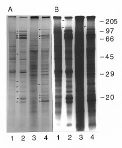
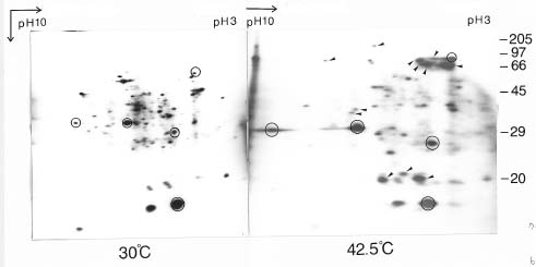
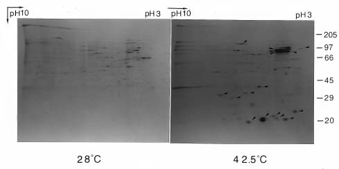
Abstract. We used a simple and highly reproducible method for the analysis of heat shock proteins in rice (Oriza sativa), maize (Zea mays), and mung bean (Vigna radiata). Cytosolic polysomes, isolated from as little as 1_4 grams of seedling biomass, were in vitro translated with the wheat germ system. SDS-PAGE or two Dimensional-PAGE produced excellent separation of the translation products. The active, translating mRNA induced by heat shock was shown by this easy and reproducible method.

Keywords: In vitro polysome translation; Heat shock proteins.

Abbreviation: HSP, heat shock proteins.

Introduction

All organisms, from bacteria to humans, respond to high temperature by inducing or enhancing the expression of a set of heat shock protein (HSP) genes (Schlesinger et al., 1982; Lindquist and Craig, 1988). In higher plants HSPs have been extensively studied (Key et al., 1985; Cooper and Ho, 1983; Tseng et al., 1993 and Vierling, 1991). However, the physiological functions of HSPs are still uncertain. For example, it is known that a proline analogue, azetidine, can induce soybean seedlings to synthesize HSPs, including low-molecular-weight HSPs; however, azetidine-treated seedlings, unlike heat-treated ones, do not acquire heat tolerance (Lee et al., 1996). So much work remains to be done in order to understand the physiological and biochemical roles of HSPs. Most of the published studies were carried out by assaying (1) in vivo heat shock protein synthesis by labeling the newly synthesized proteins in vivo followed by SDS-PAGE analysis, or (2) analyzing heat shock induced mRNA by in vitro translation or by hybridization analysis using an established heat shock gene as a probe. The second method has a chance of picking up preexisting mRNA or non-functional mRNAs, rather than the stress-induced functional transcripts.

In this report, we describe the use of in vitro cell-free translation of polysomes to investigate heat shock proteins induced in seedlings of rice, maize, and mung bean. The heat-induced functional mRNAs can be directly shown by

in vitro translation of the isolated polysomes. The same approach has been done previously in mammalian brains and other organs (Cosgrove and Brown, 1983).

Materials and Methods

Seedling Growth and Heat Shock Conditions

Rice (Oryza sativa L, cv. Tainung 67), maize (Zea mays, cv. Tainung 1), and mung bean (Vigna radiata, purchased from a local store) were used in this study. Rice and mung bean seeds were imbibed in flowing water at 28°C for 72 and 24 h, respectively, before planting in vermiculite. Maize grains were planted directly. Rice and mung bean seedlings were grown in darkness at 30°C. Maize seedlings were grown in darkness at 28°C. Ten-day old rice and maize seedlings and four-day old mung bean seedlings were heat shocked at various temperatures, ranging from 30_42.5°C for 1 h. Polysomes were isolated immediately after heat shock treatment. Exposure to 42.5°C for 1 h yielded the highest amount of heat shock proteins in all three species. Therefore, only these results will be presented.

Polysome Isolation

Seedlings were harvested, and the shoots were cut into approximately 0.5 cm sections. All procedures (modified from Blobel and Sabatini, 1971) were carried out at 4°C. Eight grams of rice seedlings and four grams of maize seedlings were each suspended in 30 ml ice-cold Mpe8 buffer (0.2 M Tris-HCl, 0.4 M KCl, 35 mM MgCl2, 25 mM EGTA, 0.1M b-mercaptoethanol, pH 9.0). Likewise, 20 grams of mung bean seedlings were suspended in 15

4Corresponding author. Fax: 886-02-7827954; E-mail: bodaihwa @gate.sinica.edu.tw.


Botanical Bulletin of Academia Sinica, Vol. 37, 1996

ml Mpe8 buffer. The tissues were homogenized in a Waring blender: rice and maize with six 5-sec high-speed bursts and mung bean with two 5-sec bursts at low speed. The homogenate was filtered through four layers of cheesecloth and then through two layers of miracloth. The filtrate was centrifuged at 14,000 g for 15 min. Triton X-100 and sodium deoxycholate were added to the supernatant to a final concentration of 1% (w/v) each. The mixture was shaken vigorously for 3_5 min and centrifuged at 25,600 g for 15 min. The supernatant was overlaid on a sucrose cushion (1.5 M in 40 mM Tris-HCl, 10 mM MgCl2, 20 mM KCl, 5 mM EGTA, 5 mM dithiothreitol, pH 8.5) and centrifuged at 90,000 g for 6.5 h at 4°C. The cytosolic polysomes were recovered from below the sucrose cushion and stored at -80°C until use. All utensils and buffers were made RNAse free according to the method of Sambrook et al., 1989.

In vitro Translation of Polysomes

The purified cytosolic polysomes (equivalent to the mass of isolated seedlings as indicated in the context) were translated with a wheat germ extract (Promega) following the manufacturer's instructions, except that 200 µM chloramphenicol was added to each reaction mixture to inhibit bacterial or organelle translation activities. The reaction mixture (containing 0.1 M Hepes, pH 7.2, enriched with 0.16 M KCl, 20 µM 20 amino acids except methionine, 225 pM of [35S] methionine, 6 mM ATP, 8 mM phosphocreatine, and 4 units of creatine phosphokinase in a final volume of 0.3 ml), was incubated at 30°C for 60 min and quick-frozen in liquid N2 to terminate the reaction. Proteins were analyzed by SDS-PAGE in a 15% gel (acrylamide:bisacrylamide 19:1) according to Laemmli (1970). For the two-dimensional PAGE the procedures of O'Farrel (1975) were followed with minor modifications. The pH for the first-dimension isoelectric focusing ranged from 3.5 to 10. Gels were stained with Coomassie Blue, dried, and exposed to Kodak XAR-5 x-ray film for 24 h at -80°C.

Results

The pattern of the in vitro polysome translation-products from 1 gram of maize seedlings, heat-stressed at 42.5°C for 1 h, shows 12 HSPs. They range from 18.5 to 97 kDa (Figure 1 A, lanes 1 & 2 , arrow heads). Four grams of mung bean seedlings, under the same experimental conditions, yielded 11 heat shock proteins ranging from 18.8 to 125 kDa (Figure 1 A, lanes 3 & 4, arrow heads). Figure 1B is the same as Figure 1A except the x-ray film was exposed for 3 days instead of 24 h.

The in vitro polysome translation-products from 4 grams of rice seedlings, heat shocked at 42.5°C for 1 h, were analyzed by two-dimensional PAGE (Figure 2). Twelve HSPs were detected (Figure 2, arrow heads).

Two-dimensional PAGE of HSPs, induced in 2 grams of maize seedlings, detected 16, mostly acidic, proteins (Figure 3, arrow heads). Our results demonstrate that as

Figure 1. Analysis of heat shock proteins by cell-free translation of polysomes from maize and mung bean seedlings induced at 42.5°C for 1 h. Controls were kept at 28°C and 30°C, respectively. Polysomes from 1 g of maize seedlings and 4 grams of mung bean seedlings were used for the in vitro translation reaction in the presence of 200 µM chloramphenicol. Total reaction mixture was loaded on each lane of a 15% SDS-PAGE gel. Heat shock proteins are indicated by arrow heads. Protein size markers are indicated on the right side of the figure. A: lanes 1 and 2: maize control and heat-shock proteins, respectively. Lanes 3 and 4: mung bean control and heat-shock proteins, respectively; B: Same as A, except x-ray film exposed for 3 days instead of 24 h.

little as 1_4 grams of seedlings, i. e., approximately 1 gram maize or 4 grams mung bean are sufficient to detect HSPs by SDS-PAGE. Two grams of maize and 4 grams of rice seedlings are sufficient to detect HSPs in two-dimensional gels.

Discussion

Isolated polysomes contain mRNAs undergoing translation. Polysomes from heat-shocked tissues, therefore, reflect the induced new gene products at both the mRNA and protein levels, not the pre-existing mRNA or the non-functional mRNA. The end products from the in vitro translation of cytosolic polysomes represents the activity of functional mRNAs that respond to heat stress. Our method of in vitro translating polysomes is faster and contains fewer steps than isolating mRNA from heat-stressed seedlings, followed by mRNA in vitro translation or Northern hybridization.


Dai et al. — Polysome translation of heat shock proteins

Figure 2. Two-dimensional polyacrylamide gel analysis on heat shock proteins (arrow heads) in rice seedlings. Four grams of rice seedlings were used for polysome isolation. The heat shock proteins are indicated by arrow heads. Protein size markers are on the right side of the figure. Circles indicate the proteins corresponding to each other existed in both control and stressed seedlings.

Figure 3. Two-dimensional polyacrylamide gel analysis of heat shock proteins (arrow heads) in maize seedlings. Two grams of maize seedlings were used for the polysome isolation. Protein size markers are on the right side of the figure.

and Figure 2). The advantage of requiring only a few seedlings and small amounts of reagents is obvious. In addition, because of the small-scale experimental design, a large variety of stress conditions can be handled in one experiment. Since the biological functions of HSPs are still unclear, a lot of research is expected. From now on, this method should be useful in stress studies in general and HSP studies in particular.

The problem frequently encountered with polysome translation is bacterial contamination. We minimized this possibility by incorporating chloramphenicol in the in vitro translation reaction mixture.

Our method is sensitive enough to use as little as 1_4 grams of seedlings. Under the conditions used, the amount of isolated polysomes was sufficient to detect even trace amounts of HSPs that are larger than 100 kDa (Figure 1B


Botanical Bulletin of Academia Sinica, Vol. 37, 1996

Acknowledgments. This work is supported by research grants from the National Science Council, Republic of China and Academia Sinica and by a research fund from the University of Chicago.

Literature Cited

Blobel, G. and D. Sabatini. 1971. Dissociation of mammalian polysomes into subunits by puromycin. Proc. Natl. Acad. Sci. USA 68: 390_394.

Cooper, P. and T. Ho. 1983. Heat shock proteins in maize. Plant Physiol. 71: 215_222.

Cosgrove, J. W. and I. R. Brown. 1983. Heat shock protein in mammalian brain and other organs after a physiologically relevant increase in body temperature induced by D-lysergic acid diethylamide. Proc. Natl. Acad. Sci. USA 80: 569_573.

Key, J.L., J. Kimpel, E. Vierling, C.-Y. Lin, R.T. Nagao, E. Czarnecka, and F.S. Schoffe. 1985. Physiological and molecular analyses of heat shock response in plants. In B.G. Atkinson and D.B. Walden (eds.), Changes in Eukaryotic Gene Expression in Response to Environmental Stress. Academic Press, New York, pp. 327_348.

Laemmli, U.K. 1970. Cleavage of structural proteins during the assembly of the head of bacteriophage T4. Nature (Lond.) 227: 680_685.

Lee, Y-R. J., R.T. Nagao, C.Y. Lin, and J.L. Key. 1996. Induction and regulation of heat shock gene expression by amino acid analogue in soybean seeding. Plant Physiol. (in press).

Lindquist, S. and E.A. Craig. 1988. The heat-shock proteins. Annu. Rev. Genet. 22: 631_677.

O'Farrel, P. H. 1975. High resolution two dimensional electrophoresis of proteins. J. Biol. Chem. 250: 4007_4021.

Sambrook, J., E. F. Fritsch, and T. Maniatis. 1989. Molecular Cloning: A laboratory Manual. Cold Spring Harbor Laboratory Press.

Schlesinger, M. J., M. Ashburner, and A. Tissiers. 1982. Heat shock proteins from Bacteria to Man. Cold Spring Harbor Laboratory press, Cold Spring Harbor, NY.

Tseng, T.S., S.S. Tzeng, K.W. Yeh, C.H. Yeh, F.C. Chang, Y. M. Chen, and C.Y. Lin. 1993. The heat shock response in rice seedlings: Isolation and expression of cDNAs that encode class I low-molecular-weight heat-shock proteins. Plant Cell Physiol. 34: 165_168.

Vierling, E. 1991. The roles of heat shock proteins in plants. Annu. Rev. Plant Physiol. Plant Mol. Biol. 42: 579_620.