Bot. Bull. Acad. Sin. (1997) 38: 109-114

Hu et al. — Ovule-enriched/specific proteins in Cycas

Identification of ovule-enriched/specific proteins in Cycas taiwaniana using two-dimensional electrophoresis and blotting

Jia-Hwey Hu, Jia-Yu Huang, and Co-Shine Wang1

Agricultural Biotechnology Laboratories, National Chung Hsing University, Taichung, Taiwan, Republic of China

(Received August 23, 1996; Accepted January 30, 1997)

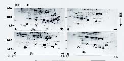
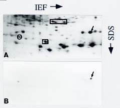
Abstract. We utilized two-dimensional polyacrylamide gel electrophoresis, coupled with silver stain, to identify five ovule-enriched and three ovule-specific polypeptides in Cycas taiwaniana Carr. (CTO). These ovule-enriched and -specific proteins are all with acidic pIs in the 4.8_6.4 range. Polyclonal antiserum to an ovule polypeptide (CTO-22) was prepared and affinity-purified to investigate the specificity of the protein. Immunoblot analyses of total protein from the vegetative organs and from organs of the strobili demonstrated that the CTO-22 polypeptide was ovule-specific. The amino terminal sequence of the CTO-22 protein exhibits no similarity with any known protein. To our knowledge, this is the first ovule-specific protein reported in gymnosperms.

Keywords: Cycas taiwaniana; 2D PAGE; Immunoblotting; Ovule-enriched/specific proteins.

Introduction

Gymnosperms were originally defined by the nature of their female reproductive organs (megastrobili), in which the seeds are naked in the carpel. The gymnosperm ovules are the precursors of seeds that are themselves complex structures. They consist of megasporangia, which except for the micropyle are entirely covered by a number of integument layers (Bold et al., 1987). Although the histological events concerning ovule anatomy and morphology are well characterized both in gymnosperms and in many angiosperms (Johri, 1984; Robinson-Beers et al., 1992), little is known about ovule development at the molecular level. With regard to angiosperms, the relative inaccessibility of the ovule within the ovary and the difficulty of collecting sufficient amounts of ovules have impeded the understanding of the molecular basis of ovule development. Taking advantage of the naked feature and the large size of the ovules, this study attempts to identify ovule-specific proteins in cycads. Previous investigators have identified several mutants affecting ovule and female gametophyte development (Robinson-Beers et al., 1992; Reiser and Fischer, 1993; Léon-Kloosterziel et al., 1994; Modrusan et al., 1994; Gaiser et al., 1995; Angenent and Colombo, 1996). Peroxidases and glycoproteins have been reported to be associated with developing ovules (Pettitt, 1977; Mellon and Triplett, 1989). Nadeau et al. (1996) recently characterized several ovule-specific cDNAs in orchid plants.

Two-dimensional polyacrylamide gel electrophoresis (2D PAGE) coupled with silver stain has been proved to be a sensitive technique for analyzing proteins of mutants

(Santoni et al., 1994), of organs at various developmental stages (Schrauwen et al., 1990; Wang et al., 1992a), and of organs responding to a variety of stresses (Hurkman and Tanaka, 1986; Vartanian et al., 1987). We report herein on the five ovule-enriched and three ovule-specific proteins in Cycas taiwaniana identified by 2D-PAGE analysis. In combination with the use of affinity-purified antibodies, we confirm CTO-22 to be an ovule-specific protein.

Materials and Methods

Plant Materials

Plants of Cycas taiwaniana Carr. were grown in the field. Maturing ovules with weights ranging from 0.5 to 0.6 gm were harvested. Microsporophylls and pollen grains at shedding stage were collected. Leaves (>10 cm) from a newborn leaf crown, entire roots (approximately 12 cm from the apex), and stems were collected separately. Material from both vegetative and reproductive organs (strobili) was dissected and frozen immediately in liquid nitrogen. All material was stored at -80°C.

Preparation, Electrophoresis, and Immunoblotting of Total Protein

Total protein was extracted according to Wang et al. (1992a). Proteins in the phenol phase were precipitated with five volumes of 0.1 M ammonium acetate in methanol at -20°C overnight. The precipitate was washed three times with 0.1 M ammonium acetate in methanol and once with 100% acetone. The pellet was dried and then resuspended in the solubilization buffer consisting of 9.5 M urea, 2% (v/v) Triton X-100, 5 mM K2CO3, 0.5% dithiothreitol (DTT), 500 µgml-1 L-lysine, 0.4%

1Corresponding author. Fax: 886-4-286-1905.


Botanical Bulletin of Academia Sinica, Vol. 38, 1997

Ampholines, pH 3-10 (Serva, Heidelberg, Germany), and 1.6% Ampholines, pH 5-7 (Serva) (Hari, 1981). When the phenol partitioning method was combined with the solubilization buffer containing lysine and Ampholines, the resolution of protein spots on the gels became sharper than when using O'Farrell (1975) solubilization buffer. Protein extractions for each tissue sample were carried out in duplicate. Protein concentration was determined by a modified Bradford method (Ramagli and Rodriguez, 1985). 2D-PAGE and SDS-PAGE were carried out according to previously published procedures (Wang et al., 1992a). Protein was either stained with Coomassie blue R-250 or silver, or electroblotted onto nitrocellulose (0.45 µm, Gelman Sciences, Ann Arbor, MI, USA). Blots were immunostained using a 1:1 dilution of eluate of affinity-purified antibodies to the CTO-22 protein.

Production and Purification of Antiserum

Production of antibody was done according to Polson et al. (1985). The ovule-specific polypeptide 43 (CTO-22) that was well isolated on 2D gels was Coomassie-stained, excised, and stored at -80°C. Approximately 20 µg of the 2D-PAGE-purified CTO-22 protein in gel slices was pooled and ground in 0.5 ml of PBS (10 mM sodium phosphate, pH 7.4, 150 mM NaCl) and emulsified with 0.5 ml of Freund's complete adjuvant (Sigma, St. Louis, MO, USA). The emulsion was intramuscularly injected into a local hen L2-BDS (kindly provided by Y.P. Lee, Department of Animal Science, National Chung Hsing University, Taichung, Taiwan).

To isolate antibodies that specifically recognize CTO-22, 30 µg of 2D-PAGE-purified CTO-22 protein was fractionated by SDS-PAGE and immunoblotted. The outside two strips of the blot were stained with Ponceau S to localize the CTO-22 protein. Sections of unstained nitrocellulose containing the CTO-22 protein were excised and incubated in blocking solution containing 3% gelatin in TTBS (20 mM Tris-HCl, pH 7.5, 500 mM NaCl, 0.05% Tween 20, v/v) for 30 min. The blot was incubated in a 1:40 dilution of the CTO-22 antiserum in TTBS for 7 h at 4°C and then washed eight times with TTBS for a total of 16 h. The blot-purified antibodies were eluted with a low pH buffer (5 mM glycine-HCl, pH 2.3, 500 mM NaCl, 100 µgml-1 BSA, 0.5% Tween 20, v/v). The eluates were immediately neutralized by the addition of 1 M sodium phosphate (pH 7.7) to a final concentration of 50 mM as described by Smith and Fisher (1984). The eluates were aliquoted, and stored at -80°C.

Sequence Analysis

Sequence analysis was performed according to Matsudaira (1987). 10 µg of purified CTO-22 protein was fractionated by SDS-PAGE, electroblotted onto Immobilon-PSQ membrane and stained with Coomassie blue R-250. The protein on the blot was excised for sequence analysis. The homology search against major sequence databases was done with the Blast programs (Altschul et al., 1990)

Results

2D-PAGE Analysis of Ovule and Vegetative Organ Proteins

More than 500 polypeptides were resolved by 2D-PAGE (Figure 1). About half of them were well resolved

Figure 1. 2D gels of total protein extracted from ovules (A) and vegetative organs of Cycas taiwaniana: stems (B), leaves (C), and roots (D). 200 µg of total protein from each sample was electrophoresed and silver-stained. Strobilus-enriched or -specific polypeptides found in ovules are indicated by arrowheads and arrows, respectively. The constitutively expressed proteins encased by the circle, oval, square, and rectangle serve as internal markers to orient each gel. Marker proteins are indicated by lines at the left.


Hu et al. — Ovule-enriched/specific proteins in Cycas

and abundant, and, therefore, their levels were monitored. For convenience of discussion, the proteins examined were serially numbered. To facilitate orientation for each 2D-PAGE analysis, five marker proteins that were detected both in ovules and in vegetative organs are indicated.

To identify strobilus-enriched and -specific polypeptides, the protein profiles of stems, young leaves, and roots were documented (Figure 1, B-D) and compared to the proteins observed in the ovule organ (Figure 1A). Strobilus-enriched polypeptides are defined as the polypeptides that were detected in one or more of the vegetative organs but were always detected at higher levels in the megastrobili. Strobilus-specific polypeptides are defined as the polypeptides detected only in the megastrobili. The profiles of vegetative protein were compared with those for the ovule identified four strobilus-enriched and four

strobilus-specific polypeptides (Figure 1 and Table 1). The four strobilus-enriched polypeptides in the ovule were also detected in leaves.

Ovule-Enriched and Ovule-Specific Polypeptides

To investigate the distribution of these organ-enriched/specific polypeptides in the strobili, total protein was extracted from mature pollen, microsporophylls, and megasporophylls. 2D-PAGE fractionation indicated that the four strobilus-enriched proteins were ovule-enriched (Figure 2 and Table 1). Of the four strobilus-specific polypeptides, polypeptides 21, 43 and 46 were ovule-specific whereas polypeptide 49 was detected in all the organs of strobili, but at a much more abundant level in the ovule than in any other organ of strobili (Table 1). Therefore, the polypeptide 49 was categorized as an ovule-enriched pro

Table 1. Ovule-enriched/specific polypeptides in Cycas taiwaniana.

Relative abundance of polypeptidesb

No.a MW pI Ovules Mega- Micro- Pollen Roots Stems Leaves

(kDa) sporophylls sporophylls

20 12.0 6.4 ++ + + _ _ _ +

44 17.0 5.4 ++ + + _ _ _ +

45 17.0 5.4 ++ + + _ _ _ +

47 17.0 5.3 ++ _ _ _ _ _ +

49 12.5 4.8 ++ + ± + _ _ _

21 14.5 6.2 + _ _ _ _ _ _

43 22.2 5.5 ++ _ _ _ _ _ _

46 17.5 5.3 + _ _ _ _ _ _

aPolypeptide numbers correspond to those shown in Figure 1.

b++, intense; +, detectable; ±, obscure; _, not detected.

Figure 2. Identification of ovule-enriched and -specific polypeptides in various organs of Cycas strobili. Total protein was extracted from various organs of the strobili: A, Ovules; B, Mature pollen; C, Megasporophylls; D, Microsporophylls. 200 µg of total protein from each sample was electrophoresed and silver-stained. Strobilus-enriched or -specific polypeptides found in ovules are indicated by arrowheads and arrows, respectively. The constitutively expressed proteins encased by the circle, oval, square, and rectangle serve as internal markers to orient each gel. Marker proteins are indicated by lines at the left.


Botanical Bulletin of Academia Sinica, Vol. 38, 1997

tein. These ovule-enriched and ovule-specific proteins are all with acidic pIs in the 4.8_6.4 range.

Characterization of the CTO-22 Protein

Of the three ovule-specific proteins, polypeptide 43, with a molecular mass of 22.2 kDa (CTO-22), was chosen for further investigation. Antiserum was raised against the CTO-22 polypeptide, and antibodies were affinity-purified from the antiserum. The specificity of the antibodies was determined by immunoblot analyses of ovule proteins (Figure 3B, lane 4). Affinity-purified CTO-22 antibodies reacted primarily with the CTO-22 polypeptide, indicating high specificity. No proteins were detected when identical blots were incubated with antibodies purified from preimmune IgG (Figure 3C, lane 4). An immunoblot of ovule proteins on the 2D gel indicated that two cross-reactive polypeptides with molecular weights lower than CTO-22 also reacted with the anti-CTO-22 antibodies, but the cross-reactivity was very weak (Figure 4B).

To confirm the organ specificity of the protein, affinity-purified antibodies were utilized in immunoblot studies of total protein extracted from vegetative organs (stems, leaves, and roots) and from organs of the strobili (pollen, microsporophylls, and megasporophylls). The CTO-22 polypeptide was detected only in ovules (Figure 3B). A small number of proteins with low molecular weights both in stems and pollen cross-reacted with affinity-purified CTO-22 antibodies, but the cross-reactivity was either very weak or resulted from reaction with proteins far more abundant than CTO-22.

The N-terminus of the CTO-22 protein was sequenced, giving the sequence: SYVLDAEGEPLK. After aligning this partial sequence with those in the sequence bank, we find that the CTO-22 protein exhibits no similarity with any known protein.

Figure 4. Immunoblot analysis of the ovule-specific CTO-22 protein in Cycas taiwaniana. Approximately 250 µg of total protein extracted from ovules was fractionated by 2D-PAGE and either (A) silver-stained, or (B) electroblotted onto nitrocellulose, and immunochemically detected using affinity-purified anti-CTO-22 antibodies. 1.6% Ampholines, pH 5-6 (Serva) was used instead of pH 5-7 in the first dimension of 2D-PAGE in order to have a better separation of ovule proteins around CTO-22. The CTO-22 protein is indicated by arrows. The constitutively expressed proteins encased by the circle, square, and rectangle serve as internal markers to orient the gel.

Discussion

We were able to identify a number of ovule-enriched and -specific proteins in Cycas taiwaniana from 2D gels because these proteins were well isolated from the others. The phenol partitioning method was used to avoid protein degradation that may occur during extraction (Schuster and Davis, 1983). Protein extractions for each tissue sample by this method were carried out in duplicate and protein gel patterns were reproducible after repeated freezing and thawing of samples.

Kamalay and Goldberg (1980) have reported that over 24,000 gene products are present in a given organ and that approximately 10,000 mRNAs in tobacco ovaries are organ-specific. Therefore, proteins encoded by these ovary-specific mRNAs would account for 40% of the total proteins, regardless of any posttranslational modification. In this study, the percentage of the ovule-specific polypeptides detected on 2D gels is far less than the above calculated average value. The detection of only a small fraction of ovule-specific proteins suggests that the level of many of the ovule-specific gene products are below the level of detection by silver staining. This phenomenon may reflect the role of these proteins in the regulation of gene expression. Alternatively, ovule-specific proteins may accumulate to high levels in selected cell types but may be significantly diluted in total ovule protein extracts.

Figure 3. Immunoblot analysis of the distribution of CTO-22 protein in Cycas taiwaniana organs. Total protein was extracted from various vegetative organs and from various organs of the strobili. 100 µg of total protein from each organ was fractionated by SDS-PAGE and then either stained by Coomassie blue (A) or electroblotted onto nitrocellulose, and immunochemically detected using affinity-purified anti-CTO-22 or preimmune antibodies (B and C). Roots (lane 1), stems (lane 2), leaves (lane 3), ovules (lane 4), mature pollen (lane 5), megasporophylls (lane 6), and microsporophylls (lane 7). M indicates lanes containing marker proteins (66.0, 45.0, 36.0, 29.0, 24.0, 20.0, 14.2 kDa).


Hu et al. — Ovule-enriched/specific proteins in Cycas

Antiserum was raised against an ovule-specific protein and affinity-purified antibodies were prepared from it. Since the amount of ovule-specific proteins collected by 2D-PAGE is limited, an induction of strong immunization may be difficult. Accordingly, the resulting antiserum with low titer yields nonspecific interactions. The anti-CTO-22 antibodies were thus purified from nitrocellulose membranes to which the CTO-22 protein was bound. The method of blot-affinity purification significantly increased the specificity of anti-CTO-22 antibodies. However, the titer of anti-CTO-22 antibodies did not markedly increase because a substantial amount of antibodies remained bound to the membrane after low pH elution. Applying different elution buffers such as 6 M urea or 5M guanidine-HCl increased unbound antibodies in the eluate, but the titer of the eluate did not proportionally increase. It is possible that IgGs may be irreversibly inactivated in the eluate at the presence of those chaotropic reagents (data not shown). The anti-CTO-22 antibodies react mainly with CTO-22 polypeptide, but the antibodies also react with two other polypeptides whose molecular weights are lower than CTO-22 (Figure 4B). Nevertheless, the relatedness of these two polypeptides to the CTO-22 protein has to be further determined.

Antiserum was also raised against the polypeptide 21 (CTO-15), another Cycas ovule-specific protein identified by 2D-PAGE. The affinity-purified CTO-15 antibodies recognize both the CTO-15 and another protein with a higher molecular weight than CTO-15 (data not shown), indicating a high specificity of the antibodies. However, its titer is lower than anti-CTO-22 antibodies. In earlier reports, we immunologically characterized several anther-specific proteins that were identified by 2D-PAGE (Wang et al., 1992b, 1996). Therefore, 2D-PAGE combining blot-purified antibodies is proved to be a powerful tool for analyzing proteins.

By the use of affinity-purified antibodies, we demonstrate that the CTO-22 is an ovule-specific protein. The same antibodies can also be employed to investigate the distribution of CTO-22 both in various species of gymnosperms and in angiosperms. Our aim is to find the CTO-22-like protein in plants of economic importance, particularly in those fruit-producing species. The presence of the CTO-22-like protein in angiosperms would suggest that the protein is evolutionary conserved and thus may play a prominent role in ovule development. The ovule-specific gene can then be manipulated in a manner that inhibits ovule development while fruit growth remains normal. Accordingly, the abnormal development of ovules may give rise to a seedless fruit.

The partial sequence of CTO-22 protein may serve a purpose in designing oligosynthetic primers. With the aid of the synthetic primers or CTO-22 antiserum, cDNA clones encoding the CTO-22 protein will be isolated and characterized in the future.

We have identified herein five ovule-enriched and three ovule-specific proteins in Cycas taiwaniana. By employing affinity-purified antibodies, we demonstrate that the

CTO-22 protein is ovule-specific. The amino terminal sequence of CTO-22 exhibits no similarity with any known protein. To our knowledge, the ovule-specific protein in gymnosperms is here reported for the first time.

Acknowledgments. This work was supported by National Science Council grant NSC84-2311-B-005-005 to Co-Shine Wang.

Literature Cited

Altschul, S. F., W. Gish, W. Miller, E. W. Mayers, and D. J. Lipman. 1990. Basic local alignment search tool. J. Mol Bilo. 215: 403_410.

Angenent, G. C. and L. Colombo. 1996. Molecular control of ovule development. Trends Plant Sci. 1: 228_232.

Bold, H. C., C. J. Alexopoulos, and T. Delevoryas (eds.). 1987. Morphology of Plants and Fungi. New York, pp. 524_545.

Gaiser, J. C., K. Robinson-Beers, and C. S. Gasser. 1995. The Arabidopsis SUPERMAN gene mediates asymmetric growth of the outer integument of ovules. Plant Cell 7: 333_345.

Hari, V. 1981. A method for the two-dimensional electrophoresis of leaf proteins. Ana. Biochem. 113: 332_335.

Hurkman, W. J. and C. K. Tanaka. 1986. Solubilization of plant membrane proteins for analysis by two-dimensional gel electrophoresis. Plant Physiol. 81: 802_806.

Johri, B. M. (ed.). 1984. Embryology of Angiosperms. Springer-Verlag, Berlin, Heidelberg, New York, Tokyo, pp. 123_157.

Kamalay, J. C. and R. B. Goldberg. 1980. Regulation of structural gene expression in tobacco. Cell 19: 935_946.

Léon-Kloosterziel, K. M., C. J. Keijzer, and M. Koornneef. 1994. A seed shape mutant of Arabidopsis that is affected in integument development. Plant Cell 6: 385_392.

Matsudaira, P. 1987. Sequence from picomole quantities of proteins electroblotted onto polyvinylidene difluoride membranes. J. Biol. Chem. 262: 10035_10038.

Mellon, J. E. and B. A. Triplett. 1989. De novo synthesis of peroxidase in cotton ovule culture medium. Physiol. Plant. 77: 302_307.

Modrusan, Z., L. Reiser, K. A. Feldmann, R. L. Fischer, and G. W. Haughn. 1994. Homeotic transformation of ovules into carpel-like structures in Arabidopsis. Plant Cell 6: 333_349.

Nadeau, J. A., X. S. Zhang, J. Li, and S. D. O'Neill. 1996. Ovule development: Identification of stage-specific and tissue-specific cDNAs. Plant Cell 8: 213_239.

O'Farrell, P. H. 1975. High resolution two-dimensional electrophoresis of proteins. J. Biol. Chem. 250: 4007_4021.

Pettitt, J. M. 1977. Detection in primitive gymnosperms of proteins and glycoproteins of possible significance in reproduction. Nature 266: 530_532.

Polson, A., T. Coetzer, J. Kruger, W. von Maltzahn, and K. J. van der Merwe. 1985. Improvements in the isolation of IgY from yolks of eggs laid by immunized hens. Immunol. Invest. 14: 323_327.

Ramagli, L. S. and L. V. Rodriguez. 1985. Quantitation of microgram amounts of protein in two-dimensional polyacrylamide gel electrophoresis sample buffer. Electrophoresis 6: 559_563.

Reiser, L. and R. L. Fischer. 1993. The ovule and the embryo


Botanical Bulletin of Academia Sinica, Vol. 38, 1997

sac. Plant Cell 5: 1291_1301.

Robinson-Beers, K., R. E. Pruitt, and C. S. Gasser. 1992. Ovule development in wild-type Arabidopsis and two female-sterile mutants. Plant Cell 4: 1237_1249.

Santoni, V., C. Bellini, and M. Caboche. 1994. Use of two-dimensional protein-pattern analysis for the characterization of Arabidopsis thaliana mutants. Planta 192: 557_566.

Schrauwen, J. A. M., P. F. M. de Groot, M. M. A. van Herpen, T. van der Lee, W. H. Reynen, K. A. P. Weterings, and G. J. Wullems. 1990. Stage-related expression of mRNAs during pollen development in lily and tobacco. Planta 182: 298_304.

Schuster, A. M. and E. Davis. 1983. Ribonucleic acid and protein metabolism in pea epicotyls. I. The aging process. Plant Physiol. 73: 809_816.

Smith, D. E. and P. A. Fisher. 1984. Identification, developmental regulation and response to heat shock of two antigenically related forms of a major nuclear envelope protein in

Drosophila embryos: application of an improved method for affinity purification of antibodies using polypeptides immobilized on nitrocellulose blots. J. Cell Biol. 99: 20_28.

Vartanian, N., C. Damerval, and D. de Vienne. 1987. Drought-induced changes in protein patterns of Brassica napus var. oleifera roots. Plant Physiol. 84: 989_992.

Wang, C.-S., L. L. Walling, K. J. Eckard, and E. M. Lord. 1992a. Patterns of protein accumulation in developing anthers of Lilium longiflorum correlate with histological events. Amer. J. Bot. 79: 118_127.

Wang, C.-S., L. L. Walling, K. J. Eckard, and E. M. Lord. 1992b. Immunological characterization of a tapetal protein in developing anthers of Lilium longiflorum. Plant Physiol. 99: 822_829.

Wang, C.-S., T.-D. Wu, C.-K. W. Chung, and E. M. Lord. 1996. Two classes of pollen-specific, heat-stable proteins in Lilium longiflorum. Physiol. Plant. 97: 643_650.