Bot. Bull. Acad. Sin. (1997) 38: 177-182

Zheng and Ko — Variation of Phytophthora cinnamomi

Continuing variation in colony morphology and fungicide

sensitivity in Phytophthora cinnamomi following exposure

to chloroneb

Xia Bo Zheng1,2 and Wen-Hsiung Ko1,3

1Department of Plant Pathology, Beaumont Agricultural Research Center, University of Hawaii at Manoa, Hilo, Hawaii 96720, U. S. A.

(Received December 9, 1996; Accepted March 8, 1997)

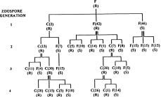
Abstract. A homothallic isolate of Phytophthora cinnamomi grown on agar medium containing fungicide chloroneb, produced fast growing resistant sectors within 3 weeks. The colony of a fungicide-resistant mutant, which was similar to the original flat (F) type, gave rise to single zoospore cultures consisting of a new cottony (C) type resistant to chloroneb and the F type either resistant or sensitive to chloroneb. The F type was continuously segregated from the C type in three successive zoospore generations. Although the chloroneb-sensitive F type revertants were stable, they produced more resistant sectors in a shorter time than did wild type when exposed again to chloroneb. Single zoospore cultures obtained from these resistant sectors were C type resistant to chloroneb and F type either resistant or sensitive to chloroneb. Both C and F type cultures resistant to chloroneb were unstable and segregated in successive zoospore generations in a fashion similar to that described above. The majority of zoospores (89 to 99%) produced by both C and F types were uninucleate.

Keywords: Chloroneb; Colony morphology; Fungicide sensitivity; Phytophthora cinnamomi; Variation.

Introduction

Phytophthora cinnamomi Rands usually produces clusters of chlamydospores, coralloid hyphae, and both spherical and irregular hyphal swellings on agar media, and forms uniform nonpapillate sporangia only under special conditions such as in nonsterile soil extract (Zentmyer, 1980). The fungus is also considered heterothallic because of the requirement for the presence of opposite mating types for production of sexual structures (Ho, 1981; Newhook et al., 1978). However some isolates of P. cinnamomi vary from the characteristic features mentioned above. Ann and Ko (1985) reported the isolation of an A2 isolate of P. cinnamomi from soil capable of producing sporangia on agar medium and A1 isolates from citrus incapable of producing chlamydospores. From black locust (Robinia pseudoacacia L.) Ho et al. (1983) obtained an A1 isolate of P. cinnamomi incapable of producing chlamydospores, but capable of producing sporangia on agar medium, and a homothallic isolate incapable of producing chlamydospores.

During the selection of fungicide resistance as a marker for studying the genetic relationship between the homothallic isolates and the heterothallic isolates of P.

cinnamomi, it was found that exposure of the fungus to chloroneb not only induced resistance to this fungicide but also altered the colony morphology. Fungi usually are inhibited by fungicides only in the presence of those chemicals, and revert to normal growth and appearance after being transferred to a medium free from these chemicals. Although development of resistance to fungicides by fungi after their exposure to these chemicals is relatively common (De Waard et al., 1993), reports on changes in other characteristics induced by fungicides are rare. We, therefore, studied the stability of the new colony type and the relationship between the new colony type and the fungicide resistance induced by chloroneb. Details of the study are reported here.

Materials and Methods

Microorganism

Isolate P630 of P. cinnamomi was obtained from a diseased tree of black locust in Jiangsu, China and was derived from a single zoospore. The fungus was homothallic, capable of producing oospores in single culture, but incapable of forming chlamydospores (Ho et al., 1983). It was also distinctive in having the ability to produce sporangia in diluted sterile soil extract.

Induction of Sector Formation

The method described by Ann and Ko (1985) was used to induce sector formation. Four culture blocks (ca.

2Present address: Department of Plant Protection, Nanjing Agricultural University, Nanjing, China.

3Corresponding author. Fax: (808) 969-7923.


Botanical Bulletin of Academia Sinica, Vol. 38, 1997

15×15×3 mm) were placed on V-8 agar (10% V-8 juice, 0.02% CaCO3 and 2% Bacto agar) amended with 100 µg/ml chloroneb (Terraneb SP, 65% active) in a Petri plate. Chloroneb was added to hot V-8 agar right after autoclaving to prevent growth of contaminants. Plates were sealed with Parafilm, incubated at 24 ºC in darkness and observed every 2_3 days. Mutants which appeared as fast growing sectors were transferred to V-8 agar amended with 200 µg/ml chloroneb. After incubation for 3_5 days, these mutants were transferred to chloroneb free medium and cultured for 3 days before being used for zoospore formation.

Isolation of Single-Zoospore Cultures

The mycelial blocks (ca. 2×2×2 mm) from a 3-day-old culture grown on V-8 agar were placed in 15 ml of 10% clarified V-8 broth in a Petri plate. V-8 juice plus 1% CaCO3 was clarified by centrifugation at 270 g for 10 min before dilution with distilled water. After incubation at 24°C in darkness for 2_3 days, the broth was drained off and mycelial mats were resuspended in 15 ml of sterile tap water amended with 0.2 ml sterile soil extract. The soil extract was prepared by mixing 300 g of garden soil with 300 ml of tap water with a glass rod. After sedimentation for 30 min, the supernatant was filtered through Whatman No. 1 filter paper and sterilized by passage through a Millipore filter (GSWP, 0.22 µm, Millipore, Bedford, MA, USA). After incubation at 24°C in darkness overnight, the soil extract was drained and 15 ml of sterile tap water was added. Plates were then incubated at 24°C under cool white fluorescent light (2,000 lux). Within 8 h, sporangia were produced and zoospores were released from sporangia. About 0.2 ml of spore suspension, containing approximately 200_400 zoospores, was spread on a Petri plate containing 0.1% clarified V-8 agar amended with 100 µg/ml ampicillin, 50 µg/ml nystatin, and 10 µg/ml pentachloronitrobenzene to prevent growth of contaminants (Ko et al., 1978). After incubation at 24°C for 24_48 h, colonies originating from single zoospores were individually transferred to V-8 agar plate.

Determination of Resistance to Chloroneb

Chloroneb resistance of single-zoospore cultures was determined by placing culture blocks (ca. 2×2×2 mm) on V-8 agar and V-8 agar amended with 100 µg/ml chloroneb (Chang and Ko, 1990). Inoculated plates were incubated at 24°C in darkness for 3 days. Those cultures which were sensitive to chloroneb did not grow in 3 days on chloroneb medium, while those which were resistant grew normally. Symbols to represent phenotypes for fungicide resistance were R for chloroneb resistance and S for chloroneb sensitivity.

Determination of Colony Morphology

Mycelial blocks (ca. 2×2×2 mm) cut from the advancing margin of each single-zoospore culture were placed equidistant from the edge of a Petri dish containing about 10 ml of V-8 agar without chloroneb. Two blocks per isolate and six blocks per plate were used. Colony morphol

ogy was determined after incubation at 24°C for 3 days. Symbols to represent phenotypes for colony appearance were F for flat, smooth colonies without conspicuous aerial growth, and C for cottony colonies with abundant aerial growth.

Nuclear Staining of Zoospores

Nuclei in zoospores were stained with 25 µg/ml acridine orange in 1% veronal acetate solution. The effectiveness of the stain was greatly improved by adjusting the pH of the solution to 8.6 rather than 4.5 as described by Yamamoto and Uchida (1982). One drop of zoospore suspension was mixed with one drop of acridine orange solution on a glass slide. The mixture was dried with a hair dryer. Another drop of acridine orange solution was placed on the dried spot and covered with a cover slip. The specimen was sealed with fingernail varnish and examined immediately with a Zeiss fluorescence microscope. Nuclei were clearly visible as bright green spherical, elliptical or irregular particles against a faint green background (Yamamoto and Uchida, 1982).

Results

Induction of Mutation

When culture blocks were incubated on agar medium containing 100 µg/ml chloroneb for 3 weeks, one fast-growing sector was obtained in the first test and two in the second test. The first chloroneb resistant mutant (P630-1) was selected for further study. The mutant had F type colony characteristics like its parent.

Variation in Successive Asexual Generations of a Resistant Mutant

All 100 single-zoospore cultures obtained from wild-type isolate P630 were of the parental F type and sensitive to chloroneb. However, among 90 zoospore cultures obtained from resistant isolate P630-1, 42 cultures retained the phenotypic characteristics of the mutant with the F type colony and resistance to chloroneb; 2 cultures were C type and resistant to chloroneb; while 46 cultures were F type and sensitive to chloroneb (Figure 1).

Fifteen zoospore cultures were obtained from each of three randomly selected F type isolates sensitive to chloroneb in the first zoospore generation. These isolates appeared to be stable as all the zoospore cultures tested expressed the parental phenotypic characteristics (Figure 1).

Thirty zoospore cultures derived from one of the two C type colonies resistant to chloroneb in the first zoospore generation were examined. The C type was found to be unstable. Although 23 cultures retained the characteristics of C type resistant to chloroneb, 7 of them reverted to F type sensitive to chloroneb (Figure 1). These revertants were also stable as all zoospore cultures maintained the parental phenotypic characteristics. When two of the 23 C type colonies resistant to chloroneb in the second


Zheng and Ko — Variation of Phytophthora cinnamomi

Variation in Successive Asexual Generations of a Resistant Mutant from a Revertant

Single-zoospore cultures were derived from three resistant sectors produced by revertant P630-1 (S1). All 30 zoospore cultures derived from resistant sector 1 were C type resistant to chloroneb. However, zoospore culture from resistant sector 2 consisted of C type resistant to chloroneb and F type either resistant or sensitive to chloroneb, while those from resistant sector 3 consisted of C type resistant to chloroneb and F type sensitive to chloroneb (Figure 2).

All the 20 zoospore cultures from a C type resistant to chloroneb in the first zoospore generation of resistant sector 1 were C type resistant to chloroneb. However, one of these second zoospore generation C types tested produced zoospore cultures consisting of C type resistant to chloroneb and F type either resistant or sensitive to chloroneb (Figure 2).

Zoospore culture from an F type resistant to chloroneb in the first zoospore generation of resistant sector 2 consisted of C type resistant to chloroneb and F type either resistant or sensitive to chloroneb (Figure 2). All 15 zoospore cultures from a revertant producing F type colony sensitive to chloroneb in the first zoospore generation of resistant sector 3 were F type sensitive to chloroneb. One of the two C types resistant to chloroneb in the first zoospore generation of resistant sector 3 produced zoospore cultures only of C type resistant to chloroneb while the other produced zoospore cultures consisting of C type resistant to chloroneb and F type sensitive to chloroneb (Figure 2).

Nuclear Number of Zoospore

Among 100 zoospores of the parental isolate P630, 99 had a single nucleus and the other one had none (Table

Figure 1. Variation in colony type and sensitivity to chloroneb among single-zoospore cultures of the chloroneb-resistant mutant (P630-1) producing the F type colony of Phytophthora cinnamomi. Each line under a phenotypic category represents a tested culture. Symbols representing phenotypes for colony appearance are F for flat and smooth colonies without conspicuous aerial growth, and C for cottony colonies with abundant aerial growth, while those representing phenotypes for chemical resistance are R for chloroneb-resistance and S for chloroneb-sensitivity. Numbers in parentheses are numbers of single-zoospore cultures under the same phenotypic category.

zoospore generation were tested, one produced C type colonies resistant to chloroneb and F type colonies sensitive to chloroneb, while the other produced only C type resistant to chloroneb. Among three C type cultures resistant to chloroneb in the third zoospore generation tested, one produced C type resistant to chloroneb and F type sensitive to chloroneb, while the other two produced only C type resistant to chloroneb (Figure 1).

The F type resistant to chloroneb in the first zoospore generation was also found to be unstable. All three isolates randomly selected produced zoospore cultures consisting of C type resistant to chloroneb and F type revertants (Figure 1). When two of the C type resistant to chloroneb in the second zoospore generation were tested, one produced C type resistant to chloroneb and F type revertants, while the other produced only C type resistant to chloroneb. Among the two C type cultures resistant to chloroneb in the third zoospore generation tested, one produced C type resistant to chloroneb and F type revertants, while the other produced only C type resistant to chloroneb (Figure 1).

Carry-Over Effect from Previous Mutation

To determine if the previous mutation to chloroneb resistance had any effect on the ability of the revertants to mutate again when reexposed to the fungicide, nine revertants from first zoospore generation and nine wild-type isolates were grown on the medium containing 100 µg/ml chloroneb. Five of the revertants produced fast growing resistant sectors within 5 days, and all of them produced resistant sectors ranging from 6 to 18 sectors after 10 days. In comparison, the wild type isolates did not produce any resistant sectors after 10 days, and only 2 resistant sectors appeared after 15 days.

Figure 2. Variation in colony type and sensitivity to chloroneb among single-zoospore cultures derived from three chloroneb-resistant sectors (RS) from revertant P630-1 (S1) with the F type colony sensitive to chloroneb of Phytophthora cinnamomi when grown on medium containing 100 µg/ml chloroneb. Each line under a phenotypic category represents a tested culture. Symbols: F for F type colony, C for C type colony, R for chloroneb resistance, and S for chloroneb sensitivity.


Botanical Bulletin of Academia Sinica, Vol. 38, 1997

sensitive isolates have F type colonies. Those F type colonies which were resistant to chloroneb were unstable and always segregated into sensitive F type and resistant C type.

Exposure of P. cinnamomi to chloroneb caused the appearance of C type colonies from F type parents and chloroneb resistant isolates from sensitive parents. These mutants were unique because the F type colonies continue to appear from the C type mutants, and chloroneb sensitive isolates also continue to appear from resistant mutants (Figures 1 and 2). Caten and Jinks (1968) observed continuous variation in growth rate in three successive zoospore generations derived from three isolates of Phytophthora infestans (Montagne) de Bary that were recovered from diseased potato leaves. Leonian (1925) also reported the persistent dissociation into two colony types over 20 sporangial generations derived from one particular isolate of Phytophthora parasitica var. rhei Godfrey. It was suggested that the continuous variation in asexual generations observed in these two cases may have originated from progeny established by selfed oospores in nature (Guo and Ko, 1995).

The mechanism of persistent segregation of F type from C type during asexual reproduction is still unknown. It was not due to heterokaryosis since the C type cultures tested originated from single zoospores which were shown to be mostly uninucleate. This is different from the variation referred to as `dual phenomenon' in imperfect fungi, which was found to be due to the heterokayotic condition of multinucleate spores with subsequent dissociation of the homotype (Hansen, 1938). It is conceivable that both sensitivity to chloroneb and colony morphology of the mutants tested were controlled by heterozygous cytoplasmic genes. Each mitochondrion is believed to contain several copies of the mitochondrial chromosome (Russell, 1992), and mitochondria are numerous in each zoospore of Phytophthora (Shaw, 1983). Based on segregation results, it is further suggested that every mutant contains many copies of these genes and that chloroneb-resistant allele is dominant to chloroneb-sensitive allele.

Chloroneb resistance in Ustilago maydis (DeCandolle) Corda (Tillman and Sisler, 1973) and P. parasitica (Chang and Ko, 1990) has been reported to be controlled by nuclear genes. The apparent discrepancy could be ex

1). The majority of zoospores produced by the chloroneb-resistant mutants tested also had a single nucleus. Only two of these zoospores contained two nuclei each. Most of the zoospores produced by the revertant tested also had only one nucleus (Table 1).

Discussion

Although development of fungicide resistance after exposure to that chemical is relatively common (De Waard et al, 1993), a fungicide seldom causes changes in other phenotypic characteristics. Alteration of the colony morphology of P. cinnamomi caused by exposure to chloroneb documented in this study represents a rare case of fungicide-induced morphological change. Previously, Durieu-Trautmann and Tavilitzki (1975) reported that chloramphenical induced a stable change in Ustilago cynodontis (Passerini) P. Henning from hyphal cells to yeast-like colonies. Shaw and Elliott (1968) obtained some stable morphological variants of Phytophthora cactorum (Lebert et Cohn) Schroeter when a wild-type was treated with streptomycin. Recently, metalaxyl has also been reported to cause variation in colony type and growth rate among single zoospore cultures of Phytophthora parasitica Dastur (Chang and Ko, 1992). In addition to induction of chloroneb resistance and change in colony morphology, exposure to chloroneb has also caused mating type change and subsequent change in hormone production and hormone reception in P. parasitica and P. cinnamomi (Ann and Ko, 1989; Ko, 1981; Ko et al., 1986).

Our study shows that chloroneb-resistant mutants of P. cimmamomi may revert to their original chloroneb-sensitive state during asexual reproduction and that some of these revertants can be induced to become chloroneb resistant in a shorter period of time than the wild-type when re-exposed to the chemical. Whether these revertants can also become resistant to other fungicides faster than the wild type when exposed to those chemicals remains to be investigated.

The pattern of segregation of the chloroneb-induced mutant of P. cinnamomi during asexual reproduction suggests that there is a correlation between colony morphology and sensitivity to chloroneb. All isolates with C type colony were resistant to chloroneb, and all the chloroneb-

Table 1. Number of nuclei in each zoospore produced by isolate P630 of Phytophthora cinnamomi and its chloroneb-resistant mutants and a revertant.

No. of zoospores with

Strain Colony type Sensitivity to chloroneb 0 1 2 (nuclei)

Wild type F S 1 99 0

Mutants and revertanta

1 F S 5 92 3

2 C R 10 89 1

3 C R 6 93 1

aFrom the first zoospore generation of Figure 1. S= sensitive to chloroneb, R= resistant to chloroneb, F= flat and smooth colony, and C= cottony colony.


Zheng and Ko — Variation of Phytophthora cinnamomi

plained by the difference in isolates used. Streptomycin resistance in Chlamydomonas reinhardi Dang. has been found to be controlled by nuclear genes in some mutant strains and cytoplasmic genes in others (Sager, 1972). Both nuclear inheritance and cytoplasmic inheritance of chloramphenical resistance have also been observed in Aspergillus nidulans (Eidam) Winter (Gunatilleke et al., 1975). Segregation of colony types can not be explained by simple dominance. Gene interaction is probably involved in the expression of colony morphology.

Acknowledgements. This research was supported in part by the U. S. Department of Agriculture under Cooperative State Research Service (CSRS) Special Grant No. 90-3435-5216, managed by the Pacific Basin Advisory Group (PBAG). We thank the generous gift of chloroneb from Kincaid Enterprises, Nitro, WV. Journal Series Paper No. 3990 of the Hawaii Institute of Tropical Agriculture and Human Resources.

Literature Cited

Ann, P. J. and W. H. Ko. 1985. Variants of Phytophthora cinnammomi extended the known limits of the species. Mycologia 77: 946_950.

Ann, P. J. and W. H. Ko. 1989. Effect of chloroneb and ethazol on mating type of Phytophthora parasitica and P. cinnamomi. Bot. Bull. Acad. Sin. 30: 207_210.

Ann, P. J. and W. H. Ko. 1992. Survey of antibiotic resistance and dependence in Phytophthora. Mycologia 84: 82_86.

Caten, C. E. and J. L. Jinks. 1968. Spontaneous variability of single isolates of Phytophthora infestans. 1. Cultural variation. Can. J. Bot. 46: 329_348.

Chang, T. T. and W. H. Ko. 1990. Resistance to fungicides and antibiotics in Phytophthora parasitica: Genetic nature and use in hybrid determination. Phytopathology 80: 1414_1491.

Chang, T. T. and W. H. Ko. 1992. Variation in growth rate and colony morphology of Phytophthora parasitica induced by metalaxyl. Ann. Phytopathol. Soc. Japan 58: 72_77.

De Waard, M. A., S. G. Georgopoulas, D. W., Hollomon, H. Ishii, P. Leroux, N. M. Ragsdale, and F. J. Schwinn. 1993. Chemical control of plant diseases: problems and prospects. Annu. Rev. Phytopathol. 31: 403_421.

Durieu-Trautmann, O. and J. Tavilitzki. 1975. Reversible and permanent effects of the carbon and various antibiotics on the morphology and metabolic properties of Ustilago cynodontis cells. J. Cell Biol. 55: 102_113.

Gunatilleke, I. A. U. N., C. Saczzicchio, and H. N. Arst, Jr. 1975. Cytoplasmic and nuclear mutations to chloramphenical re

sistance in Aspergillus nidulans. Mol. Gen. Genet. 137: 269_276.

Guo, L. Y. and W. H. Ko. 1995. Continuing variation in successive asexual generations of Pythium splendens following sexual reproduction. Mycol. Res. 99: 1339_1344.

Hansen, H. N. 1938. The dual phenomenon in imperfect fungi. Mycologia 30: 442_445.

Ho, H. H. 1981. Synoptic keys to the species of Phytophthora. Mycologia 73: 705_714.

Ho, H. H., W. Y. Zhuang, Z. R. Liang, and Y. N. Yu. 1983. Phytophthora cinnamomi on black locust (Robinia pseudoacacia) in Jiangsu Province of China. Mycologia 75: 881_886.

Ko, W. H. 1981. Reversible change of mating type in Phytophthora parasitica. J. Gen. Microbiol. 125: 451_454.

Ko, W. H., H. S. Chang, and H. J. Su. 1978. Isolates of Phytophthora cinnamomi from Taiwan as evidence for an Asian origin of the species. Trans. Brit. Mycol. Soc. 71: 496_499.

Ko, W. H., C. J. Lee, and H. J. Su. 1986. Chemical regulation of mating type in Phytophthora parasitica. Mycologia 78: 134_136.

Leonian, L. H. 1925. Physiological studies on the genus Phytophthora. Am. J. Bot. 12: 445_498.

Newhook, F. J., G. M. Waterhouse, and D. J. Stamps. 1978. Tabular key to the species of Phytophthora de Bary. Mycol. Pap. 143: 1_20.

Russell, P. J. 1992. Genetics, 3rd edn. Harper Collins Publishers, New York.

Sager, R. 1972. Cytoplasmic Genes and Organelles. Academic Press, New York.

Shaw, D. S. 1983. The cytogenetics and genetics of Phytophthora. In D. C. Erwin, S. Bartnicki-Garcia and P. H. Tsao (eds.), Phytophthora; Its Biology, Taxonomy, Ecology, and Pathology. American Phytopathological Society, St. Paul, Minnesota.

Shaw, D. S. and C. G. Elliott. 1968. Streptomycin resistance and morphological variation in Phytophthora cactorum. J. Gen. Microbiol. 51: 75_84.

Tillman, R. W. and H. D. Sisler. 1973. Effect of chloroneb on the growth and metabolism of Ustilago maydis. Phytopathology 63: 219_225.

Yamamoto, D. T. and J. Y. Uchida. 1982. Rapid nuclear staining of Rhizoctonia solani and related fungi with acridine orange and with safranin O. Mycologia 74: 145_149.

Zentmyer, G. A. 1980. Phytophthora cinnamomi and the Diseases It Causes. Monograph 10. Am. Phytopathol. Soc., St. Paul. Minnesota.