Bot. Bull. Acad. Sin. (1997) 38: 197-204

Chang — Taiwan dematiaceous hyphomycetes

Eight more dematiaceous hyphomycetes new for Taiwan

H.S. Chang

Institute of Botany, Academia Sinica, Nankang, Taipei, Taiwan 115, Republic of China

(Received October 22, 1996; Accepted March 15, 1997)

Abstract. Monodisma fragilis, Ityorhoptrum verruculosum, Berkleasmium caribense, Acrodictys queenslandica, Guedea novae-zelandiae, Cacumisporium sigmoideum, Bactrodesium longisporum and Pseudospiropes subuliferus, all dematiaceous hyphomycetes, are new records for Taiwan. In addition, an isolate of Pseudospiropes, unlike known species is briefly treated along with P. subuliferus.

Keywords: Dematiaceous hyphomycetes; Taiwan.

Eight species of dematiaceous hyphomycetes observed growing on decaying twigs collected from streams during a study of freshwater microfungi are new records for Taiwan.

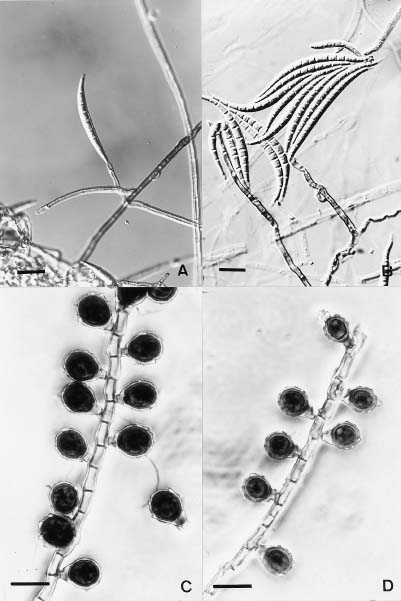
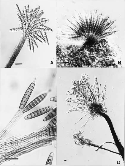
Monodisma fragilis Alcorn, Trans, Br. Mycol. Soc. 65: 139_141. 1975. (Figure 1A, B)

Conidiophores macronematous, mononematous, unbranched, brown, multiseptate, originating from the superficial hyphae, straight to flexuous or slight geniculate, smooth, up to 130 µm long, 2.5_4.5 µm and 4.5_7.0 µm wide at the middle and basal parts, respectively. Conidiogenous cells at first terminal, integrated, monoblastic, sometimes polyblastic, proliferating sympodially, conidia remaining attached laterally. Conidia dry, solitary, fusiform, hyaline, falcate, apex often rostrate, multispetate, smooth, 50_100 × 6_9 µm.

Habitat. This fungus has been isolated from blight leaves of Miscanthus spp. collected at Lusan, Nantou County (Jul. 11, 1984) and Tienchih, Taitong County (Aug. 23, 1993). Monodisma fragilis was originally described from Australia.

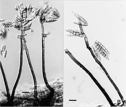
Ityorhoptrum verruculosum (M.B. Ellis) Kirk, Trans. Br. Mycol. Soc. 86: 417. 1986.

Endophragmia verruculosa Ellis, Mycol. Pap. 72: 29. 1959. (Figure 1C, D)

Conidiophores macronematous, mononematous, solitary, erect, simple, straight, smooth, septate, pale brown to brown, more than 400 µm high, 4.5_6.0 µm wide. Conidiogenous cells integrated, terminal, proliferating percurrently. Conidia acrogenous, solitary, obvoid to clavate, truncate at the base, 1-euseptate, apical cell pale brown to dark brown, verruculose, basal cell pale brown, smooth, 15_20 × 9_13 µm.

Habitat. This fungus was observed once only on an unknown decaying twig collected at Lienhwachu Forest

Branch Station at Yuitsu, Nantou County on Sep. 3, 1990. It has been recorded from Great Britain, New Zealand, & India.

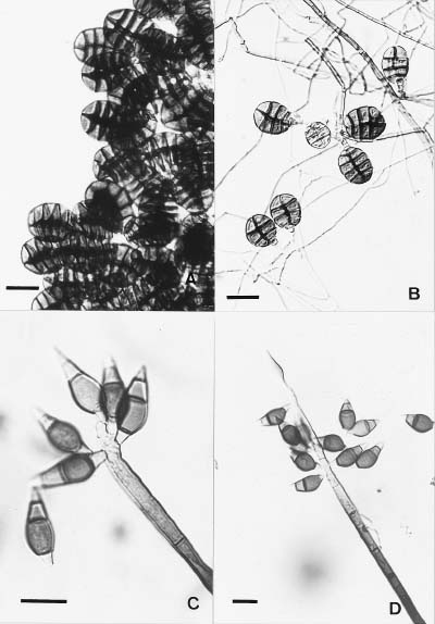
Acrodictys queenslandica Matsushima, Matsush. Myc. Mem. 6: 4. 1989. (Figure 2A)

Conidiophores macronematous, mononematous, simple, brown to dark brown, septate, up to 140 µm high, 3.0_4.0 µm at the apex, 3.5_5.0 µm wide medianly and 8.0_14 µm wide at the base. Conidiogenous cell integrated, terminal, monoblastic, often percurrent. Conidia solitary, dry, muriform, transversely ellipsoidal, brown, basal cell darker, 13.5_15.5 × 14_26 µm.

Habitat. This fungus was observed on a decaying twig collected at Wulai from a stream on Oct. 30, 1995, previously only being reported by Matsushima from material collected in Queensland, Australia in 1989.

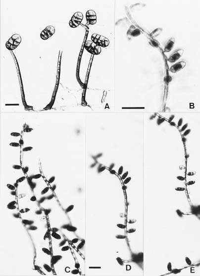
Guedea novae-zelandiae Hughes, New Zealand J. Bot. 18: 65. 1980. (Figure 2B, C, D, E)

Conidiophores macronematous, mononematous, erect, single or in groups of up to 4, simple, straight or flexuous, subcylindrical, brown to dark brown in the lower part and paler towards the upper part, up to 350 µm long, 2.5_4.0 µm wide. Conidiogenous cells integrated, indeterminate, holoblastic. Conidia holoblasticaly produced from successive penultimate cell of the conidiophore and near the septum of the terminal cell, globose, hyaline at first, becoming brown, oblong to oval, 3-celled, septa thick, resembling dark brands, terminal cell of the conidia lighter in colour, mature conidia with raised basal scars, 12_16 × 6.0_7.5 µm.

Habitat. This fungus was observed on a decaying twig collected at Wulai from a stream on Sep. 15, 1994. It was first recorded by Hughes (1980) from material collected in New Zealand and later by Wang and Sutton (1982) from New York.


Botanical Bulletin of Academia Sinica, Vol. 38, 1997

Figure 1. A_B, Monodisma fragilis; C_D, Ityorhoptrum verruculosum. Scale bar = 20 mm.


Chang — Taiwan dematiaceous hyphomycetes

Figure 2. A, Acrodictys queenslandica; B_E, Guedea novae-zelandiae. Scale bar = 20 mm.


Botanical Bulletin of Academia Sinica, Vol. 38, 1997

Figure 3. A_B, Berkleasmium caribense; C_D, Cacumisporium sigmoideum. Scale bar = 20 mm.


Chang — Taiwan dematiaceous hyphomycetes

Berkleasmium caribense Holubova-Jechova & Mercado Sierra, Ceska Mykol. 38: 99. 1984. (Figure 3A, B)

Sporodochia punctiform to effuse, granular, black. Conidiophores micronematous (or macronematious on culture), mononematous, solitary or fasciculate, erect or ascending, simple, septate, hyaline to very pale brown, smooth, indistinct on natural substrate but up to 10 × 3 µm in culture. Conidiogenous cells monoblastic, integrated, terminal, determinate. Conidia acrogenous, solitary, broadly obclavate to obpyriform in face view, ellipsoid to obclavate in lateral view, septate, with a single longitudinal septum and 3_4 transverse septa, constricted at the septa, pale brown to brown or dark brown with the pigmentation concentrated around the septa, especially the distal septa, smooth, 32_47 × 22_30 µm.

Habitat. This fungus was observed on unknown decaying wood collected from the river bed at Maysan, Chiayi County on Feb. 19, 1995. It has been recorded from Cuba and Kenya.

Cacumisporium sigmoideum Mercado Sierra & Castaneda Ruiz, Acta Bot. Cubana 50: 1. 1987.

(Figure 3C, D)

Conidiophores macronematous, mononematous, erect, straight or flexuous, unbranched, smooth-walled, dark brown at the base, pale brown towards the upper part, up to 400 µm high, 7_9 µm wide at middle region but 10_20 µm at the basal part. Conidiogenous cells polyblastic, sympodial, terminal, integrated, percurrent. Conidia obturbinate, rather asymmetrical, thin-walled, 3(-4) septate, basal cell offset, conico-truncate to somewhat curved-truncate and hyaline to subhyaline, second cell large, swollen and dark brown, third cell light brown apical cell, hyaline, tapering toward the narrowly rounded apex, thin-walled, 25_40(30_33) × 10_15 µm.

Habitat. This fungus was observed on decaying twigs collected at Wulai from a stream on Nov. 1, 1994. It was originally recorded from Cuba.

Figure 4. A, Pseudospiropes subuliferus; B, Pseudospiropes sp. Scale bar = 20 mm


Botanical Bulletin of Academia Sinica, Vol. 38, 1997

Figure 5. A_D, Bactrodesium longisporum. A, C and D with synnema; B with sporodochium. Scale bar = 20 mm.


Chang — Taiwan dematiaceous hyphomycetes

Pseudospiropes subuliferus (Corda) Ellis, More Dematiaceous Hyphomycetes: 220. 1976.

Helminthosporium subuliferus Corda, Icon. Fung. 1: 13. 1837.

Pleurophragmium subuliferum (Corda) Hughes, Can. J. Bot. 36: 798. 1958. (Figure 4A, B)

Colonies effuse, dark blackish-brown to black, hairy, thin. Conidiophores macronematous, mononematous, scattered or loosely aggregated, erect, straight or slightly flexuous, septate, thick-walled, paler towards the apex, up to 300 µm high, 3.5_5.5 µm wide, swollen at the base up to 11 µm. Conidia ellipsoidal to cylindrical or fusiform, hyaline to subhyaline, smooth, 3_7 septate, with a distinct scar at the base and rounded apex, 17_29 × 3.5_6.0 µm.

Another species of the genus was observed during the present study which is closely related to P. rousselianus but with larger conidia. It is briefly described thus: Conidiophores up to 350 µm high, 5_9 µm wide, up to 13 µm at the base. Conidia fusiform, tapering towards the apex, 45_55 × 6.5_8.5 µm.

Apparently this collection is not P. rousselianus but probably represents a new species (Figure 3B).

Habitat. These two fungi have been frequently observed on decaying twigs collected at Wulai and Puli from streams. Pseudospiropes subuliferus is distributed in Europe, including Great Britain.

Bactrodesium longisporum Ellis, More Dematiaceous Hyphomycetes: 68. 1976.

Stigmina longispora (M.B. Ellis) Hughes, New Zealand J. Bot. 16: 353. 1978.

Bactrodesium stilboideum Castaneda & Arnold, Revta Jard. Bot. Nac., Habana 6: 48. 1985.

(Figure 5A, B, C, D)

Conidiophores macronematous, fasciculate or synnematous, hyaline or pale brown, up to 600 µm high, 60 µm thick, individual hyphae 2.5 µm. Conidiogenous cells monoblastic, integrated, terminal, usually determinate, cylindrical. Conidia subulate or fusiform, truncate at the base, 6_17 septate, pale brown to brown, smooth, 43_90 µm long, 7_11 µm wide, with the apex often enveloped by a thin spherical sheath.

This fungus was described as a sporodochial fungus by Ellis (1976), Hughes (1978), Seman and Davydkina (1983; cf. Rao & de Hoog, 1986), and recently by Chen (1994) from Taiwan. However, Castaneda and Arnold (1985; cf. Rao & de Hoog, 1986) described their fungus from Cuba

showed as a synnematal species. We did collect both the synnematous type and the sporodochial type during the present study. Rao and de Hoog (1986) stated that the arrangement of conidiophores should not be over emphasized as taxonomic criterion because it does show remarkable variation under different environmental conditions. Whether the synnematous type and the sporodochial type are the same species remains to be clarified.

Habitat. This fungus was first observed on decayed twigs collected from a stream at Wulai, Taipei County on Oct. 13, 1993. Since then it has been observed frequently on the materials of the same kind collected at the same site.

Literature Cited

Alcorn, J.L. 1975. Monodisma, a new genus of Hyphomycetes. Trans. Br. Mycol. Soc. 65: 139_142.

Chen, J-L. 1994. Taxonomic study of the Hyphomycetes, Deuteromycotina from Taiwan. Ph.D. thesis, National Taiwan University. Taipei, Taiwan, ROC, pp. 100_101.

Ellis, M.B. 1959. Clasterosporium and allied Dematiaceae-Phragmosporae. II. Mycol. Pap. 72: 29.

Ellis, M.B. 1976. More Dematiaceous Hyphomycetes. CMI, Kew, Surrey England.

Holubova-Jechova, V. and A. Mercado Sierra. 1984. Studies on Hyphomycetes from Cuba II. Hyphomycetes from the Isla dela Juventud. Ceska Mycol. 38: 96_120.

Hughes, S.J. 1958. Revsiones hyphomycetum aliquot cum appendice de nominibus rejiciendis. Can. J. Bot. 36: 798.

Hughes, S.J. 1978. New Zealand fungi 25. Miscellaneous species. New Zealand J. Bot. 16: 311_370.

Hughes, S.J. 1980. New zealand Fungi. 27. New species of Guedea, Hadrosporium, and Helminthosporium. New Zealand J. Bot. 18: 65_72.

Kirk, P.M. 1986. New and interesting microfungi Miscellaneous hyphomycetes from the British Isles. Trans. Br. Mycol. Soc. 86: 409_428.

Matsushima, T. 1989. Matsushima mycological Memoirs No.6 Matsushima Fungus Collection. Kobe. Japan.

Mercado Sierra, A. and R.F. Castaneda Ruiz. 1987. Nuevoso raros hifomicetes de Cuba. I. Especies de Cacumisporium, Guedea, Rhinocladium y Veronaea. Acata Bot. Cubana 50: 1_7.

Rao. V. and G.S. de Hoog, 1986. New and critical Hyphomycetes from India. Studies in Mycology. No. 28. CBS. Baarn.

Wang, C.J.K. and B.C. Sutton. 1982. New and rare lignicolous hyphomycetes. Mycologia 74: 489_500.