Bot. Bull. Acad. Sin. (1997) 38: 245-250

Tso and Chen — Acid phosphatase isozyme in rice

Isolation and characterization of a group III isozyme of acid phosphatase from rice plants

S. C. Tso and Y. R. Chen1

Department of Botany, National Taiwan University, Taipei, Taiwan, Republic of China

(Received November 15, 1996; Accepted July 18, 1997)

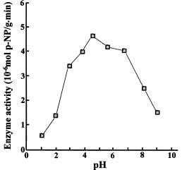
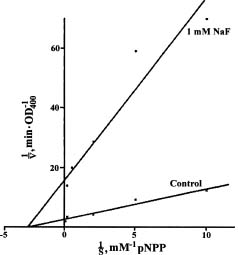
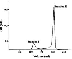
Abstract. The acid phosphatases (EC 3. 1. 3. 2.) of rice seedlings consisted of four groups of isozymes. The group III isozyme was purified through ammonium sulfate, DEAE-Sepharose, Con A-Sepharose, and chromatofocusing. A 198-fold enhancement of activity was attained. This isozyme showed only one band in native-polyacryamide gel electrophoresis. However, gel filtration revealed two peaks of enzyme activity. Their molecular weights were 130 kDa and 100 kDa. The purified isozyme showed a pH optimum of 5. Its Km for p-nitrophenyl phosphate (p-NPP) hydrolysis was 0.33 mM. Its activity was inhibited non-competitively by sodium fluoride. Mercuric chloride, sodium molybdate, and copper sulfate strongly inhibited the enzyme activity. The isozyme actively hydrolyzed adenosine triphosphate and p-NPP, and partially utilized fructose-1,6-diphosphate. It did not utilize fructose-6-phosphate, glucose-1-phosphate, glucose-6-phosphate, glycerophosphate, or adenosine monophosphate.

Keywords: Acid phosphatase; Enzyme purification; Isozyme; Oryza sativa.

Introduction

Acid phosphatases (EC 3. 1. 3. 2.) occur ubiquitously among plants and animals (Hollander, 1971; Panara et al., 1990); however, their functional significance in plants is still relatively little known. Studying acid phosphatases is difficult due to their multiform occurrence in organisms, their relative nonspecificity, their small quantity, and their instability in dilute solution (Park and van Etten, 1986). Their study is also complicated by wide variations in the activity and property of isozymes between species—and between different stages in each plant's development (Alves et al., 1994; Baes and Van Cutasem, 1993). There is also some inconsistency in nomenclature between different isozyme systems (Arnold et al., 1987; Moore et al., 1987). Only limited information is available on their specificity, localization, and physiological function (Baker and Tadakazu, 1973; Charvat and Esau, 1974; Mizuta and Suda, 1980; Chen et al., 1992).

Recent development of chromatographic methods has facilitated purification of individual isozymes of acid phosphatase. By using a combination of DEAE-Cellulose chromatography and cell fractionation, Mizuta and Suda (1980) were able to isolate twelve isozymes from bean hypocotyl. Acid phosphatase purified by a sequence of DEAE-Cellulose and/or Con A-Sepharose chromatography was also reported for tomato cultivars (Paul and Williamson, 1987), sunflower seeds (Park and Van Etten, 1986) and wheat germ (Waymack and Van Etten, 1991).

In this study, several different chromatographic methods were employed to purify one acid phosphatase

isozyme from rice seedlings, and its properties were examined.

Materials and Methods

Plant Materials

Rice (Oryza sativa Tainung 67) seeds were surface sterilized in 6% calcium hypochlorite, soaked in water for three days, and sown on plastic net moistened with nutrient solution. After germination, seedlings were transferred to the greenhouse of the Department of Horticulture, National Taiwan University. Ten-day-old seedlings were collected for enzyme extraction.

Enzyme Extraction and Purification

Rice leaves (500 g) were rapidly frozen in liquid N2, ground into powder with a mill, then soaked in 1000 ml of 50 mM buffer A (Tris-HCl, pH 7.6, 1 mM b-mercaptoethanol). The mixture was homogenized with a Polytron, filtered through four layers of cheesecloth, and centrifuged at 16,000 g for 20 min. Proteins were precipitated from the supernatant with a 40_70% saturation of ammonium sulfate, and the precipitate was dialyzed against buffer A overnight. The dialysate was loaded on to a DEAE-Sepharose Fast Flow column (Pharmacia, 26 × 700 mm) and eluted with 1000 ml of 0-1 M NaCl in a linear gradient at a flow rate of 200 ml/h. Fractions showing enzyme activity were pooled, concentrated through a YM 10 membrane (Amicon), and dialyzed against buffer B (25 mM Tris-HCl, pH=7.6, 50 ml NaCl). The dialysate was then loaded on to a Con A-Sepharose column (Bio-Rad, 12 × 40 ml). The loosely bound proteins were washed with buffer B, and the bound glycoproteins were eluted

1Corresponding author.


Botanical Bulletin of Academia Sinica, Vol. 38, 1997

with 1% a-D-methylglucoside in buffer B at a flow rate of 10 ml/h. Enzymatically active fractions of acid phosphatase were pooled and concentrated through a YM 10 membrane to about 5 ml, dialyzed against buffer C (25 ml histidine-HCl, pH = 6.2), and applied to a PBE 94 chromatofocusing column (9 × 300 mm). The column had been pre-equilibrated with buffer C and post-equilibrated with 4 ml buffer D (9-fold diluted Polybuffer 74 (Pharmacia), pH = 4.0) to create a pH gradient from 4 to 7 in the column and eluted with buffer D at a flow rate of 20 ml/h. Fractions with high acid phosphatase activity were assayed with p-nitrophenyl phosphate (p-NPP) and repooled. The purified enzyme was electrophoretically analyzed with 7.5% polyacrylamide gel as described below. The molecular weight was determined by comparing the mobility with those of protein markers (Sigma).

Acid Phosphatase Assay

Acid phosphatase activity was based on conversion of 5 mM p-NPP into p-nitrophenol in 25 mM sodium acetate at pH 5.0 (Hooley, 1984). One ml of reaction solution was incubated at 37°C for 10 min; 4 ml of stop solution containing 0.1 M NaOH and 0.6 M Na2CO3 were then added and absorbency at 400 nm was measured. One unit of enzyme activity represented the amount of enzyme required to liberate 1 µmol p-nitrophenol per min. When substrates other than p-NPP were used in the assay, the liberated inorgnic phosphate was determined by the method of Taussky and Shorr (1953). Protein content was determined by Bradford's method (1976).

Polyacrylamide Gel Electrophoresis (PAGE)

Native-PAGE and sodium dodecyl sulfate (SDS)-PAGE were performed following the method described by Hames and Rickwood (1981). After electrophoresis, the gel was incubated in a staining solution containing 0.2 M sodium acetate (pH = 5.0), 0.1% fast red TR salt, 0.1% a-naphthyl phosphate, and 5 mM MgCl2 for 3 h. Proteins separated by SDS-PAGE were silver-stained according to Merril et al. (l984).

Enzyme Characterization

Gel filtration characterization of the isozyme III was run on an Ultra gel AcA 44 column. Pooled active fractions of about 1/20 gel volume were loaded on to the column, eluted with buffer D at a flow rate of 1 ml/min and assayed.

Three buffers at pH values ranging from 1 to 9 were used for determining pH optimum. These buffers were hydrochloride (1_3), acetate (4_6) and Tris (6.5_10). The incubation medium contained 1 unit of purified isozyme, 0.5 mM phosphoric ester, and 50 mM buffer.

For substrate specificity studies, eight phosphoric esters (all in 0.5 mM) were incubated with the isolated enzyme at 35°C for 15 min and the released inorganic phosphate was measured. The incubation medium contained 1 unit of purified isozyme, 0.5 mM phosphoric ester, and 50 mM sodium acetate buffer (pH 5.0).

Enzyme kinetic studies were performed at room temperature using p-NPP in concentrations of 0.1, 0.2, 0.5, 5, and 10 mM. The incubation medium contained 1 unit of purified enzyme, 50 mM acetate buffer (pH 5.0), and p-NPP. Extrapolated plot of 1/vo against 1/(So) was used to determine the values of Km and Vmax. The Ki value of NaF was detected with the same p-NPP concentrations and adding NaF to a final concentration of 1 mM.

The effects of twelve different mineral salts on enzyme activity were tested. Assays were carried out at 37°C for 30 min with 5 mM p-NPP as substrate and 50 mM sodium acetate buffer (pH 5.0) in the incubation mixture.

Results

Four groups of acid phosphatase isozyme were electrophoretically detected in rice seedlings (Figure 1). Our previous study showed that group III isozyme occurred in various tissues of rice seedlings, showed more consistency of stainablity in gel with assay staining, and was more sensitive to sodium fluoride inhibition than that of I, II, or IV (Wang et al., 1995). Thus, this isozyme group was chosen for detailed analysis.

As shown in Table 1, the yields of group III isozyme during purification were relatively low. However, the use of Con A-Sepharose affinity chromatography was helpful in purifying the isozyme; it enhanced enzyme activity as much as 18-fold. A 198-fold increment in specific activity of acid phosphatase was obtained following purification in sequence with (NH4)2SO4 precipitation, DEAE-Sepharose, Con A-Sepharose, and chromato-focusing column chromatography. Percentage of the purified group III isozyme of acid phosphatase was 8.5%;

Figure 1. Native-PAGE zymograms of crude enzyme solution and purified isozyme III. Lanes 1 and 2 were crude enzyme solution. Lanes 3_6 were peaks I and II isozyme purified from Ultra gel AcA 44 gel filtration. Lanes 3 (10×) and 4 (1×) were obtained from peak 1 and lanes 5 (10×) and 6 (1×) were from peak II. Samples applied on lane 3 and lane 5 were concentrated with Speed-vac to a 10 fold concentration.


Tso and Chen — Acid phosphatase isozyme in rice

Table 1. Purification steps for a group III acid phosphatase in rice*.

Protein Activity Specific activity Recovery Purification-fold (mg) (unit) (unit/mg) (%)

Crude extract 3255 1190 0.4 100.0 1.0

(NH4)2SO4 ppt 1428 831 0.6 69.8 1.5

DEAE-Sepharose 244 622 2.6 52.3 6.5

Con A-Sepharose 8.3 387 46.8 32.5 117

Chromatofocusing 1.3 101 79.0 8.5 198

*Measured in triplicate.

the preparation showed a specific activity of 79 unit/mg. The isoelectric point of the isozyme was 5.5 on chromatofocusing.

Two active peaks were separated by the Ultra gel AcA 44 column, with molecular weights of 100 kDa and 130 kDa (Figure 2). However, the two fractions produced only one band on Native-PAGE (Figure 1). Meanwhile, it was difficult to observe a protein band corresponding to the purified isozyme in silver-stained gel of SDS-PAGE.

The purified acid phosphatase showed an optimal pH of around 5 (Figure 3). Enzyme kinetic study showed that its Km and Vmax were 0.33 mM and 0.34 µmol/min, respectively, when p-NPP was used as the substrate (Figure 4). Meanwhile, sodium fluoride was a non-competitive inhibitor of the isozyme and the Ki value for NaF was about 0.23 mM.

The rates of hydrolysis of several phosphorylated substrates are shown in Table 2. The acid phosphatase was a rather nonspecific enzyme. p-NPP was a good substrate. However, the dephosphorylation rate of ATP g-phosphate (143.5%) was about 50% higher than that of p-NPP. The other phosphoric esters were poor substrates. The purified isozyme was unable to hydrolyze fructose-1, 6-diphosphate, glucose-1-phosphate, glucose-6-phosphate, or adenosine monophosphate. b-Glycerophosphate, a traditional reagent for acid phosphatase cytochemical localization, was also a distinctly poor substrate (4.2%).

Figure 3. Effect of pH on the enzyme activity of isozyme III.

Figure 2. Ultra gel separation profile of acid phosphatase enzyme activity.

Figure 4. Lineweaver-Burk plot for NaF effect on purified acid phosphatase showing simple non-competitive inhibition.


Botanical Bulletin of Academia Sinica, Vol. 38, 1997

Table 2. Substrate specificity for a group III acid phosphatase in rice*.

Substrates (0.5 mM) Relative activity (%)

p-Nitrophenyl phosphate 100.0

Fructose 1,6-diphosphate 46.3 ± 0.5

Fructose-6-phosphate 0

Glucose-1-phosphate 0

Glucose-6-phosphate 0

Glycerophosphate 4.2 ± 2.7

Adenosine triphosphate 143.5 ± 1.5

Adenosine monophosphate 0

*Measured in triplicate.

observed for acid phosphatase-1 of tomato (Paul and Williamson, 1987).

Kinetic study of the acid phosphatase of Baker's yeast showed a linear Lineweaver-Burk plot, with a Km of 0.45 mM for p-NPP (Schurr and Yagil, 1971). A Km of 0.30 mM was found for the acid phosphatase of yeast-like cells of Sperothrix chenchii. The Km of the S1 acid phosphatase of barley roots was pH dependent; at pH 5.5 it was about 0.3 mM (Arnold et al., 1987). The Km values of acid phosphatases from different sources were close to 0.33 mM of those found for rice group III isozyme.

Fluoride is a non-competitive inhibitor of acid phosphatase, and its inhibitory activity is highly reversible (NagDas and Bhattacharyya, 1984). However, the inhibitory effect of NaF on acid phosphatase activity was only partial, and the degree of inhibition varied according to plant source of the enzyme (Hall, 1978; Moore et al., 1987). Meanwhile, isozymes of acid phosphatases from several plant species were also shown to be inhibited differently by NaF (Arnold et al., 1987; Chen et al., 1992; Mizuta and Suda, 1980). Sodium fluoride was also a non-competitive inhibitor of rice acid phosphatase and its Ki value was 0.23 mM (Wang et al., 1995). Moreover, its reversibility and the effectiveness of its inhibition has varied from one isozyme to another (NagDas and Bhattacharyya, 1984; Arnold et al., 1987).

The requirement of metallic ions for acid phosphatase activity has also varied according to plant species, developmental stage, and isozyme heterogeneity (Scandalois, 1974; Panara et al., 1990). A cofactor role of Mn2+ has been reported in soybean (Fujimoto et al., 1977) and yeast (Mizunaga, 1979). However, our study with the purified isozyme from rice seedlings showed no effect by CaCl2 or MnSO4. The inhibition of rice acid phosphatase by copper, zinc, molybdate, and fluoride ions has been previously reported in other species, e.g., pea (Mizuta and Suda, 1980) and tobacco (Pan et al., 1987). Rice acid phosphatase activity was also inhibited by ferrous ion. Phosphate ion has been commonly recognized as a competitive inhibitor of acid phosphatase in many plant species (Park and Van Etten, 1986; Arnold et al., 1987).

The purified isozyme of rice acid phosphatase was more substrate-specific than those of other plant species (Hasegawa et al., 1976; Panara et al., 1990). A higher rate of ATP hydrolysis by acid phosphatase was also observed in tobacco cells (Pan and Chen, 1988). A low rate of sugar-phosphate hydrolysis is a significant character of this isozyme. Meanwhile, glycerophosphate was nearly unreacted by the purified enzyme, indicating that the hydrolysis of glycerophosphate was acid phosphatase isozyme-dependent.

One problem that needs to be solved is the discrepancy between the one band in native-PAGE zymogram and two bands apparent by gel filtration. Also needed is further study on the function of the group III isozyme.

Acknowledgment. This work was financially supported by National Science Council, ROC with a grant to Y.-R. C. (NSC 82-0211-B002-325).

Table 3. Effect of salts on the activity of a group III acid phosphatase in rice*.

Salts (0.5 mM) Relative activity (%)

Control 100.0 ± 3.9

CaCl2 103.7 ± 0.3

MgCl2 104.8 ± 5.4

CuSO4 24.4 ± 2.2

HgCl2 1.9 ± 0.3

KH2PO4 90.3 ± 3.3

NaF 42.3 ± 3.6

(Na)2MoO4 2.1 ± 0.7

NH4NO3 79.5 ± 3.9

CoCl2 94.0 ± 5.8

ZnSO4 53.8 ± 1.9

MnSO4 103.5 ± 3.5

FeSO4 66.9 ± 5.4

*Measured in triplicate.

The effects of mineral salts on the activity of acid phosphatase are shown in Table 3. The activity of the purified isozyme was not significantly influenced by CaCl2 (103.7%), MgCl2 (104.8%), or MnSO4 (103.5%). However, HgCl2 and Na2MoO4 were strong inhibitors, stopping almost all enzyme activity within a short period of incubation. The enzyme activity was also suppressed by CuSO4 (24.4% remained). ZnSO4 and FeSO4 lowered enzyme activity to 53.8% and 66.9% of the initial activity, respectively. KH2PO4, NH4NO3, and CoCl2 were less-active inhibitors.

Discussion

Group III isozymes of acid phosphatase universally occur in the leaves, stems, and roots of rice plants. It is more sensitive to NaF inhibition than the other isozyme groups (Wang et al., 1995). In this study, chromatofocusing and Con A-Sepharose affinity columns proved useful in isolating a single isozyme of acid phosphatase.

The purified isozyme of acid phosphatase showed an optimum pH of 5, which was similar to that of the acid phosphatase of tobacco (Bentwood and Cronshaw, 1975). However, the constitutive and repressible acid phosphatases of baker's yeast had pH optima of 3.6 and 4.3, respectively. Also, a broader optimum pH range of 3.5_5.0 was


Tso and Chen — Acid phosphatase isozyme in rice

Literature Cited

Alves, J.M., C.D. Siachakr, M. Allot, S. Tizroutine, I. Mussio, and A. Servaes. 1994. Isozyme modifications and plant regeneration through somatic embryogenesis in sweet potato (Ipomoea batatas (L.) Lam.). Plant Cell Rep. 13: 437_441.

Arnold, W.N., K.H. Sakai, and L.C. Mann. 1987. Selective inactivation of an extra-cytoplasmic acid phosphatase of yeast-like cells of Sporothrix schenckii by sodium fluoride. J. Gen. Microbiol. 133: 1503_1509.

Baes, P.G. and P.J. Van Cutasem. 1993. Isozyme polymorphism in three gene pools of cultivated chicory (Cichorium intybus L.). Euphytica 71: 143_150.

Baker, J.E. and T. Tadakazu. 1973. Acid phosphatase in plant tissues. I. Changes in activity and multiple forms in tea leaves and tomato fruit during maturation and senescence. Plant Cell Physiol. 14: 459_47l.

Bentwood, B. and J. Cronshaw. 1975. Biochemistry and cytochemical localization of acid phosphatase in the phloem of Nicotiana tabacum. Planta 130: 97_104.

Bhargava, R. and R.C. Sachar. 1987. Induction of acid phosphatase in cotton seedlings: purification, subunit structure and kinetic properties. Phytochemistry 26: 1293_1297.

Bradford, M.M. 1976. A rapid and sensitive method for quantitation of microgram quantities of protein utilizing the principle of protein-dying. Anal. Biochem. 72: 248_254.

Charvat, I.D. and K. Easu. 1974. An ultrastructural study of acid phosphatase localization in Phaseolus vulgaris xylem by the use of an azo-dye method. J. Cell Sci. 19: 543_561.

Chen, W.S., Y.F. Huang, and Y.R. Chen. 1992. Localization of acid phosphatase in root cap of rice plant. Bot. Bull. Acad. Sin. 33: 233_239.

Fujimoto, S., T. Nakazawa, and A. Ohara. 1977. Isolation of violet-coloured acid phosphatase from soybean. Agric. Biol. Chem. 41: 599_600.

Hall, J.L. 1978. Electron Microscopy and Cytochemistry of Plant Cells. Elsevier, Amsterdam.

Hames, B.D. and D. Rickwood. 1981. Electrophoresis of Proteins: a Practical Approach. IRL Press, Oxford.

Hasegawa, Y., K.R. Lynn, and W.J. Brockank. 1976. Isolation and partial characterization of cytoplasmic and wall-bound acid phosphatase from wheat root. Can. J. Bot. 54: 1163_1169.

Hooley, R. 1984. Gibberellic acid controls the secretion of acid phosphatase in aleurone layers and isolated aleurone protoplasts of Avena fatua. J. Exp. Bot. 35: 822_828.

Hollander, V.P. 1971. Acid phosphatase. In P. D. Boyer (ed.), The Enzymes, vol. IV. Academic Press, New York, pp. 449_498.

Merril, C.R., D. Coldman, and M.L. Van Keuren. 1984. Gel protein stains: silver stain. Method. Enzymol. 104 part C:

441_447.

Mizunaga, T. 1979. Some properties of phosphate-repressible and constitutive acid phosphatase of baker's yeast. Agric. Biol. Chem. 43: 1211_1218.

Mizuta, S. and S. Suda. 1980. A comparative study of multiple acid phosphatase in cellular fractions of bean hypocotyl. Ann. Bot. 45: 369_382.

Moore, R.C., E. MeClelen, and H.S. Smith. 1987. Phosphatases. In K. C. Vaughn (ed.), Handbook of Plant Cytochemistry, vol. 1. Cytochemical localization of Enzymes, CRC Press, Boca Raton, Florida, pp. 37_65.

NagDas, S.K. and A.K. Bhattacharyya. 1984. The kinetics of inhibition of human seminal plasma acid phosphatase by sodium fluoride. Biochem. Internatl. 5: 659_668.

Pan, S.M. and Y.R. Chen. 1988. The effect of salt stress on acid phosphatase activity of Zea mays seedlings. Bot. Bull. Acad. Sin. 29: 33_38.

Pan, S.M., Y.F. Huang, and Y.R. Chen. 1987. Characterization and localization of acid phosphatase in the suspension cultured cells of Nicotiana otophora. Taiwania 32: 1_10.

Panara, F., S. Pasqualini, and M. Antonielli. 1990. Multiple forms of barley root acid phosphatase: purification and some characteristics of the major cytoplasmic isoenzyme. Biochem. Biophys. Acta 1073: 73_80.

Park, H.C. and R.L. Van Etten. 1986. Purification and characterization of homogeneous sunflower seed acid phosphatase. Phytochemistry 25: 351_357.

Paul, E.M. and V.M. Williamson. 1987. Purification and properties of acid phosphatase-1 from a nematode-resistant tomato cultivar. Plant Physiol. 83: 399_403.

Scandalois. J.G. 1974. Isozyme in development and differentiation. Annu. Rev. Plant Physiol. 25: 225_258.

Schurr, A. and E. Yagil. 1971. Regulation and characterization of acid and alkaline phosphatase in yeast. J. Gen. Microbiol. 65: 291_303.

Taussky, H.H. and E. Shorr. 1953. A microcolorimetric method for the determination of inorgnic phosphorus. J. Biol. Chem. 202: 675_685.

Treton, B.Y., M.T. Le Dall, and C.M. Gaillardin. 1992. Complementation of Saccharomyces cerevisiae acid phosphatase mutation by a genomic sequence from the yeast yarrowia lipolytica identifies a new phosphatase. Curr. Genet. 22: 345_355.

Wang, M.C., S.C. Tso, and Y.R. Chen. 1995. Inhibitory effect of sodium fluoride on activity of acid phosphatase in rice plants. Taiwania 40: 291_298.

Waymark, P.P. and R.L. Van Etten. 1991. Isolation and characterization of a homogeneous isoenzyme of wheat germ acid phosphatase. Arch. Biochem. Biophys. 288: 621_633.

Williamson, V.M. and G. Colwell. 1991. Acid phosphatase-1 from nematode resistant tomato. Plant Physiol. 97: 130_146.


Botanical Bulletin of Academia Sinica, Vol. 38, 1997

¤ô½_ªº»Ä©Ê¿M»Ä»Ã¯Àªº¯Â¤Æ»PŲ©w

¥ª¤h¹Å ³¯ºa¾U

°ê¥ß¥xÆW¤j¾Ç´Óª«¾Ç¨t

¤ô½_¥®­]§t¥|²Õ»Ä©Ê¿M»Ä»Ã¯À¦P¥\»Ã¯À¡C¸g²¸»Ä»Ï¨H¾ý¡BDEAE-Sepharose, Con A-Sepharose ¤Î Chromatofocusing µ¥ªk¯Â¤Æ©Ò±oªº²Ä¤T²Õ¦P¥\»Ã¯À¡A¨ä¬¡©Ê¼W°ª¦Ü 198 ­¿¡C¥H½¦Åé¹LÂo¦â¼h¤ÀªR¤ÀÂ÷Ų©w ¦³¨â±a¡A¨ä¤À¤l«G¤À§O¬°130 kDa ¤Î100 kDa¡AµM¦Ó¦b native-PAGE ¤W¥u§e¤@±a¡C¨ä³Ì¾A»ÄÆP«×¦b¤­¥ª¥k¡A ¹ï p-Nitrophenyl phosphate (p-NPP) ªº Km ­È¬O 0.33 mM¡C¬t¤Æ¶u¹ï¥»²Õ»Ã¯Àªº§í¨î§@¥ÎÄÝ«DÄvª§©Ê®Ä À³¡C´â¤Æ¨E¡B໻Ķu¤Î²¸»Ä»É¹ï»Ã¯À¬¡©Ê¦³«Ü±jªº§í¨î©Ê¡C¥»²Õ¦P¥\®Ä¯À¹ï ATP ¤Î p-NPP ¦³«Ü±jªº¤ô¸Ñ®Ä ªG¡A¥B¥i³¡¤À¤ô¸Ñ Fructose-1, 6-diphosphate¡A¦Ü©ó Fructose-6-phosphate, Glucose-1-phosphate, Glucose-6-phosphate, Glycerophophosphate ¤ÎAdenosine monophosphate«h«D¬°¨ü½è¡C

ÃöÁäµü¡G »Ä©Ê¿M»Ä»Ã¯À¡F¦P¥\»Ã¯À¡F¯Â¤Æ¡F ¤ô½_¡C