Chen and Chien — Some chytrids of Taiwan

Bot. Bull. Acad. Sin. (1998) 39: 47_56

Some chytrids of Taiwan (II)

Shu-Fen Chen1,3 and Chiu-Yuan Chien2

1Department of Food Health, Chia-Nan College of Pharmacy and Science, Tainan Hsien, Taiwan 717, Republic of China

2Institute of Biological Sciences, National Taiwan Normal University, Taipei, Taiwan 117, Republic of China

(Received April 11, 1997; Accepted August 28, 1997)

Abstract. This paper describes and illustrates twelve species of monocentric chytrids that were isolated and purified. They include: Rhizidium windermerense Canter, R. ramosum Sparrow, Rhizophlyctis hyalina (Karling) Sparrow, Rhizophydium biporosum (Couch) Barr, R. chlorogonii (Serbinow) Jaczewski, R. condylosum Karling, R. elyensis Sparrow, R. macrosporum Karling, R. patellarium Scholz, Spizellomyces punctatum (Koch) Barr, S. acuminatus (Barr) Barr, and S. pseudodichotomus Barr. Except for Rhizophydium elyensis, all species described above are new to Taiwan.

Keywords: Chytridiales; Chytridiomycetes; Spizellomycetales; Taiwan.

Introduction

It is clear that as early as 1846 Braun had observed chytrids on fresh-water algae (Sparrow, 1960). Sparrow's Aquatic Phycomycetes (1960) and Karling's Chytridiomycetarum Iconographia (1977) are based on observation of freshly collected material or of gross cultures. Techniques for baiting have been discussed at length by Sparrow (Barr, 1987). Many species now can be grown in axenic culture, and it has been found that nearly all morphological characterics for classifying species exhibit incredible variation (Paterson, 1963; Miller, 1968; Powell and Koch, 1977a). In the absence of a formal consensus, the isolation of chytrids and study of their growth forms under standardized conditions in pure culture has become the classification criterion (Roane and Paterson, 1974; Powell and Koch, 1977b; Barr, 1973, 1975). Based on differences in zoospore ultrastructure, which have become ordinal characteristics (Barr, 1980, 1984; Longcore, 1995), the Chytridiomycota contains four orders: Chytridiales, Spizellomycetales, Monoblepharidales, and Blastocladiales (Barr, 1990).

There have been some studies of gross culture of Chytridiales in Taiwan (Volz et al., 1976; Konno, 1984). All species of chytrids were isolated and purified in the present investigation (Chen and Chien, 1995; Chen, 1996). The following twelve monocentric chytrids were isolated from fresh water, soil and mud. They include nine species of Chytridiales and three species of Spizellomycetales.

Materials and Methods

Samples of water, soil and mud were baited with pine pollen (Barr, 1987). Emerson's 1/4 YpSs agar (containing 250 ppm penicillin G and 250 ppm streptomycin sulfate)

was used to isolate and culture the organisms. The medium consisted of soluble starch 5 g/L, yeast extract 0.25 g/L, K2HPO4 0.25 g/L, MgSO4 • 7H2O 0.125 g/L, and agar 12 g/L (or agar 1 g/L as 1/4 YpSs slush). Developmental stages and morphological characters were examined using the light microscope and scanning electron microscope. Axenic cultures were kept on slants of Emerson's 1/4 YpSs agar in screw-cap tubes and transferred every three months. All pure cultures have been deposited at the mycology laboratory of the Institute of Biological Sciences, National Taiwan Normal University, Taipei, Taiwan, ROC.

Species Descriptions

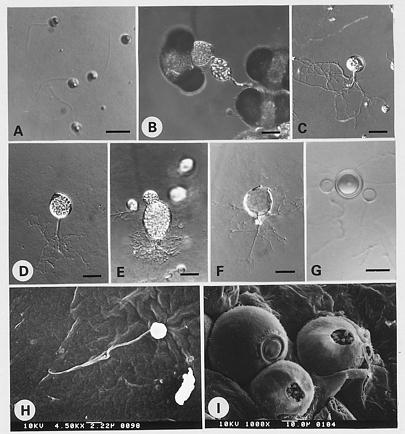
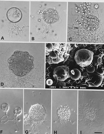
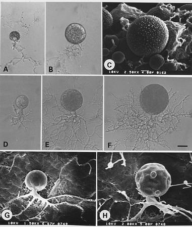
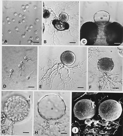
Rhizidium windermerense Canter, Ann. Bot. London (N. S.) 14: 268, 1950. Figure 1A_G

On pine pollen: Sporangium epibiotic or interbiotic, ovate or pyriform, 23.3_40 × 33.3_73.3 µm.

On 1/4YpSs agar: Sporangium ovate or pyriform, 13.8_17.5 × 112.5_120 µm; with one, occasionally two, apical or subapical gelatinous papillae, 5_37.5 µm diam. Rhizoids arise from a strong, long, or several branched main axis. Small sporangia contain four to five zoospores. Zoospore globose, 5_7.5 µm diam., emerging in a hyaline vesicle, simultaneously motile before swimming away. Resting spore spherical, about 20 µm diam.; with a large, subcentric oil globule; wall smooth, about 2.5 µm thick; formed by rhizoidal anastomosis of two thallus. Color of colony khaki.

Specimen examined. TAIPEI CITY: Botanical garden, pond water, 9 May 1994, NTNU402a. Isolated on pine pollen from water.

Notes. Although the main character for Rhizidium is the rhizoid that arises from a strong main axis, this criterion was not considered to be sharply defined by Karling

3Corresponding author. Fax: (06) 266-6411


Botanical Bulletin of Academia Sinica, Vol. 39, 1998

(1977). However, the important character of R. windermerense is the resting spore formed by conjugation of the rhizoidal tips from two thalli.

Rhizidium ramosum Sparrow, Proc. Amer. Phil. Soc. 78(1): 44, 1937. Figure 1H_I

On 1/4YpSs agar: Sporangium spherical, 25_62.5 µm diam., or ellipsoidal, with a slightly thickened wall; discharge papillae single, occasionally 2_3, leaving a 7.5_12.5 µm pore following discharge. Main rhizoid stout, 5_7.5 µm diam. with a system of branched rhizoids. Zoospores emerge in a globular mass which is enveloped by a vesicle, then swim away. Zoospores numerous, globose, 4_5 µm diam.; emerging from the sporangium after the deliques

cence of papillae. Resting spore not observed. Color of colony buff.

Specimen examined. TAIPEI HSIEN: Taishan, mud from lake, 2 Dec 1993, NTNUS05b. Isolated on pine pollen from mud.

Notes. Rhizidium ramosum with a very short subsporangial main axis is similar to Sparrow's (1960) description. Sparrow and Karling (1967b) reported that this species is chitinophilic in soil samples, whereas, our culture was obtained using pine pollen from soil, and it is uncertain what the natural substrate is in nature. Further, our isolate differs from Sparrow's description by many sporangia having a subapical or lateral, instead of a terminal exit pore.

Figure 1. A_G, Rhizidium windermerense. A, uniflagellate zoospores; B, on pine pollen, epibiotic and interbiotic sporangium; C, young sporangium; D, mature sporangium with a subapical papilla; E, vesicular zoospores releasing; F, an empty sporangium; G, resting spore between two empty contributing thalli. H_I, Rhizidium ramosum. H, uniflagellate zoospore; I, mature sporangium with a lateral discharge pore. (Bar = 10 µm in A and G; 20 µm in B_F; 2.22 µm in H; 10 µm in I)


Chen and Chien — Some chytrids of Taiwan

Rhizophlyctis hyalina (Karling) Sparrow, Aquatic Phycomycetes p. 445, 1960.

ºKarlingia hyalina Karling, Mycologia, 39: 63, 1947. Figure 2A_E

On 1/4YpSs agar: Sporangium spherical or subspherical, 12.5_87.5 µm diam., wall smooth, with 1_6 exit papillae, 12.5_22.5 µm diam. each filled with a gelatinous plug, 5_10 µm high. Rhizoids arising from several places on the sporangial base, much branched and extensive. Zoospore globose, 4_5 µm diam., with a lateral refractive granule. Color of colony white to offwhite.

Specimen examined. ILAN HSIEN: Harpen, pond water, 11 Apr 1993, NTNU811c. Isolated on pine pollen from water.

Notes. The sporangium of Rhizophlyctis hyalina is smooth-walled, with evenly dispersed exit papillae. This species can be distinguished from R. variabile and R. mastigotrichis by the number and dispersed phase of exit papillae. Further, R. variabile has crystal granules on the sporangial wall (Chen, 1996).

Rhizophydium biporosum (Couch) Barr, Can. J. Bot. 51: 969_970. 1973.

ºPhlyctochytrium biporosum Couch, J. Elisha Mitchell Sci. Soc., 47: 254, 1932.

=Phlyctidium bumilleriae Couch, J. Elisha Mitchell Sci. Soc., 47: 256, 1932. Figure 3A

On 1/4YpSs agar: Sporangium spherical, 12.5_27.5 µm diam., or ovate with two to three papillae on opposite sides. Rhizoids either finely tubular or swollen into globular shapes, 2.5_5 µm diam. Zoospores globose, 2_4 µm diam., or tapering, 3_5 µm in length, escaping from discharge pore, movement hopping. Color of colony white.

Specimen examined. YUNLIN HSIEN: garden soil, 22 Jul 1992, NTNUS01c. Isolated on pine pollen from soil.

Notes. The absence of an apophysis and ultrastructure of zoospore demonstrates that this species belongs to the Rhizophydium type (Chen, 1996), and we agree with Barr (1973) on revising Phlyctochytrium biporosum to R. biporosum.

Rhizophydium chlorogonii (Serbinow) Jaczewski, Opredelitel Gribov. Sovershennye Griby (Diploidnye Stadii). I. Fikomitsety (Phycomycetes), p. 38, 1931.

=Phlyctidium chlorogonii Serbinow, Bot. Zap. 24: 156, 1907. Figure 3B_D

On 1/4YpSs agar: Sporangium spherical, 20_37.5 µm diam., with one to six papillae. Rhizoids fine, arising from an ovate or peg-like extension. Zoospores globose, about 3 µm diam., escaping individually from discharge pore,

Figure 2. Rhizophlyctis hyalina. A, B and C, young sporangium with several rhizoidal axes and extensive rhizoids; D, nearly mature sporangium with three conical exit papillae; E, sporangium with three exit pores. (Bar = 20 µm in A_D; 10 µm in E)


Botanical Bulletin of Academia Sinica, Vol. 39, 1998

remaining motileless before swimming away with a hopping motion. Color of colony orange yellow.

Specimen examined. HWALIEN HSIEN: Hsiukuluan, creek water, 25 Aug 1993, NTNU1601; TAITUNG HSIEN: Pahsientung, creek water, 25 Aug 1993, NTNU1602; Chialapan, creek water, 30 Oct 1993, NTNU1703. Isolated on pine pollen from all water samples.

Notes. Rhizophydium chlorogonii differs from R. laterale in the number of discharge papillae. In addition, the colony color of R. laterale is distinctly yellow (Chen and Chien, 1995).

Rhizophydium condylosum Karling, Arch. f. Mikrobiol. 61: 118_120. 1954. Figure 3E_H

On pine pollen: Sporangium epibiotic or interbiotic, spherical, 10_30 µm diam., or pyriform; wall smooth, with 2_6 papillae. Rhizoidal system arising from a single point, usually with one subsporangial swelling. Zoospores oval, 2.5_3 µm diam., emerging away from the pores of sporangium.

On 1/4YpSs agar: Sporangium spherical, 12.5_37.5 µm diam., or suboval, 25_30 µm diam., rarely fusiform, 22.5 × 30 µm, with 3_12 papillae or discharge tube, 5_20 µm in length. Rhizoidal system developed from one, frequently

Figure 3. A, sporangia of Rhizophydium biporosum. B_D, Rhizophydium chlorogonii. B, young sporangium with a peg-like rhizoidal axis; C, sporangium with several papillae; D, discharging zoospores. E_H, Rhizophydium condylosum. E, young sporangium with an apophysis; F, sporangium with several sickle-shaped discharge tubes; G and H, sporangium with several papillae or discharge pores. (Bar = 2.86 µm in A; 20 µm in B_D; 10 µm in E and F; 3.33 µm in G and H.)


Chen and Chien — Some chytrids of Taiwan

several, unbranched or branched apophysis. Zoospores globose, 3_4 µm diam., swimming away individually from discharge pores, movement hopping. Resting spore resembling the sporangium, 17.5_30 µm diam., but with long papillae, 15_20 µm in length, frequently sickle-shaped; upon germinating, the papillae deliquesce and discharge zoospores. Color of colony chrimic yellow.

Specimen examined. ILAN HSIEN: Shuanglienpyi, soil, 3 Oct 1992, NTNU601a; Shuanglienpyi, pond water, 3 Oct 1992, NTNU602b. Isolated on pine pollen from soil and water.

Notes. This species frequently has a branched apophysis. The very long, sickle-shaped, discharge tubes on the resting spore is the main character for this species.

Rhizophydium elyensis Sparrow, Trans. Brit. Mycol. Soc. 40: 525. 1957. Figure 4

On pine pollen: Sporangium epibiotic or interbiotic, spherical, 20_45 µm diam., ovate or ellipsoid, 20 ×

27_22.5 × 25 µm. Zoospores globose, about 5 µm diam. Rhizoidal system extensive, up to 250 µm, with branches reaching several pollen grains.

On 1/4YpSs agar: Immature sporangia spherical, subspherical or ovoid; mature sporangia becoming irregularly polygonal, 30_65 × 77.5 µm diam.; wall smooth, with 1_12 exit papillae filled with a spherical plug. Main rhizoidal axis tubular, 3.2_7 µm diam., branched and extensive up to 112.5_212.5 µm. Zoospores globose, 3_3.5 µm diam., often emerging from several papillae simultaneously in an amoeboid manner, then darting away. Color of colony white to offwhite.

Specimen examined. ILAN HSIEN: Shuanglienpyi, pond water, 23 Feb 1994, NTNU604a. Isolated on pine pollen from water. This species has been reported by Konno (1984), but our isolate is the only strain obtained in pure culture.

Notes. The sporangium has several exit papillae which are filled with a spherical plug. When mature, the sporan

Figure 4. Rhizophydium elyensis. A, uniflagellate zoospores; B, on pine pollen, interbiotic sporangium; C and D, young sporangium; E, F and G, simultaneous discharge of zoospores from several papillae; H, an empty sporangium; I, sporangium with several exit papillae surmounted with spherical plugs. (Bar = 10 µm in A; 20 µm in B_H; 6.67 µm in I.)


Botanical Bulletin of Academia Sinica, Vol. 39, 1998

gium becomes irregularly polygonal. The above character is similar to Sparrow's (1957) and Karling's (1967a) descriptions.

Rhizophydium macrosporum Karling, Bull. Torrey. Bot. Club 64: 439_442, 1938. Figure 5A_E

On pine pollen: Sporangium usually sessile, epibiotic or interbiotic; spherical 18.3_23.3 µm diam., or ovoid 13 × (15_22) µm, with one to three (or more) apically or subapically exit papillae; discharge pore 3.33 µm diam. Rhizoidal system consists of an elongated tubular stalk or axis, 2.5_3.3 µm diam. giving the sporangium a distinctly stalked appearance.

On 1/4YpSs agar: Sporangium spherical, 17.5_113 µm diam., oval or urceolate, with 1_5, but occasionally as many as 10 or more exit papillae. Rhizoidal system usu

ally arising from one, occasionally several axes, delimited distinctly from the sporangia by a cross wall. Zoospores spherical, 4_5 µm diam., occasionally becoming amoeboid, with a large refractive globule, discharging simultaneously though several pores. Color of colony white.

Specimen examined. ILAN HSIEN: Harpen, lake water, 13 Jan 1993, NTNU802b. Isolated on pine pollen from water.

Notes. One or more of the exit papillae deliquesce simultaneously with 3_10 zoospores escaping into globules of hyaline fluid and lying quiescent for a few moments; as the globules dissolve, the zoospores begin to separate and soon dart away, others become actively motile; eventually some zoospores degenerate or germinate in situ; that is the main character for this species.

Figure 5. A_E, Rhizophydium macrosporum. A, young sporangium; B and C, discharging zoospores; D and E, sporangium with several papillae. F_I, Rhizophydium patellarium. F, young sporangium; G, mature sporangium with several papillae; H, discharging zoospores; I, zoospores discharge after the upper portion of sporangial wall dissolved. (Bar = 10 µm in A_D and F_I; 6.67 µm in E.)


Chen and Chien — Some chytrids of Taiwan

Rhizophydium patellarium Scholz, Arch. f. Mikrobiol. 29: 354_362, 1958. Figure 5F_I

On 1/4YpSs Agar: Sporangium spherical, 12.5_25 µm diam., occasionally ovoid or ellipsoidal; wall smooth, delicate with one to several papillae through which some of the zoospores emerge followed by deliquescence of the upper 2/3 of the wall. Rhizoids short, unbranched or branched thread. Zoospores spherical, 2_2.5 µm diam., with a lateral globule. Resting spore not observed. Color of colony white.

Specimen examined. CHIAYI HSIEN: Alishan, mud, 13 Oct 1992, NTNU302; ILAN HSIEN: Harpen, pond water, 11 Apr 1993, NTNU812a. Isolated on pine pollen from mud and water.

Notes. After some zoospores escaping, the upper portion of the sporangial wall dissolves as in Barr's (1971) description.

Spizellomyces punctatum (Koch) Barr, Can. J. Bot. 58: 2384, 1980.

ºPhlyctochytrium punctatum Koch, J. Elisha Mitchell Sci. Soc. 73: 109, 1957.

=Phlyctochytrium verruculosum Knox, 1971, p. 55, not valid. publ. (in a thesis)

=Phlyctochytrium peruvianum Knox, 1971, p. 60, not valid. publ. (in a thesis) Figure 6A_C

Figure 6. A_C, Spizellomyces punctatum. A and B, young sporangium with a branched apophysis; C, mature sporangium wall, minutely warted; D_H, Spizellomyces acuminatus. D_G, young sporangium with trunk-like apophysis; H, mature sporangium with discharge papillae. (Bar = 20 µm in A, B and D_F; 4 µm in C; 6.67 µm in G; 5 µm in H.)


Botanical Bulletin of Academia Sinica, Vol. 39, 1998

On pine pollen: Sporangium epibiotic or interbiotic; spherical, 35_55 µm diam.

On 1/4YpSs agar: Sporangium spherical, 23_55 µm diam.; wall minutely warted, with 3-6 papillae. Rhizoidal system moderately extensive up to 137.5 µm, arising from a taproot-like main rhizoid, 5_10 µm diam., or branched apophysis. Zoospores globose, about 4 µm diam., or amoeboid; lipid globules numerous and inconspicuous. Resting spore spherical, about 28 µm diam., wall smooth, 1.7 µm thick. Color of colony white.

Specimen examined. YUNLIN HSIEN: farm soil, 22 Jul 1992, NTNUS02a. Isolated on pine pollen from mud.

Notes. Based on zoospore ultrastructure, P. punctatum was transfered to the genus Spizellomyces (Barr, 1980).

This is the type species of Spizellomyces. The minutely warted sporangial wall is the main character.

Spizellomyces acuminatus (Barr) Barr, Can. J. Bot. 62: 1187, 1984.

ºPhlyctochytrium acuminatum Barr, Can. J. Bot. 47: 993, 1969.

=Phlyctochytrium africanum Gaertner, Arch. f. Mikrobiol., 21: 120, 1954. Figure 6D_H

In 1/4YpSs slush: Sporangium spherical, 45_95 µm diam.; wall smooth or occasionally minutely warted, with 8_9 discharge papillae. Rhizoidal system arising from trunk-like or taproot-like unbranched or branched axis, 7.5_15 µm diam., with a slightly constricted neck; rhiz

Figure 7. Spizellomyces pseudodichotomus. A, zoospores; B, interbiotic sporangium among pine pollen; C, on pine pollen, an empty sporangium; D and E, young sporangium; F, sporangium with three papillae; G, individual zoospore escapes from pore (arrow); H, an empty sporangium; I, two sporangia with bulbous apophysis. (Bar = 10 µm in A and C_H; 20 µm in B; 8.33 µm in I)


Chen and Chien — Some chytrids of Taiwan

oids extensive richly branched and with blunt ends. Zoospores globose, about 3 µm diam., or amoeboid, 3_4 × 7 µm, with a lateral or posterior lipid droplet; emerging from exit pores.

On 1/4 YpSs agar: Sporangium spherical, 32_83.5 µm diam., or ovoid; rhizoidal system arising from inflated, 7.5_15 µm diam., main axis. Color of colony white.

Specimen examined. YUNLIN HSIEN: garden soil, 22 Jul 1992, NTNUS01a. Isolated on pine pollen from soil.

Notes. Barr (1984) suggested that Spizellomyces acuminatus complex could be divided into four groups. Based on physiological (Chen, 1996) and morphological characteristics, our isolate belongs to Group I.

Spizellomyces pseudodichotomus Barr, Can. J. Bot. 62: 1194, 1984. Figure 7

On pine pollen: Sporangium interbiotic among the pollens, spherical, 12.5_85 µm diam., or subspherical, with several discharge pores, 3 µm diam. Apophysis tubular or bulbous, 5_15 µm diam. with extensive rhizoidal system up to 250 µm, tapering to blunt ends.

On 1/4 YpSs agar: Sporangium spherical or subspherical, 30_75 µm diam. with 3_10 discharge papillae, 2.5 µm high, 5 µm diam. Main rhizoidal neck isthmus-like, below tubular, 5_7.5 µm diam., inflated or bulbous, 10_17.5 µm diam.; rhizoids irregularly branched or occasionally somewhat dichotomously branched, even or uneven and tapered to blunt ends. Zoospore globose, 4_5 µm diam., or amoeboid with a conspicious lipid globule; zoospores escaping upon the dissolution of the papillae. Resting spore spherical, about 31 µm diam.; wall smooth, 2.2 µm thick. Color of colony white.

Specimen examined. YUNLIN HSIEN: garden soil, 22 Jul 1992, NTNUS01b. Isolated on pine pollen from soil.

Notes. This species is characterized by mature thallus with bulbous main rhizoid, and an open and short rhizoidal system that is occasionally somewhat dichotomously branched (Barr, 1984).

Literature Cited

Barr, D.J.S. 1971. Rhizophydium patellarium and R. capillaceum (Chytridiales). Can. J. Bot. 49: 1497_1502.

Barr, D.J.S. 1973. Six Rhizophydium species (Chytridiales) in culture. Can. J. Bot. 51: 967_975.

Barr, D.J.S. 1975. Morphology and zoospore discharge in single-pored, epibiotic Chytridiales. Can. J. Bot. 53: 164_178.

Barr, D.J.S. 1980. An outline for the reclassification of the Chytridiales, and for a new order, the Spizellomycetales. Can. J. Bot. 58: 2380_2394.

Barr, D.J.S. 1984. The classification of Spizellomyces, Gaetneriomyces, Triparticalcar, and Kochiomyces (Spizellomycetales, Chytridiomycetes). Can. J. Bot. 62: 1171_1201.

Barr, D.J.S. 1987. Isolation, culture, and identification of Chytridiales, Spizellomycetales, and Hyphochytriales. In M.S. Fuller and A. Jaworski (eds.), Zoosporic Fungi in Teaching and Research, Southeastern Pub. Co., Athens, Georgia, pp. 118_120.

Barr, D.J.S. 1990. Phylum Chytridiomycota. In L. Margulis, J.O. Corliss, M. Melkonian, and D.J. Chapman (eds.), Handbook of Protoctista, Jones and Bartlett, Boston, Massachusetts, pp. 454_466.

Chen, S.F. 1996. Studies on Morphology and Taxonomy of Chytridiales in Taiwan. Ph.D. thesis, National Taiwan Normal University, Taipei, Taiwan, ROC.

Chen, S.F. and C.Y. Chien. 1995. Some chytrids of Taiwan (I). Bot. Bull. Acad. Sin. 36: 235_241.

Karling, J.S. 1967a. Some Zoosporic Fungi of New Zealand. III. Phlyctidium, Rhizophydium, Septosperma, and Podochytrium. Sydowia 20: 74_85.

Karling, J.S. 1967b. Some Zoosporic Fungi of New Zealand IV. Polyphlyctis gen. nov., Phlyctochytrium and Rhizidium. Sydowia 20: 86_95.

Karling, J.S. 1977. Chytridiomycetarum Iconographia. Luberecht J Cramer. Monticello, New York, 414 pp.

Konno, K. 1984. Water molds from Taiwan. Bull. Natl. Sci. Mus. Tokyo, Ser. B. 10(2): 87_99.

Longcore, J.E. 1995. Morphology and zoospore ultrastructure of Entophlyctis luetolus sp. nov. (Chytridiales): Implications for chytrid taxonomy. Mycologia 87(1): 25_33.

Miller, C.E. 1968. Observations concerning taxonomic characterics in chytridiaceous fungi. J. Elisha Mitchell Sci. Soc. 84: 100_107.

Paterson, R.A. 1963. Observation on two species of Rhizophydium from northern Michigan. Trans. Brit. Mycol. Soc. 46(4): 530_536.

Powell, M.J. and W.J. Koch. 1977a. Morphological variations in a new species of Entophlyctis I. The species concept. Can. J. Bot. 55: 1668_1685.

Powell, M.J. and W.J. Koch. 1977b. Morphological variations in a new species of Entophlyctis II. Influence of growth conditions on morphology. Can. J. Bot. 55: 1686_1695.

Roane, M.K. and R.A. Paterson. 1974. Some aspects of morphology and development in the Chytridiales. Mycologia 66: 147_164.

Sparrow, F.K.Jr. 1957. British Phycomycetes. Trans. Brit. Mycol. Soc. 40: 525_535.

Sparrow, F.K.Jr. 1960. Aquatic Phycomycetes. 2nd edn. The University of Michigan Press, 1187 pp.

Volz, P.A., Y.C. Hsu, and C.H. Lin. 1976. The Thraustochytriaceae and other intertidal fungi of Taiwan. Taiwania 21(2): 1_5.