Hou et al. Chitinase of sweet potato

Bot. Bull. Acad. Sin. (1998) 39: 93-97

Chitinase activity of sweet potato (Ipomoea batatas [L.] Lam var. Tainong 57)

Wen-Chi Hou1, Ying-Chou Chen2 and Yaw-Huei Lin1,3

1Institute of Botany, Academia Sinica, Nankang, Taipei 115, Taiwan, Republic of China

2Department of Bioengineering, Tatung Institute of Technology, Taipei 115, Taiwan, Republic of China

(Received September 19, 1997; Accepted February 12, 1998)

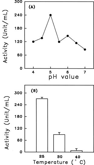
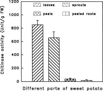
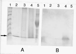
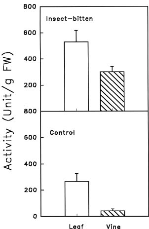
Abstract. Chitinase activity was detected in the crude extracts of different parts of sweet potato (Ipomoea batatas [L.] Lam var. Tainong 57) using commercial deacetyl glycol chitin as a substrate. The activity of chitinase in crude extracts, which was expressed as units/g fresh weight, was arranged in decending order of leaves > sprouts > peels of storage roots > peeled storage roots. Under field conditions, the chitinase activity of leaves was higher than vines. The chitinase activity of insect-bitten leaves was higher than that of the controls, and the chitinase activity of vines attached to insect-bitten leaves was also higher than that of vines attached to intact leaves. The molecular weight of main chitinase determined by activity staining was about 16 kDa. The optimum pH was 5.0 and the optimum temperature of chitinase activity was 25°C.

Keywords: Chitinase; Defense against pests; Ipomoea batatas.

Abbreviations: CBB R-250, coomassie brilliant blue R-250; GlcNAc, N-acetylglucosamine; PAGE, polyacrylamide gel electrophoresis; PVP, polyvinyl polypyrrolidone; SDS, sodium dodecyl sulfate; TCA, trichloroacetic acid

Introduction

Chitinases (EC 3.2.1.14) catalyze the hydrolysis of chitin, a linear homopolymer of b-1,4-linked N-acetylglucosamine (GlcNAc) residues. No substrates for this group of enzyme have been identified in plants, whereas chitin is commonly a component of fungal cell walls and the exoskeleton of arthropods, organisms which include many important pathogens and pests. Plants respond to attack by microorganisms, insects, and animals by the induction of sets of genes encoding diverse proteins, many of which are believed to play a role in defense (Bol et al., 1990; Bowles, 1990; Collinge et al.,1993; Dixon and Harrison, 1990; Linthorst, 1991; Punja and Zhang, 1993). The most marvelous of these proteins is chitinase. It has been demonstrated that enhanced chitinase in transgenic plants can indeed reduce the damage caused by pathogens (Broglie et al., 1991).

Most plant chitinases belong to endochitinases that randomly hydrolyze chitin polymers. They have been purified from barely (Leah et al., 1991; Swegle et al., 1989), bean (Abeles et al., 1970; Boller et al., 1983; Broglie et al., 1986), cabbage (Chang et al., 1992), corn (Lin et al., 1982), pea (Vad et al., 1991), potato (Gaynor, 1988), rice (Huang et al., 1991; Zhu and Lamb, 1991), rye (Yamaggami and Funatsu, 1993), soybean (Wadsworth and Zikakis, 1984), tobacco (Shinshi et al., 1990), wheat (Molando et al., 1979), and yam (Tsuyoshi et al., 1984). Plant chitinases fall into three classes based on primary structures (Shinshi et al., 1990). Some plant chitinases

also show lysozyme activity, which hydrolyzes the b-1,4-linkages between N-acetylmuramic acid and GlcNAc residues in peptidoglucan (Roberts and Selitrennikoff, 1988). In this preliminary report, we detected chitinase activities in different parts of sweet potato (Ipomoea batatas [L.] Lam var. Tainong 57), including leaves, sprouts, vines, peels and peeled storage roots. These results will be helpful for further investigations of sweet potato chitinases.

Materials and Methods

Materials

Fresh storage roots of sweet potato (Ipomoea batatas [L.] Lam var. Tainong 57) were purchased from a local market. After being cleaned with water, the roots were placed in a thermostated (30°C ) growth chamber in the dark and sprayed with water twice a day. Etiolated sprouts of 3_5 cm length were collected for further use. Leaves and vines of the same variety were kindly provided by Dr. C. H. Cheng (Provincial Agricultural Research Institute, Chia-Yi branch). Cut vines of sweet potato variety Tainong 57 were planted with five other varieties in Sep. 1996 in a randomized block design (4 repeat blocks, 10 m2/block). Number 39 complex fertilizer (N : P2O5 : K2O = 12 : 18 : 12) of the Taiwan Fertilizer Company was applied (0.28 kg per block) both basally and two months after planting. Both intact and insect-bitten leaves with attached vines were collected (May 19, 1997). Leaves and vines were weighed immediately and put in an envelope and frozen in liquid nitrogen in situ. The frozen samples were kept at -70°C for further use.

3Corresponding author. Fax: +886-2-2782-7954.


Botanical Bulletin of Academia Sinica, Vol. 39, 1998

Crude Extraction of Chitinase

Each sample was ground immediately to a fine powder in liquid nitrogen with a mortar and pestle. The powders were suspended in an extraction buffer consisting of 100 mM acetate buffer (pH 5.0) containing 1% (W/V) ascorbic acid and 1% (W/V) PVP. After centrifugation at 16,700 g for 30 min, the supernatant was saved and designated as crude extract.

Chitinase Activity Determination

Commercial deacetyl glycol chitin (G-7753, Sigma Chemical Co., St. Louis, MO, USA) was used as a substrate. A volume of 0.1 mL crude extract was added to 1.0 mL of 0.05% deacetyl glycol chitin in 50 mM acetate buffer (pH 5.0) at room temperature to start the enzyme reaction. After 30 min, the reaction was stopped by heating at 100°C for 5 min. The increased reducing ends, which were released by sweet potato chitinase, were determined according to the method of Imoto and Yagishita (1971). The preheated crude extracts (100°C for 5 min) were used as controls. One unit of chitinase activity was defined as an amount capable of releasing reducing ends corresponding to 1 µg GlcNAc from deacetyl glycol chitin at pH 5.0 in one hour. The GlcNAc (5_40 µg) was used to plot the standard curve.

Optimum pH and Temperature of Chitinase Activity

The optimum pH of chitinase activity of crude extracts was determined using 100 mM phosphate buffer (pH 6.0, 6.5 and 7.0) and 100 mM acetate buffer (pH 4.0, 4.5, 5.0 and 5.5). The optimum temperature of the chitinase activity was tested at 25°C, 30°C, and 40°C in 100 mM acetate buffer (pH 5.0).

Electrophoresis and Activity Staining of Chitinase

A discontinuous SDS slab gel electrophoresis of 15 % acrylamide was performed using a vertical mini-gel system (Bio-Rad Inc., USA) with a 0.75 mm thickness. The gel was prepared mainly according to Weber and Osborn (1969). When SDS-PAGE was finished, gels were cut into two parts. One was fixed with 12.5% TCA and stained with CBB R-250 dye. The other was immersed and shaken twice in 25% (V/V) isopropanol in 10 mM acetate buffer (pH 5.0) for 10 min, and equilibrated in 10 mM acetate buffer (pH 5.0) for 15 min before activity staining. The activity staining of chitinase was mainly according to the method of Trudel and Asselin (1989). The 1 % (W/V) agarose gel (pH 5.0) containing 0.01% (W/V) deacetyl glycol chitin was overlaid with the equilibrated polyacrylamide gel. After incubation at 37°C for 1 h, the agarose gel was dipped in 0.01% (W/V) Fluorescent brightener 28 (designated as Calcofluor white M2R in the original paper) fluorescent dye (F-3543, Sigma Chemical Co., St. Louis, MO, USA) in 50 mM Tris buffer (pH 8.3) for 5 min and then washed with distilled water several times to remove excess fluorescent dye. The active chitinase in an agarose gel appeared dark on a bright background under UV light.

Results

In preliminary experiments, we detected high chitinase activity in the crude extract of sweet potato leaves using deacetyl glycol chitin as substrates. The optimum pH (5.0) and temperature (25°C ) of chitinase activity was shown in Figures 1A and 1B, respectively.

The specific chitinase activities, which were expressed as units/g fresh weight, in crude extracts of different parts of sweet potato were arranged in decending order of leaves > sprouts > peels of storage roots > peeled storage roots (Figure 2).

The chitinase activities in leaves and vines of field-grown sweet potato collected at Provincial Agricultural Research Institute, Chiayi branch are shown in Figure 3. It is clear that chitinase activities of leaves are higher than those of vines. Insect-bitten leaves have higher chitinase activity than the controls. The chitinase activities of vines attached to insect-bitten leaves are also higher than those of vines attached to intact leaves.

After electrophoresis one part of the gel was fixed with 12.5% TCA and stained for protein with CBB R-

Figure 1. The optimum pH (A) and optimum temperature (B) of chitinase activity in crude extract of sweet potato leaves.


Hou et al. Chitinase of sweet potato

250 dye (Figure 4A), and the other one was used for chitinase activity staining (Figure 4B). In Figure 4, lane 1 contained prestained markers (See BlueTM pre-stained standard for SDS-PAGE from NovexTM, San Diego, CA, USA). Lane 2 contained purified lysozyme (Hou and Lin, 1997) as a positive control according to the report of Trudel and Asselin (1989). Lanes 3 and 4 were crude extracts of sprouts and leaves, respectively. Lane 5 was ovalbumin as a negative control. Comparing Figure 4B with Figure 4A, it was found that lane 5 did not have fluorescence; and lane 4 had a higher fluorescence than either lane 2 or lane 3. The prestained marker of 16 kDa (lysozyme) also had fluorescence. It is clear that the main chitinase from leaves or sprouts of sweet potato has a molecular weight of about 16 kDa as determined by activity staining.

Figure 2. The chitinase activities in crude extracts of different parts of sweet potato at pH 5.0. Each mean and error bar were obtained from samples of four repeated blocks.

Figure 4. Protein staining (A) on a 15% polyacrylamide gel and activity staining (B) on an agarose gel of chitinase from sweet potato after SDS-PAGE. Lane 1, the See BlueTM pre-stained standard for SDS-PAGE, and the lysozyme (16 kDa) was indicated by the arrow; lane 2, purified lysozyme from egg white as a positive control (Trudel and Asselin, 1989); lane 3, crude extract of sprouts; lane 4, crude extract of leaves; lane 5, ovalbumin as a negative control. 25 µg protein was loaded in each well.

Discussion

Because so far no substrates are available for chitinase in plant tissues, plant chitinases are believed to play a role in defense (Bol et al., 1990; Bowles, 1990; Collinge et al., 1993; Dixon and Harrison, 1990; Linthorst, 1991). It has been demonstrated that enhanced chitinase in transgenic plants can indeed reduce the damage caused by pathogens and pests (Broglie et al., 1991).

Many chitinase activities have been found in different plants, and most of them belong to endochitinases, which randomly hydrolyze chitin polymers. The optimum pH of chitinase from leaves of sweet potato was 5.0 (Figure 1A ), the same as chitinase purified from cabbage (Chang et al., 1992), corn (Lin et al., 1982), and rye (Yamaggami and Funatsu, 1993). The optimum temperature was 25°C

Figure 3. The chitinase activities in field-grown leaves and vines of sweet potato. Both intact leaves and insect-bitten ones were collected. Vines attached to intact leaves or those attached to insect-bitten leaves were also collected. Each mean and error bar were obtained from samples of four repeated blocks.


Botanical Bulletin of Academia Sinica, Vol. 39, 1998

(Figure 1B). The optimum temperature of chitinase purified from corn and cabbage was 45°C or 55°C (Lin et al., 1982) and 40 °C to 50°C (Chang et al., 1992), respectively.

The chitinase activities in leaves were the highest among different parts of sweet potato (Figure 2). High chitinase activities have already been found to be induced by ethylene or pathogens in leaves of several plants, including bean (Boller et al., 1983) and pea (Vad et al., 1991). The possible defense roles of high chitinase activity in leaves of sweet potato reported in this study need further elucidation.

Chitinase activities in both leaves and vines of field-grown sweet potato are shown in Figure 3. Leaves have higher chitinase activity than vines. Insect-bitten leaves have higher chitinase activity than the controls. Vines attached to insect-bitten leaves also have higher chitinase activity than vines attached to intact leaves. Higher trypsin inhibitor activities in sweet potato leaves and vines were induced systematically by insect-bitting (Lin, Y. H., unpublished data). Our data now suggest that chitinase activities may also be induced systematically by insect-bitting.

The molecular weight of main chitinase activity in sprouts and leaves of sweet potato is about 16 kDa (Figure 4). Although the molecular weight of most plant chitinases is about 30 kDa (Boller et al., 1983; Chang et al., 1992; Molanol et al., 1979; Vad et al., 1991; Wadsworth and Zikakis 1984; Yamaggami and Funatsu, 1993); chitinases purified from corn have molecular weights of 11.5 kDa and 10 kDa (Lin et al., 1982). The difference in molecular weight needs further elucidation.

In summary, our preliminary results regarding chitinases in sweet potato will be helpful for further investigation of their physicochemical properties, physiological significance, and role in defense mechanisms of the sweet potato.

Acknowledgments. The authors want to thank the financial support (NSC87- 2311-B-001-0053) from the National Science Council, Republic of China (R.O.C.) and Dr. C. H. Cheng of the Provincial Agricultural Research Institute, Chiayi branch for providing leaf and vine samples.

Literature Cited

Abeles, F.B., R.P. Bosshart, L.E. Forrence, and W.H. Habig. 1970. Preparation and purification of glucanase and chitinase from bean leaves. Plant Physiol. 47: 129_134.

Bol, J.F., H.J. Linthorst, and B.J. Cornelissen. 1990. Plant pathogenesis-related proteins induced by virus infection. Ann. Rev. Phytopathol. 28: 113_138.

Boller, T., A. Gehri, F. Mauch, and U. Vogeli. 1983. Chitinase in bean leaves:induction by ethylene, purification, properties, and possible function. Planta 157: 22_31.

Bowles, D.J. 1990. Defense-related proteins in higher plants. Ann. Rev. Biochem. 59: 873_907.

Broglie, K. E., J. J. Gaynor, and R. M. Broglie. 1986. Ethyl

ene-regulated gene expression: molecular cloning of the genes encoding an endochitinase from Phaseolus vulgaris. Proc. Natl. Acad. Sci. USA. 83: 6820_6824.

Broglie, K., I. Chet, M. Holliday, R. Cressman, P. Biddle, S. Knowlton, C. J. Mauvais, and R. Broglie. 1991. Transgenic plants with enhanced resistance to the fungal pathogen Rhizoctonia solani. Science 254: 1194_1197.

Chang, C. T., H. F. Lo, C. J. Wu, and H. Y. Sung. 1992. Purification and properties of chitinase from cabbage. Biochem. Int. 28: 707_715.

Collinge, D.B., K.M. Kragh, J.D. Mikkelsen, K.K. Nielsen, U. Rasmussen, and K. Vad. 1993. Plant chitinases. Plant J. 3: 31_40.

Dixon, R.A. and M. Harrison. 1990. Activation, structure and organization of genes involved in microbial defence in plants. Adv. Genet. 28: 165_234.

Gaynor, J.J. 1988. Primary structure of an endochitinase mRNA from Solanum tuberosum. Nucl. Acids Res. 16: 5210.

Hou, W.C. and Y.H. Lin. 1997. Egg white lysozyme purification with sweet potato (Ipomoea batatas [L.] Lam) leaf preparations. J. Agric. Food Chem. 45: 4487_4489.

Huang, J.K., L. Wen, M. Swegle, H.C. Tran, T.H. Thin, H.M. Naylor, S. Muthukrishnan, and G.R. Reeck. 1991. Nucleotide sequence of a rice genomic clone that encodes a class I endochitinase. Plant Mol. Biol. 16: 479_480.

Imoto, T. and K. Yagishita. 1971. A simple activity measurement of lysozyme. Agric. Biol. Chem. 35: 1154_1156.

Leah, R., H. Tommerup, I. Svendsen, and J. Mundy. 1991. Biochemical and molecular characterization of three barley seed proteins with antifungal properties. J. Biol. Chem. 226: 1564_1573.

Lin, Z.F., D. Wu, A. Luo, and W. Zhang. 1982. Chitinase from seed of Zea mays and Coix lachrymajobi L., purification and some properties. Process Biochem. 27: 83_88.

Linthorst, H.J.M. 1991. Pathogenesis-related proteins of plants. Crit. Rev. Plant Sci. 10: 123_150.

Molando, J., I. Polacheck, A. Duran, and E. Cabib. 1979. An endochitinase from wheat germ. J. Biol. Chem. 254: 4901_4907.

Punja, Z.K. and Y.Y. Zhang. 1993. Plant chitinases and their roles in resistance to fungal disease. J. Nematol. 25: 526_540.

Roberts, W.K. and C.P. Selitrennikoff. 1988. Plant and bacterial chitinases differ in antifungal activity. J. Gen. Microbiol. 134: 169_176.

Shinshi, H., J. M. Neuhaus, J. Ryals, and F. Meins. 1990. Structure of a tobacco endochitinase gene: evidence that different chitinase gene can arise by transposition of sequences encoding a cysteine-rich domain. Plant Mol. Biol. 14: 357_368.

Swegle, M., J.K. Huang, G. Lee, and S. Muthukrishnan. 1989. Identification of an endochitinase cDNA clone from barely aleurone cells. Plant Mol. Biol. 12: 403_412.

Tsuyoshi, T., D. Koga, A. Ide, T. Ishibashi, M. Horino-Matsushige, K. Yagishita, and T. Imoto. 1984. Purification and some properties of chitinase from yam, Dioscorea opposita Thunb. Agric. Biol. Chem. 48: 931_939.

Trudel, J. and A. Asselin. 1989. detection of chitinase activity after polyacrylamide gel electrophoresis. Anal. Biochem. 178: 362_366.


Hou et al. Chitinase of sweet potato

weight determination by dodecyl sulphate-polyacrylamide gel electrophoresis. J. Biol. Chem. 244: 4406_4412.

Yamaggami, T. and G. Funatsu. 1993. Purification and some properties of three chitinases from the seeds of rye. Biosic. Biotech. Biochem. 57: 643_647.

Zhu, Q. and C.J. Lamb. 1991. Isolation and characterization of rice gene encoding a basic chitinase. Mol. Gen. Genet. 22: 289_296.

Vad, K., J.D. Mikkelsen, and D.B. Collinge. 1991. Induction, purification and characterization of chitinase isolated from pea leaves inoculated with Ascochyta pisi. Planta 184: 24_29.

Wadsworth, S.A. and J.P. Zikakis. 1984. Chitinase from soybean seeds: Purification and some properties of the enzyme system. J. Agric. Food Chem. 32: 1284_1288.

Weber, K. and M. Osborn. 1969. The reliability of molecular

¥ÌÁ¦¤¤ªº´X¤B½èú@

«J¤åµX1 ³¯¬Õ¦{2 ªLÄ£½÷1

1¤¤¥¡¬ã¨s°|´Óª«¬ã¨s©Ò

2¤j¦P¤u¾Ç°|¥Íª«¤uµ{¾Ç¨t

¥H°Ó«~¤A¤G¾J´X¤B½è¬°°ò½è¡A§Ú­ÌÀË´ú¨ì¥ÌÁ¦¤£¦P³¡¦ì»Ã¯À²Ê©â²G¤¤ªº´X¤B½èú@ªº¬¡©Ê¡C­Y¥H ¨C§J·sÂA­«¶q¤¤ªº³æ¦ì¬¡©Êªí¥Ü¡A«h²Ê©â²G¤¤ªº´X¤B½èú@¬¡©Êªº¤j¤p¦¸§Ç¬°¡G¸­¤l¡ÖªÞ¡Ö¶ô®Úªº¥Ö ¡Ö¥h¥Öªº¶ô®Ú¡C¦b¥Ð¶¡ºØ´Óªº¥ÌÁ¦¤¤¡A¸­¤lªº´X¤B½èú@¬¡©Ê°ª©ó½¯¡C³QÂΫr¹Lªº¸­¤lÅãµM¤ñ¹ï·Ó²Õ ¡]§¹¾ãªº¸­¡^ ¡A¦³¤ñ¸û°ªªº´X¤B½èú@ªº¬¡©Ê¡C³s±µ©ó³QÂΫr¹Lªº¸­¤lªº½¯¤ñ³s±µ©ó¹ï·Ó²Õªº½¯¡A¦³ ¸û°ªªº´X¤B½èú@ªº¬¡©Ê¡C¨Ì¾Ú¬¡©Ê¬V¦â¡A¥ÌÁ¦¸­¤l¤¤ªº¥D­n´X¤B½èú@ªº¤À¤l¶q¬° 16 kD¡A³Ì¾A¤ÏÀ³ pH ¬° 5.0¡A³Ì¾A¤ÏÀ³·Å«×¬° 25°C¡C

ÃöÁäµü¡G ´X¤B½èú@¡F¥ÌÁ¦¡F¨¾¿m¨¤¦â¡C