Bot. Bull. Acad. Sin. (1998) 39: 147_152

Griffiths — Senescence plasmid

(Invited review paper)

The kalilo family of fungal plasmids

Anthony J. F. Griffiths

Department of Botany, University of British Columbia, Vancouver, Canada V6T 1Z4

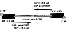
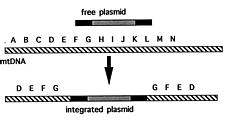
Abstract. A. J. F. Griffiths (1997). The prototypic kalilo plasmid is approximately 9 kb long and has the structure of an invertron. There are 5'-bound terminal proteins, a long terminal inverted repeat (TIR) and two ORFs corresponding to viral RNA and DNA polymerases. Kalilo plasmids have been found in several different heterothallic and one pseudohomothallic species of Neurospora from around the world. No examples have been found in true homothallic species. The prototypic plasmid was found in N. intermedia on the Hawaiian island of Kauai. It causes death by integration into the mtDNA, creating an inserted form flanked by long symmetrical repeats of the mtDNA flanking one side of the insertion point. The plasmid can be transmitted horizontally through heterokaryosis or sexually via the maternal parent. Spread is potentially explosive and can be prevented by cell incompatibility or by suppressor mutations. The related plasmid LA-kalilo has been found in N. tetrasperma from Louisiana, USA. This is essentially the same as kalilo but shows a short deletion in the middle of each TIR. This plasmid has been found not to insert into mtDNA. In a related genus Gelasinospora, also from Louisiana, a relative called Gel-kalilo has been found. This plasmid shows many differences from kalilo, but is still obviously homologous over large regions. This plasmid also does not integrate into the mtDNA. Lack of integration can be due to structural differences or to host genome suppression. The plasmids have arrived in their present hosts by a combination of vertical descent from a common ancestor and horizontal asexual transmission.

Keywords: Mitochondria; mtDNA; Neurospora; Plasmid, Senescence.

Contents

Introduction 147

The Kalilo Prototype 148

Discovery 148

Structure 148

Distribution 148

Transmission 149

Expression 149

Structural Variants 150

Suppressors 150

LA-Kalilo 150

Gel-Kalilo 151

Evolution 151

Literature Cited 152

Introduction

Plasmids are replicating DNA molecules that are additional to the normal genomic components. They contain few genes and so are dependent on the normal operation of the genomic machinery for their propagation and survival. In this sense, they are parasitic molecules, drawing on not only the information of the host but also on its energy reserves. Bacterial plasmids carry genes that seem to

benefit the host and are of adaptive value. Genes for resistance to environmental toxins are the best example of this. However, it must be said that little is known of the cost/benefit ratios or the evolutionary dynamics of bacterial plasmids in natural populations. Plasmids are not as commonly encountered in eukaryotic organisms. No plasmids have ever been found in animals, though several have been found in plants, and many in fungi (Griffiths, 1995). The reasons for this `kingdom specificity' are not known.

As in bacteria, fungal plasmids are either linear or circular. Linear plasmids are more common than circular plas

Fax: 604-822-9179; E-mail: agriff@unixg.ubc.ca


Botanical Bulletin of Academia Sinica, Vol. 39, 1998

mids in the model filamentous fungi that have been extensively used in routine genetic research, for example Neurospora and Podospora. In Saccharomyces cerevisiae the 2 micron plasmid that has been the basis of several cloning vectors used in that organism is a circular plasmid found in the nucleus. However in the filamentous fungi all plasmids are mitochondrial in location. Arganoza et al. (1994) and Yang and Griffiths (1993a) surveyed natural populations of several Neurospora species and found that there are distinct `homology groups' or `families' of plasmids. It is not possible to give a precise number of known families because the appropriate cross-homologies have not been tested, but it is probably on the order of ten. The most extensively researched of these families is the kalilo family, the subject of this review.

The Kalilo Prototype

Discovery

The prototypic kalilo plasmid was discovered in natural isolates of N. intermedia from the Hawaiian island of Kauai (Bertrand et al., 1988; Griffiths and Bertrand, 1984). Interest in this discovery was twofold: a) it was the first linear plasmid to be discovered in filamentous fungi and b) it caused senescence and death of its host fungus. Kalilo is a Hawaiian word meaning "dying" or "hovering between life and death." The senescent phenotype is called Kalilo, and the causative plasmid is called kalilo or kalDNA.

Structure

The general structural organization of the kalilo plasmid is shown in Figure 1. The kalilo plasmid consists of 8643 bp of double-stranded DNA (Chan et al., 1991). The A+T content of the DNA is 70%, close to that of mtDNA (67%). The 5' termini at each end of the plasmid are occluded by a covalently-bound protein (Vierula et al., 1990). This protein makes the plasmid resistant to 5' exonuclease digestion, but susceptible to attack at the 3' termini. Virtually all fungal and plant linear plasmids carry such a protein. The ends of the plasmid are a perfect terminal inverted repeat (TIR) 1366 bp long. The presence of a TIR is also typical of linear plasmids generally. A linear structure with TIRs and terminal proteins, found in many lin

ear plasmids and linear viral genomes, has been called an `invertron' (Sakaguchi, 1990).

The central region of the plasmid contains two large open reading frames (ORFs) that seem to be the only potential genes in the structure (Chan et al., 1991). ORF-1 is 2433 bp long, encoding a putative protein of 811 amino acids. The amino acid sequence shows blocks of 40_60% similarity to phage and yeast RNA polymerases. Hence, this is presumed to be a gene for RNA polymerase although no enzyme activity has been associated with this sequence.

ORF-2 is 2910 bp long and codes for a putative protein of 970 amino acids. This protein shows similarities to DNA polymerases of viruses and phages (Chan et al., 1991). However, no enzyme activity has been demonstrated for this gene either.

It is interesting to note that the two ORFs both begin slightly inside the TIRs, so if this is indeed a genuine translational start site, then the two proteins will have identical amino acid sequences at the amino terminus.

These features are common in eukaryotic linear plasmids. Some have only one ORF encoding a DNA polymerase (Meinhardt et al., 1990), but most have two also apparently corresponding to RNA and DNA polymerases. The various plasmids with blocks of amino acid similarity to polymerases do not, nevertheless, hybridize at the DNA level.

Distribution

The prototypic kalilo was originally found in N. intermedia on Kauai and has since been found on other Hawaiian islands (Griffiths and Bertrand, 1984). A survey of other global sites by Yang and Griffiths (1993a) found many plasmids but none that hybridize with kalilo. However a survey of different strains by Arganoza et al. (1994) revealed numerous kalilo-binding plasmids not only in N. intermedia from Hawaii, but in N. crassa from Haiti and the Ivory Coast, N. discreta from Florida (USA), the Ivory Coast, Papua-New Guinea, and Thailand, and N. tetrasperma from Tahiti. These plasmids were not characterized so it is not known how similar they are to the prototypic kalilo. No kalilo-homologous plasmids were found in N. sitophila. All the above species are heterothallic with the exception of N. tetrasperma, which is pseudohomothallic. Isolates of five true homothallic species were screened too, but these showed no plasmids of any type.

Arganoza et al. (1994) also made the following interesting observations concerning distribution:

a) The circular Fiji group of plasmids and kalilo plasmids are dramatically over-represented in the Hawaiian Islands in relation to their mean incidence worldwide.

b) Although N. intermedia isolates from around the world were sampled, only in Hawaii was kalilo found in this species.

c) The kalilo plasmid shows a tendency to be paired with the Fiji family of plasmids. This might be a result of

Figure 1. Structural organization of the kalilo plasmid. The total size of the plasmid is 8.6 kb. ORF = open reading frame; TIR = terminal inverted repeat; TP = terminal protein.


Griffiths — Senescence plasmid

their common geographical ranges, but also might reflect a functional interrelationship.

Transmission

In crosses, the kalilo plasmid and the senescence phenotype are by and large transmitted maternally (Bertrand et al., 1988; Griffiths and Bertrand, 1984). Some plasmid-free ascospore progeny are found if the maternal parent is relatively juvenile, and this might reflect cytoplasmic heterogeneity at this stage, or even the possibility of curing as a result of the sexual process. However, the progeny of dying cultures are never rejuvenated.

Rare cases of "paternal leakage" have been observed (Yang and Griffiths, 1993b). These are unlikely to be instances of the paternal mitochondria travelling down the trichogyne, and more likely to be invasions of maternal tissue by fusion with paternal cells.

The kalilo plasmid is also transmitted via cell-cell contact. If a forced heterokaryon is made between two compatible strains of N. crassa, one of which carries kalilo, then from the heterokaryon the other nuclear phenotype can be recovered, also now carrying kalilo (Griffiths et al., 1990). Debets et al. (1994) showed that if a kalilo strain is grown together with a compatible plasmid-free strain, then the plasmid is aggressively transferred to the other strain, and even in cases in which the mixture of input strains is heavily skewed in favour of the normal strain, the culture rapidly dies.

If a heterokaryon is made between a kalilo strain and one carrying another plasmid, then homokaryotic cells carrying both plasmids can be derived from the heterokaryon. Experiments of this type in conjunction with the distribution studies show that there seems to be little incompatibility between plasmid types.

It is possible to transfer kalilo across cell incompatibility barriers. Debets et al. (1994) showed that incompatibility for the het genes A/a, D/d and E/e could not prevent transfer of plasmids, albeit at a somewhat slower rate. However, incompatibility for the C/c het gene did prevent transfer. Kalilo has even been transferred between species: a transient forced heterokaryon made between a kalilo strain of N. intermedia and a plasmid-free strain of N. crassa enabled the plasmid to be transferred to N. crassa (Griffiths et al., 1990). It was interesting to note that in this experiment the plasmid had jumped into N. crassa mitochondria, and no N. intermedia mtDNA could be detected. Hence, the plasmid must be able to travel through the cytosol to its new location, or mitochondria of the two species may have the ability to fuse and then disjoin.

Arganoza et al. (1994) found that the association and distribution of different plasmids, including kalilo, were essentially random. Similarly, Debets et al. (1994) showed that the co-distribution of kalilo and a circular plasmid Hanalei-2 was random on the Hawaiian Islands. These observations argue for free travel of plasmids through populations, overcoming many of the incompatibility barriers known to exist between haplotypes.

Expression

The phenotypic expression of the plasmid is as senescence and death. This can be followed by serial subculturing; at some strain-specific subculture the growth of the fungus slows, and both growth and fertility decline in subsequent subcultures until death occurs. At death and just before death the cells become highly vacuolated and mitochondria degenerate. The same process can be followed in growth tubes (Griffiths et al., 1986). In some cases a culture that is apparently dying recovers temporarily, an effect named the Lazarus Effect after the Biblical character who arose from the dead. Cultures die faster by serial conidial transfer than by continuous growth, an effect probably related to the fact that death at growing front of a continuous culture can be revived by an infusion of juvenile cytoplasm from behind the front.

As the culture approaches death, mitochondrial physiology changes dramatically. Cytochrome c increases in amount while cytochromes aa3 and b decline. Simultaneously there is a switch from cyanide-sensitive respiration to cyanide-insensitive salicyl hydroxamide-sensitive respiration, probably reflecting a switch to an alternative oxidase system (Bertrand et al., 1985; Rieck et al., 1982). These changes reflect profound disturbance in mitochondrial function. Indeed, it was found that the cause of this disturbance is the integration of the kalilo plasmid into the mtDNA (Bertrand et al., 1985, 1988) apparently acting as an insertional mutagen. Apparently the initial event that results in death is a single insertion at one site because at death a clone of this type of mtDNA predominates and wild type mtDNA is rare. Sometimes the initial insertion is not the one that causes death, and the culture tolerates its insert until another lethal insertion occurs (Myers et al., 1989).

The mode of insertion is novel for DNA integrative events generally, and is summarized in Figure 2. The inserted DNA is an almost full-length version of the plasmid, lacking a small number of base pairs equally at each end. When the integration site and the truncated segments are compared, the integration site shows homology between five base pairs in the plasmid and mtDNA, but this homology can be from anywhere within the last 20 or so base pairs (Bertrand and Griffiths, 1989).

Figure 2. Integration of the kalilo plasmid into mitochondrial DNA (mtDNA). The letters A through N represent regions of part of the mtDNA, which has been linearized for the purpose of the illustration.


Botanical Bulletin of Academia Sinica, Vol. 39, 1998

Sequencing outwards from the inserted plasmid showed that the mtDNA flanking the plasmid is identical on both ends. Hence integration duplicates the mtDNA to one side of the integration site. One way of explaining this is that a single-stranded replication intermediate of the plasmid pairs at the TIRs to form a racquet-like structure. The TIR then undergoes crossing over with some target site within the mtDNA. When the plasmid-containing recombinant molecule replicates, the plasmid becomes flanked by a symmetrical repeat of the mtDNA to one side of the insertion point.

The phenotypic features of senescence are reminiscent of those of mtDNA mutations, many of which are suppressive, that is, they can take over a culture. This process is still poorly understood. Intuitively it would seem that an integration event would incapacitate that molecule or mitochondrion causing it to be selected against, never to affect phenotype. However, what actually happens is that the defective inserted mtDNA becomes renegade and somehow outcompetes its normal siblings, possibly because the inserted type replicates better; if true this is unlikely to be through the possession of an origin of replication because kalilo contains no sequences similar to replication origins, and in any case the suppressivity of the regular mitochondrial mutations cannot be explained this way. Another possible explanation is that mitochondria have a system for detecting oxidative damage, and this induces the mitochondrial replication machinery, in a strategy that is supposed to compensate for the oxidative defect (Bertrand and Griffiths, 1989). This model is shown in Figure 3.

The kalilo plasmid is transcribed (Vickery and Griffiths, 1993). The start sites are in the TIRs 101 bp from the termini. Two transcripts are seen of 4.4 and 4.6 kb, which correspond to the two ORFs. No translational products have been sought. It would be desirable to engineer mutations in various regions of the plasmid to investigate their functions, but this cannot be achieved by recombinant techniques because there is no known way of introducing the plasmid back into a mitochondrion.

Structural Variants

Variants of the prototypic kalilo plasmid have arisen in culture. Vierula and Bertrand (1992) found two deleted types in cultures that had been treated with chloramphenicol. One type was essentially a pair of TIRs with little intervening material. The other type was a hairpin structure that seemed to have arisen as a fold-back of a single-stranded double TIR structure. Such a single strand might have arisen as a displaced strand during an adenovirus-like replication primed by the 5' terminal protein. Hence, this constitutes indirect evidence of a viral-like replication mechanism. Indeed there are no data concerning other possible modes of replication.

Yang and Griffiths (1993a) observed a variety of novel kalilo derivatives occasionally arising in continuous culture of N. intermedia. Deletions were observed but these were complex and seemed to be nested sets of molecules in which small fragments had been lost from throughout the linear sequence. "Sibling" plasmids were also observed which which were approximately the same size as the kalilo plasmid, but these were not characterized. Also plasmid-homologous structures were observed near the well of the gel; some acted as though circular and some linear. These could be concatamerized forms of the plasmid, but restriction analysis showed that there was not a simple head-to-tail concatamerization. Alternatively, these could be complex replication intermediates that sometimes become visible on the gel.

Suppressors

Since plasmids are common in natural populations, and some of these (such as kalilo) are potentially harmful, it seems possible that a fungal host could mutate to generate alleles that constitute plasmid suppressors. Such alleles seem to be reasonably common in laboratory stocks. Griffiths et al. (1992) showed that such suppressors result in 4:4 segregations of senescent and nonsenescent ascospores in asci. Two suppression modes were identified in this limited study; in the first the replication and insertion of the plasmid are inhibited; in the second the plasmid and insertion composition seem typical for Kalilo strains but no senescence occurs, and hence somehow the suppressor works to circumvent the insertional cascade. A large set of such suppressors would be useful in dissecting the contribution of the host fungus to plasmid maintenance.

No studies have been attempted to try to find suppressor alleles in natural populations, although this would be a profitable line of research.

LA-Kalilo

Several cultures of the pseudohomothallic N. tetrasperma isolated from Louisiana (USA) have been found to contain a plasmid that hybridized to a kalilo probe (Marcinko-Kuehn et al., 1994). This plasmid showed a restriction map identical to kalilo in the ORF-containing region, but shorter by approximately 100 bp. This resulted

Figure 3. Model for suppressivity of mtDNA containing a kalilo insert. Mitochondria containing the insert (black dots) become renegade eventually dominating the mycelium when death occurs.


Griffiths — Senescence plasmid

in a smaller overall size which could be visualized on the gel. This plasmid was named LA-kalilo (Louisiana-kalilo).

Many of the Louisiana isolates senesced and died, so a study was undertaken to determine if this was caused by the plasmid. A total of 48 strains were categorized regarding plasmid content and senescent phenotype. Out of 16 senescent strains 15 bore the plasmid and one did not, whereas out of 28 nonsenescent strains 19 bore the plasmid and 9 did not. Hence, there was no strong support for the idea that the plasmid caused senescence. Therefore a search was undertaken for inserts of the LA plasmid into mtDNA. Since kalilo and LA-kalilo plasmids contain no Pst-1 restriction sites, inserts into the mtDNA can be visualized in a Pst-1 digest as kalilo elements bearing mtDNA tails and slightly larger than the free element. No such Pst-1 structures could be detected in senescent cultures bearing the plasmid. Lack of integration could be due to the difference in structure, but this seems small and does not involve the presumed crossover region. It is possible that this small region of deletion is an important binding site for a protein that promotes integration. Alternatively, lack of integration might be due to host suppression.

Ascospores issuing from the pseudohomothallic cycle do contain the plasmid so the breeding system of this fungus is compatible with long term propagation of this plasmid. When homokaryotic derivatives of the LA-kalilo strains were crossed to other plasmid-free homokaryons of opposite mating type, then maternal inheritance was observed. Hence the plasmid can be used to show that even though fertilization within the pseudohomothallic mycelium must be isogametic, the fungus can still form structures that allow it to adopt a heterothallic-like anisogametic system. This discovery also constitutes evidence that the pseudohomothallic cycle is secondarily derived from the heterothallic.

Gel-Kalilo

Gelasinospora is a genus closely related to Neurospora. The main taxonomic criterion for this genus is the presence of pitted ascopores instead of the striated ones of Neurospora. From Louisiana soil two isolates of a homothallic species of Gelasinospora were found to contain a kalilo-homologous plasmid, which was named Gel-kalilo (Yuewang et al., 1996). This discovery made the kalilo plasmid family the first to be shown to have an intergeneric distribution.

Whereas the LA-kalilo plasmid is remarkably similar to the prototypic kalilo, Gel-kalilo, although clearly homologous, shows numerous differences in sequence. The plasmid is shorter and 8231 bp in length. However, it shows the basic invertron structure, with 5'-bound terminal proteins, identical TIRs, and two large ORFs that are presumptive RNA and DNA polymerases.

The TIRs are of length 1137 bp, approximately 140 bp shorter than those of kalilo. The inner third of the TIR shows 93% similarity to the equivalent position of kalilo. The middle third shows numerous differences from kalilo.

The outer third shows a large deletion running from kalilo position 8163 to 8322. The terminal 30 bases, believed to be important in kalilo insertion, show a high level of similarity with kalilo.

The ORFs probably start 60 bp further inside the TIR than in kalilo, but it is not certain if this methionine is a true initiation site. If true, it would make the polymerases 20 amino acids larger. ORF-1 is 95% similar to kalilo at the DNA level, whereas ORF-2 shows 93% similarity. The fact that both these long reading frames are open suggests that they are indeed functional. The intergenic region between the ORFs is longer than the equivalent region in kalilo and shows numerous small substitutions and additions or deletions.

All the Gelasinospora isolates from the Louisiana population senesced in growth tubes, so because not all contained the plasmid, it is not possible to associate the plasmid with senescence in this fungus. Neither was it possible to demonstrate integration of Gel-kalilo into mtDNA.

Evolution

The high level of DNA similarity shown by these plasmids makes it highly likely that they are related through a common ancestral plasmid. Even though this seems incontrovertible, it is by no means clear how the plasmid attained its present global distribution. One possibility is that the plasmid was transmitted vertically over the time period during which the Neurospora and Gelasinospora species evolved. If this is true, then a concordance could be expected between the phylogenetic trees of the hosts and of the plasmids. Generally there is: Gelasinospora is the taxonomic outgroup, and so is the Gel-kalilo plasmid. Hence the data are compatible with the idea that the kalilo plasmid family is older than the divergence of the two genera and has been passed down each of these major branches of evolution. Under this model the plasmid must have been lost in many sub-branches because today it is not present in all isolates of Neurospora or Gelasinospora. Loss may have occurred by chance, by sexual curing, or by suppressor activity.

Horizontal transmission between different isolates is also a possibility. Certainly this seems possible between isolates in one geographical area because for example the distribution of kalilo and Hanalei-2 plasmids in Kauai seems random. It has been demonstrated that the plasmid has an explosive potential to spread, and can only be slowed by cell incompatibility. Therefore over the long term it seems likely that the plasmid could spread extensively. Since N. tetrasperma is capable of acting as a heterothallic species, transmission in and out of this species seems to present no conceptual problem.

Several different mechanisms seem capable of accomplishing horizontal transmission. First, mycelial contact and full or transient heterokaryon formation has been shown to be a viable possibility. Second, male transmission effects a type of horizontal transfer between vertical lineages. Third, it is possible that unknown transfer vec


Botanical Bulletin of Academia Sinica, Vol. 39, 1998

tors are at work. Possibly undiscovered types of viruses might effect a type of transduction. Herbivorous invertebrates might transmit plasmids by feeding on fungi (mites are a proven method of transposon transfer in Drosophila cultures). Finally, it is conceivable that fungi might be able to absorb DNA from the substrate.

The activities of the kalilo family illustrate well the Darwinian type of struggle for existence that must occur not only at the organismal level but at the molecular level. If indeed these plasmids are parasites, then they must play out the same finely-balanced interactions as conventional parasites. Plasmids possibly represent the minimal parasitic unit of life. Paramount is the strategy of how much harm can be done to the host. The perfect parasite can survive in harmony with the host, and indeed this seems to be the strategy of kalilo in N. tetrasperma and in Gelasinospora. It is possible that the plasmid even confers some type of advantage to the host in these cases in a natural setting. The virulent (prototypic) form either represents an aberration of the harmonious parasitic strategy or an aggressive strategy of kill and pass on to the next host.

The origin of plasmids is still a mystery. There is such a diversity of types and reproductive strategies among eukaryotic plasmids that it seems unlikely they are all descended from one ancestral type. Since bacteria have plasmids it is tempting to postulate that plasmids accompanied bacteria in the endosymbiotic fusion event that created the first eukaryotic cell. If this is true, then there must have been multiple endosymbiotic events, not one, as some suppose.

Acknowledgement. Funding by a Research Grant from the Canadian Natural Sciences and Engineering Research Council is gratefully acknowledged.

Literature Cited

Arganoza, M.T., J. Min, Z. Hu, and R.A. Akins. 1994. Distribution of seven homology groups of mitochondrial plasmids in Neurospora: evidence for widespread mobility between species in nature. Curr. Genet. 26: 62_73.

Bertrand, H. and A.J.F. Griffiths. 1989. Linear plasmids that integrate into mitochondrial DNA in Neurospora. Genome 31: 155_159.

Bertrand, H., A.J.F. Griffiths, D.A. Court, and C.K. Cheng. 1988. An extrachromosomal plasmid is the etiological precursor of kalDNA insertion sequences in the mitochondrial chromosome of senescent Neurospora. Cell 47: 829_837.

Bertrand, H., B.S-S. Chan, and A.J.F. Griffiths. 1985. Insertion of a foreign nucleotide sequence into mitochondrial DNA causes senescence in Neurospora intermedia. Cell 41: 877_884.

Chan, B.S.-S., D.A. Court, P.J. Vierula, and H. Bertrand. 1991 The kalilo linear senescence-inducing plasmid of Neurospora is an invertron and encodes DNA and RNA polymerases. Curr. Genet. 20: 225_237.

Court, D.A., A.J.F. Griffiths, S.R. Kraus, P.J. Russell, and H. Bertrand. 1991. A new senescence-inducing linear plasmid

in field-isolated Neurospora crassa strains from India. Curr. Genet. 19: 129_137.

Debets, F., X. Yang, and A.J.F. Griffiths. 1994. Vegetative incompatibility in Neurospora: its effect on horizontal transfer of mitochondrial plasmids and senescence in natural populations. Curr. Genet. 26: 113_119.

Griffiths, A.J.F. 1995. Natural plasmids of filamentous fungi. Microbiol. Rev. 59: 673_685.

Griffiths, A.J.F. and H. Bertrand. 1984. Unstable cytoplasms in Hawaiian strains of Neurospora intermedia. Curr. Genet. 8: 387_398.

Griffiths, A.J.F., S.R. Kraus, and H. Bertrand. 1986. Expression of senescence in Neurospora intermedia. Can. J. Genet. Cytol. 28: 459_467.

Griffiths, A.J.F., S.R. Kraus, R. Barton, D.A. Court, C.J. Myers, and H. Bertrand. 1990. Heterokaryotic transmission of senescence plasmid DNA in Neurospora. Curr. Genet. 17: 139_145.

Griffiths, A.J.F., X. Yang, R. Barton, and C.J. Myers. 1992. Suppression of cytoplasmic senescence in Neurospora. Curr. Genet. 21: 479_484.

Marcinko-Kuehn, M., X. Yang, F. Debets, D.J. Jacobson, and A.J.F. Griffiths. 1994. A kalilo-like linear plasmid in Louisiana field isolates of the pseudohomothallic fungus Neurospora tetrasperma. Curr. Genet. 26: 336_343.

Meinhardt, F., F. Kempken, J.K. Kamper, and K. Esser. 1990. Linear plasmids among eukaryotes: fundamentals and applications. Curr. Genet. 17: 89_95.

Myers, C.J., A.J.F. Griffiths, and H. Bertrand. 1989. Linear kalilo DNA is a Neurospora mitochondrial plasmid that integrates into the mitochondrial DNA. Mol. Gen. Genet. 220: 113_120.

Rieck, A., A.J.F. Griffiths, and H. Bertrand. 1982. Mitochondrial variants of Neurospora intermedia from nature. Can. J. Genet. Cytol. 24: 741_759.

Sakaguchi, K. 1990. Invertrons, a class of structurally and functionally related genetic elements that includes linear DNA plasmids, transposable elements, and genomes of adenoviruses. Microbiol. Rev. 54: 66_74.

Vickery, D.B. and A.J.F. Griffiths. 1993. Transcription of the kalilo linear senescence plasmid from Neurospora intermedia. Plasmid 29: 180_192.

Vierula, P.J., C.K. Cheng, D.A. Court, R.W. Humphrey, D.Y. Thomas, and H. Bertrand. 1990. The kalilo senescence plasmid of Neurospora intermedia has covalently-linked 5' terminal proteins. Curr. Genet. 17: 195_201.

Vierula, J.P. and H. Bertrand. 1992. A deletion derivative of the kalilo senescence plasmid forms hairpin and duplex DNA structures in the mitochondria of Neurospora. Mol. Gen. Genet. 234: 361_368.

Yang, X. and A.J.F. Griffiths. 1993a. Plasmid diversity in senescent and nonsenescent strains of Neurospora. Mol. Gen. Genet. 237: 177_186.

Yang, X. and A.J.F. Griffiths. 1993b. Male transmission of linear plasmids and mitochondrial DNA in the fungus Neurospora. Genetics 134: 1055_1062.

Yuewang, W., X. Yang, and A.J.F. Griffiths. 1996. Structure of a Gelasinospora linear plasmid closely related to the kalilo plasmid of Neurospora intermedia. Curr. Genet. 29: 150_158.