Bot. Bull. Acad. Sin. (1998) 39: 199_203

Chang et al. — Daylength and flowering in tuberose

Daylength affects protein pattern and flowering in tuberose (Polianthes tuberose L.)

Yu-Pei Chang1, Shih-Fen Ding1, Chin-Chih Chou1, Bo-Shiun Du2 and Wen-Shaw Chen1,3

1Department of Biological Sciences, National Sun Yat-Sen University, Kaohsiung City, Taiwan, Republic of China

2Department of Horticulture, National Chia-Yi Institute of Technology, Chia-Yi City, Taiwan, Republic of China

(Received December 8, 1997; Accepted April 2, 1998)

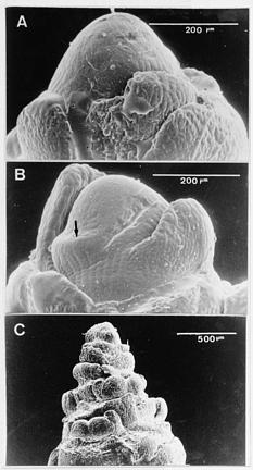
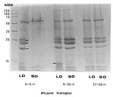
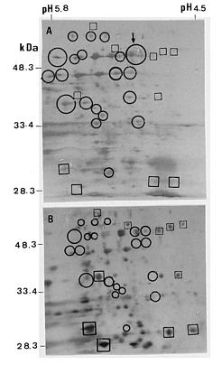
Abstract. Tuberose (Polianthes tuberose L. cv. Double) was exposed to 8 h and 16 h daylengths, to investigate the effects of these conditions on floral initiation and development. Corm apices remained vegetative when plant height was 0_5 cm at 8 h daylength. Floral initiation was attained at five-thirteenth (38%) of this stage at 16 h daylength. However, no significant difference in floral initiation or development of tuberose was found between 8 and 16 h daylengths when plants were over 5 cm in height. When tuberose seedlings were exposed to 16 h daylength, differences in protein pattern of the corm tissues were observed at 19, 23, 27, and 57 kDa. Furthermore, a significant protein band at 11 kDa was found in 16 h daylength but not in 8 h daylength, when plant heights were 6 to 30 cm. However, no significant difference in protein patterns was found between daylengths when plant heights were over 30 cm. The electrophoregrams obtained by two-dimensional electrophoresis allowed characterization of about 185 protein spots in corm tissues when plants were 0_5 cm in height and exposed to 8 and 16 h daylengths. At this stage, 20 newly synthesized proteins were observed in 16 h daylength, and 4 other polypeptides showed significant increase in 16 h, as compared to 8 h, daylength. The most significant peptides were 57 kDa protein with isoelectric point 5.1. These results are discussed in connection to flowering.

Keywords: Daylength; Flowering; Polianthes tuberose; Polypeptide.

Introduction

The effects of photoperiod and temperature on apical meristem development and the transition to reproductive growth have been studied extensively over the past 70 years (Bernier, 1988; Bolling, 1977; Gibby and Salisbury, 1971; Kadman-Zahavi and Peiper, 1987; Lumsden et al., 1982; McMillan, 1974; Salisbury, 1981). However, little study has focused on the correlation between the perception of environmental signals leading to flower induction and biochemical and molecular changes within the plants (Koorneef et al., 1995). Tuberose is an economically important ornamental crop in tropical and subtropical areas. However, how its flowering behavior is affected by photoperiod remains unknown. Visually, flowering in tuberose begins with floral initiation and then accelerates and sustains rapid elongation of the flower stalk. Investigation of the control of floral initiation and development by photoperiod is expected to be useful information. In this paper, we report the effects of different photoperiod conditions on protein synthesis in corm tissues and the flowering response of tuberose.

Materials and Methods

Plant Material

Tuberose (Polianthes tuberosa L. cv. Double) corms about 1.8 × 2.3 cm in diameter were planted in plastic pots (30 cm wide and 20 cm deep) containing vermiculite: domestic peat: sand (3/2/1 by volume). Seedlings were irrigated with tap water weekly and fertilized with a commercial mixture of 15N-7P-14.2K in two irrigations out of four. Corms were grown in chambers, and kept at 8 and 16 h photoperiods (Philips cool-white fluorescent tubes, 120 µE m-2s-1). Temperature was maintained constantly at 30°C.

Investigation of Flower Bud Development

The experiments were conducted using a randomized complete-block design. Thirteen seedlings from each plant height (0 to 5 cm, 6 to 30 cm, and 31 to 50 cm) under SD and LD conditions, respectively, were used for investigating apical development. The experiment was done three times. Each corm apex was dissected, and replicated (Green and Linstead, 1990) in the form a firm, elastic mold. For the entire procedure, the outer scaly leaves were entirely removed, and two polymer mixtures were used. The cements used were: (A) Mirror-wash 3 polymers (two

3Corresponding author. Tel: 886-7-5252000 ext 3612; Fax: 886-7-7251903.


Botanical Bulletin of Academia Sinica, Vol. 39, 1998

polymers), from Kerr's Mfg. Co., Romulus, MI., USA; and (B) Reflect dental-impression polymers (two polymers), from Devcon Co., Danver, MA., USA. The scheme for non-destructive observation of the corm apex involved attaching a mixture of (A). Once hardened, the molds were removed with a pair of tweezers, inverted, and affixed to a slide using a mixture of (B). The replica mixture (C) Master Mend Epoxy (from Duro Locitite Co., Automotive Consumer Group, Cleveland, OH, USA) was used to fill the depression in the molds. Casts were allowed to harden overnight and were then mounted on a stub for SEM. The casts were coated with gold, examined in a Hitachi 2400 (Tokyo, Japan) SEM, and photographed. The developmental stages of the corm apices are shown in Figure 1. The same apex continues to make leaf pairs, but floral buds are made in the axils of the leaves. The sepal is the first floral organ formed (Figure 1B). An analysis of the variance was carried out on data of floral initiation (Table 1) using the SAS statistical package (SAS Institute, 1990), and significant differences between the means were identified using Fisher's test at P£0.05. Five corms of each of the three developmental stages shown in Figure 1 were selected under the dissecting microscope and were then stored at -70°C for protein extraction. All corm tissues were harvested at 9:30 AM.

Protein Extraction

Samples were taken at different developmental stages as indicated in Table 1 and Figure 1. Proteins were analysed in bulked tissue samples which contained both meristematic and corm tissues in unknown proportions. The tissues were homogenized with 10 ml of extraction buffer at pH 9.2 [0.7 M sucrose, 0.5 M Tris, 30 mM HCl, 0.1 M KCl, 2% (v/v) 2-mercaptoethanol, 1 mM PMSF, 0.1 mM leupeptin and 0.1 mM N-tosyl-L-phenylalanine chloromethyl ketone]. The homogenate was centrifuged at 2,000 g for 10 min at 4°C. The supernatant contained predominately soluble proteins. The supernatants were further treated with an equal volume of phenol (content less than 0.1% of hydroxyquinoline), shaken for 10 min and centrifuged at 10,000 g for 10 min at 4°C. The proteins in the phenolic phase were washed with 5 volumes

Figure 1. Different developmental stages of tuberose corm apices. A, non-floral initiation (undifferentiation); B, initial stage of floral initiation. Arrow indicates the early stage of flower primordium. The same apex continues to make leaf pairs, but floral buds are made in the axils of the leaves; C, flower differentiation.

Table 1. Effects of different photoperiods on floral initiation and development in tuberose.

Item Floral initiation and development

Seedling height 0_5 cm 6_30 cm 31_50 cm

Mean length of the plants at harvest 4.3 cm 17.5 cm 42.8 cm

Photoperiod Long-day Long-day Long-day

Floral differentiation NFa EFb NF EF EF FDc

8 5 3 10 8 5

Photoperiod Short-day Short-day Short-day

Floral differentiation NF EF NF EF EF FD

13 0 5 8 9 4

Statistical significance * * NS NS NS NS

aNF: Non-floral initiation.

bEF: Early stage of floral initiation.

cFD: Flower differentiation.

NS, *Nonsignificant or significant at P£0.05.


Chang et al. — Daylength and flowering in tuberose

of 0.1 M ammonium acetate in methanol at -20°C overnight. After centrifugation, the pellets were washed with 0.1 M ammonium acetate in methanol and centrifuged again at 10,000 g at 4°C for 5 min. The solution was then dried with an N2 stream and dissolved in a sample buffer. The sample buffer consisted of 9.5 M urea, 2% (v/v) Nonidet P-40, 5% (v/v) 2-mercaptoethanol, 2% (v/v) ampholytes [0.4% (v/v) pH 3_10 Pharmalyte (Sigma) and 1.6% (v/v) pH 5_8 (Bio-Rad)]. The total protein levels were determined with the dye-binding method using BSA as a standard (Bradford, 1976).

One-Dimensional Gel Electrophoresis

Equal amounts of proteins from corms grown under either 8 h or 16 h photoperiod were analyzed using SDS-PAGE according to the procedure of Laemmli (1970).

Two-Dimensional Gel Electrophoresis

2-D gel electrophoresis was performed as described in Pharmacia LKB Biotech (Uppsala, Sweden) instruction manual using the Multiphor II electrophoresis system. For the first dimension, Immobiline DryStrip, precast polyacryamide gels containing an immobilized pH gradient from 4 to 7 (size 110 × 3 mm), were used. After rehydration, the gel thickness of the strip was 0.5 mm. The temperature on MultiTemp was set to 20°C. Eighty µg of total protein were loaded on the upper high pH end of the focusing gel and an isoelectric focusing was performed for 1 h at 300 V, and 25 h at 1200 V. For the second dimension run, the strips were equilibrated and drained, and the ExcelGel was used for the second dimension. Each consisted of a stacking gel zone followed by a polyacrylamide gradient 8_18% (size 245 × 110 mm). The temperature on MultiTemp was set to 15°C. Silver staining was performed as described by the Pharmacial Biotech manual.

Results and Discussion

Currently, tuberose has major commercial significance as a field-grown plant and is becoming more popular because it has attractive foliage, beautiful flowers, and is tolerant to high temperatures. Our objective was to determine the daylength requirements for tuberose flowering to aid growers in the scheduling of blooming to coincide with the optimum sale period. The use of controlled environments in this study minimized any temperature effects so that photoperiod would be the critical factor in tuberose flowering. In general, there is a strong linear correlation between flowering time and number of leaves on the tuberose plants. In this report, for both SD and LD conditions, the number of tuberose leaves were almost the same at all growth stages (data not shown). However, the results obtained showed that LD (16 h) promoted floral initiation as compared to SD (8 h) when the plant was 0-5 cm in height (Table 1). Floral initiation was attained at five-thirteenth (38%) of this stage at LD, whereas no floral iniation was found at SD. The involvement of protein-associated processes in the photoperiodic reaction of plants has long

been postulated (Bernier, 1988; Kohli et al., 1980; Pierard et al., 1977; Sawhney et al., 1976; Sherwood et al., 1971). To obtain an overview of the changes in total protein patterns during floral induction, it was necessary to analyze tuberose corms at different stages of development using SDS-polyacrylamide gel electrophoresis. The analysis of these differences was especially difficult in corm tissues because of the presence of large amounts of hydrolytic enzymes (W. S. Chen, unpublished results). In 1-D gel, increases were observed in bands at 19, 23, 27, and 57 kDa under LD as compared to SD conditions when the seedling was 0_5 cm in height (Figure 2). The electrophoregrams obtained by 2-D gel electrophoresis allowed to characterize 185 protein spots ranging from 10 to 80 kDa at the stage of plant height 0_5 cm. At this developmental stage 20 polypeptides appeared and 4 were found in considerably increased quantity under LD as compared to SD conditions. The most significant increases in LD conditions concerned 57 kDa proteins at pI 5.1, which were barely detectable in SD (Figure 3). However, 11 polypeptides were suppressed under LD as compared to SD conditions. The most important changes in protein patterns were observed in conditions favorable for flowering (plant heights 0_5 cm in LD) (Table 1), reflecting the role of these proteins in the floral morphogenesis process (corm apices are also included in plant materials). Also, tuberose leaves are greener in LD as compared to SD plants (data not shown). These observations suggest that the effect of LD on plant growth and protein synthesis may be rapid in the early stage of development. Changes occurring at later stages were minor. Although a significant band of 11 kDa protein was found in 1-D gel under LD conditions when the plant heights reached 6 to 30 cm (Figure 2), no significant difference in flower bud development was found between the photoperiods (Table 1). It is concluded

Figure 2. Protein profiles of one-dimensional gel electrophoresis of corm tissues of tuberose at different developmental stages. Arrows indicate specific proteins in LD. Polypeptides at 19, 23, 27, and 57 kDa were prominent in intensities when plants were 0_5 cm in height in LD as compared to SD conditions.


Botanical Bulletin of Academia Sinica, Vol. 39, 1998

included (at the site of floral morphogenesis). The mechanisms of control for floral initiation relating to photoperiod defy easy explanation. In this experiment, under the SD and LD conditions tested, tuberose is almost photoperiod insensitive for floral induction. Thus, it is interesting to speculate that floral induction in tuberose is regulated by some other elements. Weigel (1995) reported recent advances in the understanding of the genetic control of floral initiation and determination of flower-meristem identity in Arabidopsis thaliana. He summarizes that several mutants showing altered flowering time have been found, but most of the studies involved late-flowering specimens. He also indicates in Arabidopsis that although LDs are not absolutely required for flowering, they will greatly accelerate the transition to it. Screen processes based on flowering time yielded mutations in about 20 different genes (Weigel, 1995). For tuberose, individual genes and their products in relation to flowering could also be identified by using the techniques of molecular genetics. These works are now in progress at our laboratory.

Acknowledgements. This work was supported by a student grant (87-2815-C-110-022-B) to Y-P Chang from the National Science Council, Taipei, Taiwan, Republic of China.

Literature Cited

Bernier, G. 1988. The control of floral evocation and morphogenesis. Ann. Rev. Plant Physiol. Plant Mol. Biol. 39: 175_219.

Bolling, I. 1977. Different circadian rhythms regulate photoperiodic flowering response and leaf movement in Pharbitis nil L. Choisy. Planta 135: 137_142.

Bradford, M.M. 1976. A rapid and sensitive method for the quantition of microgram quantities of protein utilizing the principle of protein-dye binding. Anal. Biochem. 72: 248_254.

Gibby, D.D. and F.B. Salisbury. 1971. Participation of long-day inhibition in flowering of Xanthium strumarium L. Plant Physiol. 47: 784_789.

Green, P.B. and P. Linstead. 1990. A procedure for SEMs of complex shoot structures applied to the inflorescence of snapdragon (Antirrhinum). Protoplasma 158: 33_38.

Kadman-Zahavi, A. and D. Peiper. 1987. Effects of moonlight on flower induction in Pharbitis nil, using a dark period. Ann. Bot. 6: 621_623.

Kohli, R.K., N. Sawhney, and S. Sawhney. 1980. Photo-induced changes in proteins associated with floral induction in Amaranthus. Plant Cell Physiol. 21: 1483_1490.

Koornneef, M., C. Hanhart, V. Loenen-Martinet, and H.B. Vries. 1995. The effect of daylength on the transition to flowering in phytochrome-deficient, late-flowering and double mutants of Arabidopsis thaliana. Physiol. Plant. 95: 260_266.

Laemmli, U.K. 1970. Cleavage of structural proteins during the assembly of the head of bacteriophage T4. Nature 227: 680_685.

Lumsden, P., B. Thomas, and D. Vince-Prue. 1982. Photoperiodic control of flowering in dark-grown seedlings of Pharbitis nil Choisy. Plant Physiol. 70: 277_282.

Figure 3. Protein profiles of two-dimensional gel electrophoresis of corm tissues when plants were 0_5 cm in height under LD (A) and SD (B) conditions, respectively. Twenty protein spots appeared and 4 were found considerably enhanced (circle) in LD compared to SD conditions. However, 11 polypeptides were reduced or suppressed (square) in LD conditions. Arrow indicates polypeptides of 57 kDa at pI 5.1, which were prominent in intensity in LD conditions.

that tuberose is a day-neutral species (flowering is not strictly controlled by photoperiod), even though floral development was slightly inhibited in SD at the early stage of growth as compared to LD conditions. Vince-Prue (1975) reported that the events that lead to floral induction take place in the leaves. The activities of photoperiodic induction for floral evocation, such as enlargement and mitotic activity in the meristematic apex during transition from vegetative to reproductive growth, are still temporally and spatially separated from the inductive events in the leaves. In the present study we used tuberose corms as plant materials, and about half of the corm tissues were exposed to light. We hypothesized that the tissues harvested were heterogeneous, although corm apices were


Chang et al. — Daylength and flowering in tuberose

Sawhney, S., N. Sawhney, and K.K. Nanda. 1976. Gel electrophoretic studies of proteins in photo-induced and vegetative plants of Impatients balsamina. Plant Cell Physiol. 17: 751_755.

Sherwood, S.B., J.O. Evans, and C. Ross. 1971. Gel electrophoresis studies of proteins from leaves of photoperiodically induced and vegetative cocklebur plants. Plant Cell Physiol. 12: 111_116.

Vince-Prue, D. 1975. Photoperiodism in Plants. MaGraw Hill, London.

Weigel, D. 1995. The genetics of flower development: From floral induction to ovule morphogenesis. Annu. Rev. Genet. 29: 19_39.

McMillan, C. 1974. Photoperiodic adaptation of Xanthium strumarium in Europe, Asia Minor, and northern Africa. Can. J. Bot. 52: 1779_1791.

Pierard, D., A. Jacqmard, and G. Bernier. 1977. Changes in the protein composition of the shoot apical bud in Sinapis alba in transition to flowering. Physiol. Plant. 41: 254_258.

Salisbury, F. 1981. Response to photoperiod. In O.L. Lang, P.S. Nobel, C.B. Osmond, and H. Ziegler (eds.), Physiological Plant Ecology I. vol. 12A. In A. Pirson, M.H. Zimmermann (eds.), Encyclopedia of Plant Physiology, New Series. Springer-Verlag, Berlin, Heidelberg, pp. 135_167.

SAS Institute. 1990. SAS/STAT User's Guide Version 6, 4th ed., vol. 2. SAS Inst., Cary, N. C.