Bot. Bull. Acad. Sin. (1998) 39: 251_254

Lee and Ko — Macadamia quick decline

Phytoplasmas are not associated with quick decline of

macadamia trees in Hawaii

Ing-Ming Lee1 and Wen-Hsiung Ko2,3

1Molecular Plant Pathology Laboratory, Agriculture Research Service, USDA, Beltsville, Maryland 20705, USA

2Department of Plant Pathology, Beaumont Agricultural Research Center, University of Hawaii at Manoa, Hilo, Hawaii 96720, USA

(Received January 5, 1998; Accepted April 9, 1998)

Abstract. Association of phytoplasma with quick decline of macadamia trees in Hawaii was reevaluated. Twenty samples were collected from symptomatic and symptomless trees of the two susceptible cultivars cv. 333, and cv. 344 grown at Keaau and Kaiwiki on the island of Hawaii. Ultrasensitive nested-PCR assays using two universal primer pairs failed to detect phytoplasmas in all samples tested, contradicting the recent reports that phytoplasma may play a role in macadamia decline or quick decline. In contrast, infection of trunks by two fungal pathogens, Nectria rugulosa and Xylaria arbuscula were consistently associated with declining but not healthy macadamia trees, supporting the previous suggestion that sudden death of macadamia trees is primarily caused by girdling of trunks resulting from infection by fungal pathogens.

Keywords: Macadamia integrifolia; Nectria rugulosa; Phytoplasma; Quick decline; Xylaria arbuscula.

Introduction

Quick decline of macadamia (Macadamia integrifolia Maiden & Betche) trees has become a serious problem in Hawaii in recent years. The infected trees die within 2_3 months after the appearance of initial foliage symptoms (Ko and Kunimoto, 1991a). This is different from the dieback and gradual decline of macadamia trees resulting from root rot caused by Kretzschmaria clavus (Fr.) Sacc. (Ko et al., 1977) and Ganoderma lucidum (W. Curt. ex Fr.) Karsten (Ann and Ko, 1988). Several macroscopic fungi frequently observed on trees showing quick decline symptoms were tested following Koch's rules and found to be among the causal organisms of the disease (Ko and Kunimoto, 1991a,b; 1996). Microscopic Phytophthora capsici Leonian (Ko and Kunimoto, 1994) and Acremonium recifei (Leao & Lobo) Gams (Ko and Kunimoto, 1997) frequently isolated from trunks of declining trees in certain orchards were tested with the same procedure and were also found to be the causal organisms of the disease. Recently, phytoplasmas (previously known as mycoplasmalike organisms) were reported to be associated with macadamia trees showing symptoms of quick decline (Borth et al., 1994a,b). Since symptoms of zonal lesions and girdling of trunks on declining macadamia trees (Ko and Kunimoto, 1991a,b; 1994; 1996) have never been reported to be induced by phytoplasma infection, the association of phytoplasmas with quick decline of macadamia trees was reevaluated in this study using the ultrasensitive methods for general detection of

phytoplasmas. A brief account of this work has been published (Lee and Ko, 1997).

Materials and Methods

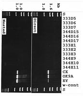
Asymptomatic (H) and symptomatic (D) (with quick decline syndrome) macadamia trees were sampled from two cultivars, cv. 333 and cv. 344 grown at Keaau and Kaiwiki on the island of Hawaii (Table 1). Direct and nested-polymerase chain reaction (PCR) assays using the two universal primer pairs, R16mF2/R16mR1 and R16F2n/R16R2, previously designed (Gundersen and Lee, 1996; Lee at al., 1993b) for general detection of phytoplasmas were employed to detect the presence of phytoplasmas in afftected leaf midribs. Total nucleic acid was extracted as previously described (Lee et al., 1993a) from apparently healthy (asymptomatic) or symptomatic macadamia leaves (midribs) collected from two cultivars on the island of Hawaii. Nucleic acid samples were diluted in sterile deionized water to give a final concentration of 20 ng/µl. Direct-PCR assays using primer pair R16mF2/R16mR1 were performed as previously described (Lee et al., 1995), with 20 ng of total nucleic acid, 200 µM each dNTP and 0.4 to 1.0 µM primer pair. Thirty-five PCR cycles were conducted in an automated thermocycler (Perkin-Elmer Cetus, Norwalk, CT). The following parameters were used: 1 min (2 min for the first cycle) denaturation step at 94°C, annealing for 2 min at 60°C (55°C for nested-PCR), and primer extension for 3 min (10 min in final cycle) at 72°C. Nucleic acid samples prepared from X-diseased phytoplasma (strain CX)- and aster yellows phytoplasma (strain OK9A)-infected periwinkle plants (Catharanthus roseus [L.] G. Don ) and elm

3Corresponding author. Tel: (808) 974-4105; Fax: (808) 974-4110.


Botanical Bulletin of Academia Sinica, Vol. 39, 1998

Table 1. Detection of phytoplasmas in leaf veinal tissues and of fungal pathogens on trunks of macadamia trees.

Plant species Conditions of Detection of phytoplasmab Detection of

plantsa Direct-PCR Nested-PCR fungal pathogensc

Macadamia

cv. 333 1 H _ _ _

2 H _ _ _

3 H _ _ _

4 H _ _ _

5 D _ _ N. r.

6 D _ _ N. r.

7 D _ _ N. r.

8 D _ _ N. r., X. a.

cv. 344 9 H _ _ _

10 H _ _ _

11 H _ _ _

12 H _ _ _

13 H _ _ _

14 H _ _ _

15 D _ _ X. a.

16 D _ _ X. a.

17 D _ _ X. a.

18 D _ _ N. r., X. a.

19 D _ _ N. r.

20 D _ _ X. a.

Periwinkled 1 XDP + +

2 AYP + +

Elmd EYP + +

aH, asymtomatic; d, with quick decline syndrome; XDP, infected with X-disease phytoplasma; AYP, infected with aster yellows phytoplasma; EYP, infected with elm yellows phytoplasma.

b_, phytoplasma not detected; +, phytoplasma detected. Direct-PCR was performed using a universal primer pair R16mF2/R16mR1; nested-PCR was performed using R16mF2/R16mR1, followed by using another universal primer pair R16F2n/R16R2.

c_, fungal pathogen not detected; N. r., Nectria rugulosa; X. a., Xylaria arbuscula.

dPhytoplasma-infected plants.

yellows phytoplasma (strain EY)-infected elm (Ulmus americana L.) were included as positive controls. Tubes with the reaction mixture devoid of DNA templates were included in each experiment as negative controls. In the nested-PCR assay (Lee et al., 1995), PCR products initially amplified using the universal primer pair R16mF2/R16mR1 were diluted (1/30) with sterile deionized water and used as template DNA for a subsequent series of 35 PCR cycles in which reaction mixtures contained the universal primer pair R16F2n/R16R2. PCR products were analyzed by electrophoresis through a 1% agarose gel followed by staining in ethidium bromide and visualization of DNA bands using a UV transilluminator.

The trunks of both declining and healthy macadamia trees tested were inspected for the presence of macroscopic fungal fruiting bodies. Pieces of bark and wood tissues were removed with a chisel from healthy and diseased trunks for comparison.

Results and Discussion

Neither direct-PCR nor nested-PCR assays detected the presence of phytoplasmas in any asymptomatic or symptomatic macadamia samples (Table 1, Figure 1). Both direct- and nested-PCR assays detected phytoplasmas in the

Figure 1. Direct and nested polymerase chain reaction (PCR) amplification of phytoplasma 16SDNA sequences from symptomless and symptomatic macadamia trees showing quick decline syndromes. Direct PCR amplification with the universal primer pair R16mF2/R1 was followed by R6F2n/R2. S=DNA fragment sizes standard; w-cont=water control; lane 2_12; representative samples (D. symptomatic and H, asymptomatic) of macadamia cv. 333 and cv. 344; X-disease phytoplasma strain CX and aster yellows phytoplasma strain OK9A are positive control samples from infected periwinkles; elm yellows phytoplasma strain EY is a positive control sample from an infected elm.


Lee and Ko — Macadamia quick decline

positive control samples. In contrast, fruiting bodies of Nectria rugulosa and Xylaria arbuscula were consistently found on trunks of symptomatic, but not asymptomatic, trees (Table 1). Bark tissues under reddish perithecia of N. rugulosa were dead and dry in appearance, while healthy bark tissues appeared moist and exuded reddish brown latex on the cut surface (Ko and Kunimoto, 1991a). Under the black club-shaped stromata of X. arbuscula, the bark tissues showed extensive decay and the wood tissues showed light to dark brown discoloration and black zone lines (Ko and Kunimoto, 1991b).

Phytoplasmas have been associated with decline symptoms in several shade and fruit trees, including stunting, brittle leaves, general chlorosis and off-season discoloration of foliage, premature defoliation and death of affected trees within one to several years (Ahrens et al., 1993; Sinclair et al., 1994). In nature, phytoplasma-induced diseases are transmitted and spread by insect vectors (McCoy et al., 1989). Generally, phytoplasma infection starts locally on above-ground branches and leaves and becomes systemic throughout the whole tree including the root system. Phytoplasmas are confined in phloem tissues in affected trees and induce systemic symptoms. Symptoms like zonal lesions or girdling of lower trunk consistently associated with macadamia quick decline have never been reported to be induced by phytoplasma infection. Macadamia quick decline has been suspected to be caused by phytoplasma infection because some symptoms induced on affected macadamia trees resemble those induced by phytoplasma infection in other tree crops. Inability to detect the presence of phytoplasmas with the direct-PCR and ultrasensitive nested-PCR assays in all symptomatic samples collected from the two most susceptible cultivars of macadamia in Hawaii, however, contradicts the previous reports (Borth et al., 1994a,b) in which a possible role of phytoplasmas in macadamia decline was suggested. Ultrasensitive nested-PCR assays using the two universal primer pairs have been shown to be able to detect the presence of phytoplasma in very low titers in several fruit crops which are normally undetectable using direct-PCR assays (Lee et al., 1995). Failure to confirm the presence of phytoplasmas in all the tested macadamia trees that showed decline syndrome raises a doubt that phytoplasmas actually play a role in the disease development. Phytoplasma association, if any, may be an occasional event unlikely to be directly correlated with quick decline of macadamia trees.

In contrast, fruiting bodies of fungal pathogens have been readily found on trunks of symptomatic, but not healthy, macadamia trees in the same fields. These fungi generally cause zonal lesions or girdling of the trunks, which are consistently observed in macadamia with the quick decline syndrome. Their pathogenicity has been appropriately demonstrated elsewhere (Ko and Kunimoto, 1991a,b; 1994; 1996). Little is known about the predisposing factors for the onset of the disease. However, the girdling of trunks by fungal pathogens was thought to be the major factor causing sudden death of affected macad

amia trees (Ko and Kunimoto, 1991a,b; 1994; 1996; 1997). Presumably, quick decline of trees could have resulted from a mixed infection, first by phytoplasma and followed by fungal pathogens. This does not seem to be the case for macadamia quick decline because of the failure to detect the presence of phytoplasmas in any of the symptomatic trees.

Acknowledgment. We thank Irene M. Bartoszyk for the excellent technical assistance.

Literature Cited

Ahrens, U., K.H. Lorenz, and E. Seemuller. 1993. Genetic diversity among mycoplasmalike organisms associated with stone fruit diseases. Mol. Plant-Microbe Interact. 6: 686_691.

Ann, P.J. and W.H. Ko. 1988. Root rot of macadamia caused by Ganoderma lucidum and Kretzschmaria clavus. J. Agric. Res. China 7: 424_429.

Borth, W.B., J.S. Hu, D. Ullman, and V. Jones. 1994a. PCR detection of mycoplasma-like organisms in Hawaii. Phytopathology 84: 1127. (Abstr.)

Borth, W.B., J.S. Hu, D. Ullman, and V. Jones. 1994b. Detection of MLOs from MQD-affected macadamia trees. In Proc. HMNA 34th Annu. Conf., Hawaii Macadamia Nut Association, Kona, Hawaii, pp. 72_73.

Gundersen, D.E. and I.M. Lee. 1996. Ultrasensitive detection of phytoplasmas by nested-PCR assays using two universal primer pairs. Phytopathol. Medditerr. 35: 144_151.

Ko, W.H. and R.K. Kunimoto. 1991a. Quick decline of macadamia trees: association with Nectria rugulosa. Plant Prot. Bull. (Taiwan) 33: 204_209

Ko, W.H. and R.K. Kunimoto. 1991b. Quick decline of macadamia trees: association with Xylaria arbuscula. Plant Pathol. 40: 643_644.

Ko, W.H. and R.K. Kunimoto. 1994. Quick decline of macadamia trees; association with Phytophthora capsici. J. Phytopathol. 141: 386_389

Ko, W.H. and R.K. Kunimoto. 1996. Quick decline of macadamia trees; association with Phellinus gilvus. Ann. Phytopathol. Soc. Japan 62: 37_39.

Ko, W.H. and R.K. Kunimoto. 1997. Acremonia recifei as one of the causal organisms of macadamia quick decline. Phytopathology 87: S53. (Abstr.)

Ko, W.H., R.K. Kunimoto, and I. Maedo. 1977. Root decay casused by Kretzschmaria clavus: its relation to macadamia decline. Phytopathology 67: 18_21.

Lee, I.M. and W.H. Ko. 1997. Lack of evidence that phytoplasmas are associated with quick decline of macadamia trees in Hawaii. Phytopathology 87: S56. (Abstr.).

Lee, I.M., A. Bertaccini, M. Vibio, and D.E. Gundersen. 1995. Detection of multiple phytoplasmas in perennial fruit trees with decline symptoms in Italy. Phytopathology 85: 728_735.

Lee, I.M., R.E. Davis, W.A. Sinclair, N.D. DeWitt, and M. Conti. 1993a. Genetic relatedness of mycoplasmalike organisms detected in Ulmus spp. in the United States and Italy by means of DNA probes and polymerase chain reactions.


Botanical Bulletin of Academia Sinica, Vol. 39, 1998

Phytopathology 83: 829_833.

Lee, I.M., R.W. Hammond, R.E. Davis, and D.E. Gundersen. 1993b. Universal amplification and analysis of pathogen 16SrDNA for classification and identification of mycoplasmalike organisms. Phytopathology 83: 834_842.

McCoy, R.E., A. Caudwell, C.J. Chang, T.A. Chen, L.N. Chiykowski, M.T. Cousin, G.T.N. Dale de Leeuw, D.A. Golino, K.J. Hackett, B.C. Kirkpatrick, R. Marwitz, H.

Petzold, R.H. Sinha, M. Sugiura, R.F. Whitcomb, I.L. Yang, B.M. Zhu, and E. Seemuller. 1989. Plant diseases associated with mycoplasmalike organisms. In R.F. Whitcomb and J.G. Tully (eds.), The Mycoplasmas. vol. 5. Academic Press, New York, pp. 545_640.

Sinclair, W.A., H.M. Griffiths, and I.M. Lee. 1994. Mycoplasmalike organisms as causes of slow growth and decline of trees and shrubs. J. Arboric. 20: 176_179.