Bot. Bull. Acad. Sin. (1998) 39: 269_277

Ho and Chen — Zygospore wall ontogeny in Rhizopus stolonifer

Ultrastructural study of wall ontogeny during zygosporogenesis in Rhizopus stolonifer (Mucoraceae), an amended model

Hsiao-Man Ho1,2,3 and Zuei-Ching Chen1

1Department of Botany, National Taiwan University, Taipei, Taiwan, Republic of China

(Received September 1, 1997; Accepted April 16, 1998)

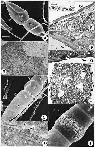
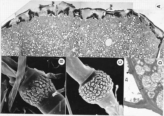
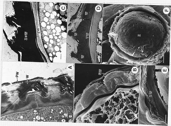
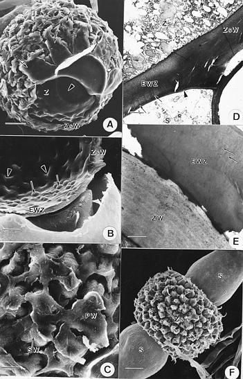
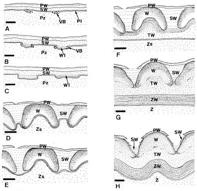
Abstract. The ultrastructural changes of the zygospore and zygosporangial walls of Rhizopus stolonifer during zygosporogenesis were studied by both scanning and transmission electron microscopy. The mature zygosporangial wall is composed of a primary, secondary, and tertiary layers. The mature zygospore wall is also multilayered. It was observed, in contrast to earlier reports, that the inner surface of the tertiary zygosporangial wall and zygospore wall are originally smooth and become warty at maturity. We suggest that this feature of wall development be added to the conventional model of zygosporogenesis in Rhizopus.

Keywords: Rhizopus stolonifer; Ultrastructure; Zygosporangium; Zygospore; Zygosporogenesis.

Introduction

Rhizopus stolonifer (Ehrenberg : Fries) Vuillemin is the type species of the genus Rhizopus, the so-called black bread mold. The existence of homothallic and heterothallic strains in the lower fungi was discovered in this fungus by Blakeslee in 1907. Since then, the structure of its sexual form has attracted the attention of many investigators. Previous studies include McCormick's (1912) and Wu's (1989) observations of the process of zygospore formation with light microscopy; Cutter's (1942) investigation of nuclear behavior during zygosporogenesis with light microscopy; Schipper's (1984) observation of zygospore surface structure using the scanning electron microscope; and Ho's (1988) scanning electron microscopic observation of changes in surface structure during zygosporogenesis. While internal changes during zygosporogenesis have been studied on the ultramicroscopic level in the homothallic species Rhizopus sexualis (Smith) Callen (Hawker and Gooday, 1967, 1968, 1969; Hawker and Beckett, 1971), no observations have been reported in any other species of Rhizopus. In this study, scanning and transmission electron microscopy were used to investigate the ultrastructural changes of the zygospore and zygosporangial walls of R. stolonifer.

Materials and Methods

Compatible strains of R. stolonifer were isolated in Taiwan and stored in the Culture Collection and Research

Center of FIRDI (Food Industry Research and Developing Institution) as CCRC 32449(+) and CCRC 32450(-).

Cultural Conditions

Zygosporic cultures of R. stolonifer were obtained by growing compatible strains on Difco potato dextrose agar medium in 5 cm petri dishes. Inocula were placed 1_2 cm apart. All cultures were grown at 22°C in constant darkness.

Transmission Electron Microscopy

All stages of zygospore ontogeny were selected under a dissecting microscope and fixed with 2% KMnO4 in distilled water for 30_40 min followed by a distilled water wash for 30 min. The material was dehydrated in a graded acetone series as follows: 30, 50, 70, 90%, 15 min at each step; 100% acetone for 15 min, followed by 1 h in fresh 100% acetone. The specimens were embedded in low viscosity epoxy resin (Spurr, 1969). A thin layer of resin (~1 mm) was polymerized in a mold at 70°C for 10_12 h. Selected stages were removed and individually mounted for sectioning using a glass or diamond knife on a Reichert Ultracut E microtome. Thin sections (~80_90 nm) were examined by means of a Hitachi H-600 or JEOL 1200EX-2 transmission electron microscope at 75 KV.

Scanning Electron Microscopy

All stages of zygospore ontogeny were selected under a dissecting microscope and fixed for 1 h with 2.5% glutaraldehyde and post-fixed for 1 h with 1% OsO4. The materials were washed and dehydrated in a graded acetone series. Specimens were dried in a critical point dryer, coated with gold, observed, and photographed with a Hitachi S-520 scanning electron microscope at 20 KV.

2Present address: Department of Natural Science Education, National Taipei Teachers College, No. 134, Sect. 2, Ho-Ping E. Rd., Taipei, Taiwan, Republic of China

3Corresponding author. Tel: (02) 2732-1104 ext. 321; Fax: (02) 2737-5419; E-mail: ho@tea.ntptc.edu.tw


Botanical Bulletin of Academia Sinica, Vol. 39, 1998

Cryofracturing

The techniques used for cryofracturing of the zygosporangia were similar to those of O'Donnell (1977). The observations were made with a Hitachi S-520 scanning electron microscope at 20 KV.

Results

When the vegetative hyphae of compatible strains of R. stolonifer approached each other, stout zygophores grew forth and fused at the apical regions to produce a fusion wall and two fused progametangia. Progametangia continued to grow and became carrot-shaped (Figure 1A). Subsequently, a transverse gametangial septum developed centripetally from the lateral wall and ultimately divided each progametangium into a gametangium and a suspensor cell (Figure 1B_C ). Plasmodesmata were formed at intervals along the septum with vesiculate bodies on either side. The gametangial septum was comprised of two electron-opaque layers and a central electron-transparent layer (Figure 1D). The septum continued to increase in thickness.

Upon formation of the gametangial septa, the fusion wall began to disintegrate. As a result of the rapid increase in volume in the equatorial region, the prozygosporangium became cylindrical. At the same time, its lateral wall developed a secondary wall beneath the primary wall. The onset of warting was recognized by the localized appearance of vesiculate bodies on the inner side of the secondary lateral wall. Then discrete, regularly-spaced, saucer-shaped blocks, the margins of which projected inward, developed. Vesiculate bodies frequently occurred along the margins of the blocks (Figure 1E). These blocks, designated as warts initials, were more noticeable soon after completion of the gametangial septa. They became electron-opaque and appeared allantoid in sections (Figure 1F_G). The fusion wall continued to degrade until only a peripheral fringe remained (Figure 1G_H). Expansion of the prozygosporangium caused the primary wall to rupture equatorially (Figure 1H_I).

As the zygosporangium became barrel-shaped, the secondary wall continued to thicken, and inverted, bowl-shaped warts formed in the inner side of the secondary wall. These events were more obvious in the equatorial region than in other areas. At this stage, the inner margin of the zygosporangial wall was irregular. The fusion wall was gone (Figure 2A). Owing to the continuous increase in volume, both primary and secondary lateral walls gradually ruptured near the equatorial region. Eventually, almost the entire outer surface was sloughed off (Figure 2A_C). Scanning electron micrographs showed that the remnants of the primary wall remained attached to the apices of the warts (Figure 2B_C). The original gametangial septa now formed the end walls of the zygosporangium. Secondary thickening of the end walls occurred on both zygosporangial and suspensor sides. Hemispherical protrusions were also found on the zygosporangial side of the end wall (Figure 2D).

After the warts matured, tertiary wall material was deposited along the interior margin of the secondary wall, filling in the cavities beneath the warts. The boundary between tertiary wall and zygosporangial cytoplasm appeared flat and smooth. In micrographs, the tertiary layer was more uniform and less electron-dense than the warts (Figure 3A), while the secondary wall was electron-transparent and fibrous. The zygosporangial wall now consisted of a primary wall, a secondary wall containing the warts, and a tertiary wall (Figure 3A_B). The slight waviness of the interior boundary of zygosporangial wall as seen in the micrograph (Figure 3B), might be an artifact resulting from desiccation during sample processing.

Additional layers of wall material were then laid down along the smooth inner surface of the tertiary wall, forming the zygospore wall. Micrographs of ultrathin sections and cryofractured zygospores showed a stratified zygospore wall composed of about 20 layers at maturation (Figure 3C_E). Meanwhile, the tertiary wall became more electron-opaque and hard to distinguish from the warts, as observed by transmission electron microscopy. As maturation progressed, the margins of the warts approached each other (Figure 3D).

The surface of the young zygospore was relatively smooth (Figure 3F) while the mature, globose zygospore surface had conical to hemispherical protuberances (Figure 4A), which corresponded to the hollow cavities of the warts (Figure 4A_B). In contrast, the two end walls of the zygospore remained flattened and smooth. The outermost primary wall had completely ruptured. Only the portion of the wall next to the suspensor remained attached, forming a short irregular skirt (Figure 4A). The fibrous material between the warts was the remnant of the ruptured secondary zygosporangial wall (Figure 4C).

Following its formation, the zygosporangial end wall became considerably thicker. Wall material was laid down more rapidly and deposition continued longer on the zygosporangial side rather than on the suspensor side. Ultrathin sections showed that the appearance of this end wall was uniform at first and became electron-opaque after thickening (Figure 4D). The electron-transparent central layer observed in the original gametangial septum and plasmodesmata were still observed in the end wall, even after zygospore wall formation (Figure 4D_E). Wall thickness was up to 4.7 µm at maturation. Occasionally, the inside of the end wall surface exhibited hemispherical protuberances (Figure 4B). Upon maturation, the zygosporangium became almost spherical with a warty surface. The two suspensors were usually equal (Figure 4F).

Discussion

Formation of the gametangial septum of R. stolonifer follows the general mucoraceous pattern in that it develops centripetally (Hawker and Beckett, 1971; O'Donnell et al., 1976). Multiple-perforated gametangial septa are found in other mucoraceous fungi, such as Gilbertella


Ho and Chen — Zygospore wall ontogeny in Rhizopus stolonifer

Figure 1. Scanning and transmission electron micrographs of R. stolonifer from progametangial to prozygosporangial stage. A: SEM, surface view of two carrot-shaped progametangia (Pg) fused at the fusing wall (FW). The collapsed left-sided progametangium might be a result of desiccation. bar=25 µm; B: TEM, longitudinal section through part of a progametangium during the stage of gametangial septum formation. Gametangial septum (arrowheads) tapering from the lateral wall toward the center. bar= 1 µm; C: SEM, surface view of two fused gametangia (G) separated from suspensors (S) by gametangial septa (arrowheads). bar=25 µm; D: TEM, longitudinal section through gametangium (G) and adjacent suspensor (S), showing newly formed gametangial septum (GS) with plasmodesma (P) and vesiculate body (VB). bar=200 nm; E_H: TEM, longitudinal sections of prozygosporangia during early stage of wart formation; E: Portion of a prozygosporangium (Pz) near gametangial septum (GS) showing an outer primary wall (PW) and an inner secondary wall (SW). Vesiculate bodies (arrows) appear on the inner side of secondary wall and at the margins of wart initial (arrowhead). bar=1 µm, inset bar=1 µm; F: The lateral prozygosporangial wall showing electron-opaque wart initial (WI) projecting inward. Also note the primary (PW) and secondary (SW) layers of prozygosporangial wall. bar=2 µm; G: A later stage of the prozygosporangial wall near remnant of fusing wall (FW). Note the discrete allantoid wart initials (arrowheads) on the secondary wall (SW). bar=2.9 µm; H: The prozygosporangium (Pz) showing ruptured primary lateral wall (PW), thickening secondary wall (SW) and wart initials (arrowheads). Fusion wall (FW) nearly dissolved. bar=7.7 µm; I: SEM, a doliform prozygosporangium (Pz) showing ruptured primary wall in equatorial region. bar= 15 µm.


Botanical Bulletin of Academia Sinica, Vol. 39, 1998

Figure 2. Scanning and transmission electron micrographs during early zygosporangial wall formation. A: TEM, longitudinal section of half of a zygosporangium (Zs) during wart (arrowheads) formation in secondary wall. Primary (PW) and secondary lateral walls (SW) rupture equatorially. bar=3.3 µm; B: SEM, surface view of developing zygosporangium (Zs), showing partially ruptured primary wall (arrowheads) attached to the apex of the warts. bar=15 µm; C: SEM, surface view of a barrel-shaped zygosporangium (Zs) at a later stage of development, the entire outer surface is nearly sloughed off. bar=25 µm; D: TEM, longitudinal section of zygosporangium (Zs) during wart formation. The end wall of zygosporangium (EWZ) showing secondary thickening along both zygosporangial (Zs) and suspensor (S) sides. Note the hemispherical protrusion (arrowhead). bar=1 µm.


Ho and Chen — Zygospore wall ontogeny in Rhizopus stolonifer

Figure 3. Scanning and transmission electron micrographs during zygosporangial and zygospore wall formation. A: TEM. longitudinal section of zygosporangium (Zs) during the formation of tertiary zygosporangial wall (TW). Zygosporangial wall comprises primary outer wall (PW), secondary wall (SW) containing electron-opaque warts (W), and an innermost tertiary wall (TW) layer that is smooth along the surface of the zygosporangial cytoplasm. bar=1 µm; B: SEM, cryofractured zygosporangium (Zs), at the same stage as in Figure 3A. Fractured zygosporangial wall showing wart (W) formation between tertiary (TW) and primary wall (PW). Note the torn fibrous secondary wall (SW). bar= 2 µm; C_D: TEM, longitudinal sections through parts of zygosporangia during zygospore wall development; C: A layered zygospore wall (ZW) developing along the smooth inner surface of the zygosporangial wall (ZsW). bar=1 µm; D: Part of the zygosporangium at later stage showing stratified zygospore wall (ZW), warts (W), tertiary wall (TW) and torn primary (PW) and secondary walls (SW). bar=5 µm; E: SEM, part of a fractured, multilayered, nearly mature zygospore wall (ZW). bar=2 µm; F: SEM, young, smooth-surfaced zygospore (Z) inside the fractured zygosporangial wall (ZsW). bar=21 µm.


Botanical Bulletin of Academia Sinica, Vol. 39, 1998

Figure 4. Scanning and transmission electron micrographs during the maturation of zygosporangial and zygospore walls. A_C: Scanning electron micrographs of nearly mature zygosporangium; A: Fractured zygosporangium with suspensor and part of the zygosporangial wall (ZsW) removed showing the warty zygospore (Z) inside. The truncated end wall of the zygospore (arrowhead) is smooth. Arrows indicate ruptured primary wall. bar=21 µm; B: Cryofractured zygosporangium showing the inner surface of the zygosporangial wall (ZsW). Note the protuberances on the lateral wall pointing outward (arrowheads) and several hemispherical protrusions (arrows) on the end wall of zygosporangium (EWZ) pointing inward. bar=10 µm; C: Surface view of part of a mature zygosporangium showing pieces of the primary wall (PW) on the apex of the zygosporangial wart. The amorphous material between warts is the remains of secondary wall (SW). bar=5 µm; D: TEM, longitudinal section of a developing zygosporangium (Zs) showing the thickening end wall of the zygosporangium (EWZ) continous with the lateral zygosporangial wall (ZsW). Note the plasmodesmata (arrows) penetrating the end wall and the central electron-transparent layer (arrowhead). bar=2 µm; E: TEM, longitudinal section of a mature zygosporangium showing the layered zygospore wall (ZW) and the adjacent zygosporangial end wall (EWZ). Note plasmodesmata (arrows) on the end wall. bar=1 µm; F: SEM, surface view of a mature zygosporangium (Zs) with two suspensors (S). bar=25 µm.


Ho and Chen — Zygospore wall ontogeny in Rhizopus stolonifer

persicaria (Eddy) Hesseltine (O'Donnell et al., 1977), Phycomyces blakesleeanus Burgeff (O'Donnell et al., 1976), R. sexualis (Hawker and Gooday, 1967; Hawker and Beckett, 1971) and Zygorhynchus heterogamus Vuillemin (Edelmann and Klomparens, 1995). O'Donnell et al. (1977) suggested that this is a common characteristic of mucoraceous fungi. Vesiculate bodies also occur along the developing gametangial septum and the young zygosporangial lateral wall and are associated with wart initials early in wart formation. In Rhizopus, similar structures occur along the developing columella wall in R. stolonifer (Ho and Chen, 1994) and along the fusion wall and zygosporangial wall of R. sexualis (Hawker and Beckett, 1971). In Z. heterogamus vesiculate bodies ap

pear throughout the entire zygosporangium and are later replaced by wart initials (Edelmann and Klomparens, 1995). Secondary thickening of the gametangial septum in Rhizopus is mainly on the gametangial side, but in Phycomyces, it is on the suspensor side (O'Donnell et al., 1976).

Ultrastructural studies on mucoraceous fungi showed that the mechanism underlying the formation of the zygosporangial lateral wall is more or less similar among its members. The most detailed observation was made on R. sexualis by Hawker and Beckett (1971), who proposed a model for wall ontogeny during zygosporogenesis in Rhizopus.

Figure 5. Diagrammatic illustration of wall development during zygosporogenesis in R. stolonifer. A: Primary and secondary walls, showing vesiculate bodies scattered along inner side of secondary wall and outside of the plasmalemma; B: Two wart initials developing on inner surface of secondary wall with vesiculate bodies occurring at the margins of wart initials; C: Wart initials appearing allantoid in section and more electron-opaque; D: Warts growing at the margins of wart initials and becoming inverted bowl-shaped; E: The electron-transparent secondary wall continuing to swell, the inner surface of zygosporangium is irregular; F: Tertiary wall developing along the inner side of warty secondary wall resulting in a smooth inner surface along the zygosporangial cytoplasm; G: A multilayered zygospore wall developing adjacent the smooth inner side of tertiary wall; H: The nearly mature, elastic tertiary zygosporangial and zygospore walls protrude outward conforming the inner surface of the warts. Primary and secondary walls are mostly ruptured at this stage. A_F, bar= 2 µm; G_H, bar=5 µm. Key to lettering: Pl: plasmalemma; PW: Primary wall; Pz: Prozygosporangium; SW: Secondary wall; TW: Tertiary wall; VB: Vesiculate bodies; W: Wart; WI: Wart initial; Z: Zygospore; Zs: Zygosporangium; ZW: Zygospore wall.


Botanical Bulletin of Academia Sinica, Vol. 39, 1998

Hawker and Beckett observed that the tertiary zygosporangial wall and the zygospore wall are laid down within the secondary wall, conforming to its irregular shape. Thus, both tertiary zygosporangial wall and zygospore wall are irregular from the beginning. In contrast, our study of over twenty zygosporogenic structures under SEM and TEM showed that in R. stolonifer, the inner surface of the tertiary zygosporangial wall and the newly formed zygospore wall are initially smooth (Figure 3A, C, D, F). The smooth zygospore wall becomes warty only upon maturation and conforms to the warty internal surface of the secondary zygosporangial wall (Figure 4A_B). Ho (1995) reported that the zygospore wall of R. sexualis is actually smooth at first and becomes warty upon maturation. Based on combined observations, we propose an amended model of zygosporogenesis of Rhizopus (Figure 5). In this model, we use the term "secondary wall" instead of Hawker and Beckett's "gelatinized inner primary wall."

The smooth-to-warty change of the zygospore wall (Figure 5G_H) indicates that the newly formed tertiary zygosporangial wall and zygospore wall are somewhat elastic. Over time, they distend to fill the cavities beneath the inverted, bowl-shaped warts, resulting in the warty outer surface of zygospore wall. The zygosporangial wall is permeable to fixatives, and the contents of the zygosporangium can be observed. As the zygospore wall is laid down and increases in thickness, it becomes impermeable to fixatives, and the contents of the zygospore are difficult to see. However, the multi-layering of the zygospore wall is clearly visible.

Under SEM, the zygospore wall of R. sexualis, like that of R. stolonifer, has been observed to consist of several thin layers (Hawker and Beckett, 1971). In Mortierella indohii Chien, the zygospore wall has a primary layer and a zonate secondary layer (Penelope and Young, 1988). There is no discernible stratification in the zygospore wall of P. blakesleeanus (O'Donnell et al., 1976) or P. nitens (Kunze) van Tieghen et Le Monnier (O'Donnell et al., 1978). In Z. heterogamus (Edelmann and Klomparens, 1995), the mature zygospore wall appears as three structurally distinct regions: an electron-opaque, outer warty layer, an intermediate laminate layer, and an inner electron-transparent layer. Because these investigators defined the whole structure within the primary wall of zygosporangium as the zygospore, the electron-opaque, outer warty layer is equal to the zygosporangial wall, and the intermediate laminate layer is the same as the zygospore wall of R. stolonifer and R. sexualis. However, an inner electron-transparent layer was not observed in R. stolonifer or R. sexualis. This indicates that the nature of the zygospore wall may vary among taxa.

While glutaraldehyde and osmium tetraoxide were used as fixatives for SEM, neither can penetrate the thick zygosporangial wall of R. stolonifer well, and each proved unsatisfactory for TEM. KMnO4 is known to induce artifacts and distortion in TEM, and certain cytoplasmic de

tails cannot be well fixed. However, it provided excellent membrane and wall preservation in this investigation. In fact, for the first time the multilayered character of the mature zygospore wall of Rhizopus was clearly observed with TEM. The observations under TEM correlate with those from SEM of internal surfaces exposed by cryofracture and previous reports on Rhizopus (Hawker and Beckett, 1971; Ho, 1988; Ho and Chen, 1994; Ho, 1995). Therefore, the authors believe the use of unbuffered KMnO4 can be justified and the amended model is supported.

Acknowledgements. We would like to thank Drs. G. L. Benny of the Department of Plant Pathology, University of Florida, and S. C. Jong, American Type Culture Collection, for reading the manuscript and making many helpful suggestions.

Literature Cited

Blakeslee, A.F. 1907. Heterothallism in bread mold, Rhizopus nigricans. Bot. Gaz. 43: 415_418.

Cutter, V.M.Jr. 1942. Nuclear behavior in the Mucorales-II. The Rhizopus, Phycomyces and Sporodinia patterns. Bull. Torrey Bot. Club 69: 592_616.

Edelmann, R.E. and K.L. Klomparens. 1995. Zygosporogenesis in Zygorhynchus heterogamus, with a proposal for standarization of structural nomenclature. Mycologia 87: 304_318.

Hawker, L.E. and A. Beckett. 1971. Fine structure and development of the zygospore of Rhizopus sexualis (Smith) Callen. Philos. Trans. Ser. B. 263: 71_100.

Hawker, L.E. and M.A. Gooday. 1967. Delimitation of the gametangia of Rhizopus sexualis (Smith) Callen: an electron microscope study of septum formation. J. Gen. Microbiol. 49: 371_376.

Hawker, L.E. and M.A. Gooday. 1968. Development of the zygospore wall in Rhizopus sexualis (Smith) Callen. J. Gen. Microbiol. 54: 13_20.

Hawker, L.E. and M.A. Gooday. 1969. Fusion, subsequent swelling and final dissolution of the apical walls of the progametangia of Rhizopus sexualis (Smith) Callen: an electron microscope study. New phytol. 68: 133_140.

Ho, H.M. 1988. The study of Rhizopus stolonifer isolated in Taiwan II. Morphological study of Rhizopus stolonifer: Asexual apparatus and zygosporognesis. Trans. Mycol. Soc. ROC 3: 73_81.

Ho, H.M. 1995. Zygospore wall in early stage of Rhizopus sexualis (Mucoraceae): smooth or warted? Fung. Sci. 10: 29_31.

Ho, H.M. and Z.C. Chen. 1994. Post cleavage transformation of columella in Rhizopus stolonifer (Mucoraceae). Bot. Bull. Acad. Sin. 35: 249_259.

McCormick, F.A. 1912. Development of the zygospore of Rhizopus nigricans. Bot. Gaz. 53: 67_68.

O'Donnell, K.L. 1977. Cryofracturing as a technique for the study of fungal structures in the scanning electron microscope. Mycologia 69: 309_320.

O'Donnell, K.L., G.R. Hooper, and W.G. Fields. 1976. Zygosporogenesis in Phycomyces blakesleeanus. Can. J. Bot. 45: 2573_2586.


Ho and Chen — Zygospore wall ontogeny in Rhizopus stolonifer

O'Donnell, K.L., J.J. Ellis, and C.W. Hesseltine. 1977. Zygosporogenesis in Gibertella persicaria. Can. J. Bot. 55: 662_675.

O'Donnell, K.L., S.L. Flegler, and G.R. Hooper. 1978. Zygosporangium and zygospore formation in Phycomyces nitens. Can. J. Bot. 56: 91_100.

Penelope, J.A. and T.W.K. Young. 1988. The zygospore of Mortierella indohii. Trans. Brit. Mycol. Soc. 91: 221_226.

Schipper, M.A.A. 1984. A revision of the genus Rhizopus I. The Rhizopus stolonifer-group and Rhizopus oryzae. Stud. Mycol. 25: 1_19.

Spurr, A.J. 1969. A low viscosity epoxy resin embedding medium for electron microscopy. J. Ultrastruct. Res. 26: 31_43.

Wu, M.L. 1989. Zygospore formation of Rhizopus stolonifer from Taiwan. Trans. Mycol. Soc. ROC 4: 59_64.