Bot. Bull. Acad. Sin. (1999) 40: 101_106

Huang et al. — Infection of pollen by Botrytis cinerea

Infection of alfalfa pollen by Botrytis cinerea

H.C. Huang1, E.G. Kokko, and R.S. Erickson

Research Centre, Agriculture and Agri-Food Canada, P. O. Box 3000, Lethbridge, Alberta, Canada T1J 4B1

(Received May 6, 1998; Accepted July 24, 1998)

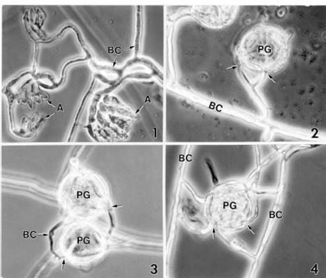
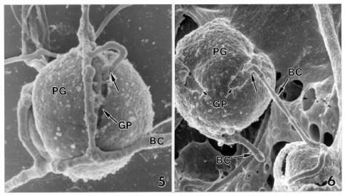
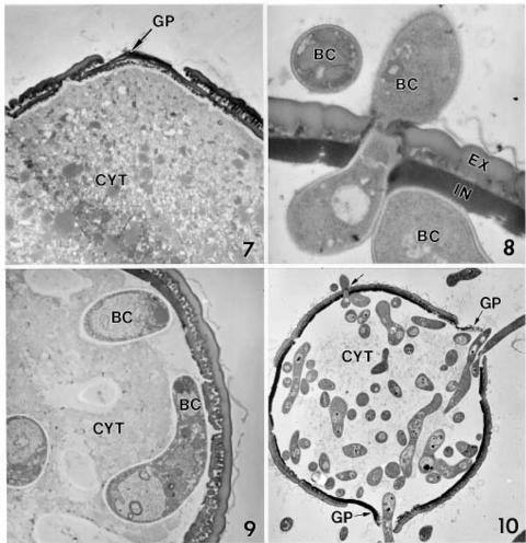
Abstract. A microscopic study proved that pollen grains of alfalfa are susceptible to infection by Botrytis cinerea, an important pathogen for blossom blight of alfalfa in western Canada. Pollen grains were inoculated by mixing pollen with drops of a B. cinerea spore suspension on glass slides. Infection occurred by direct hyphal penetration of the pollen cell walls without the formation of appressoria. Infection through the germ pores was observed more frequently than through other parts of the pollen walls. Plasmolysis of the plasma membrane and destruction of the cytoplasm occurred as a result of intensive growth of hyphae in the cell lumen of infected pollen grains. The impact of pollen infection by B. cinerea on the epidemiology of blossom blight of alfalfa is discussed.

Keywords: Alfalfa; Blossom blight; Botrytis cinerea; Epidemiology; Infection of pollen.

Introduction

Botrytis cinerea Pers.:Fr. is an important pathogen causing blossom blight, pod rots, and fruit rots on many crops, including alfalfa (Medicago sativa L.) (Gossen et al., 1996; 1997). Numerous reports indicate that high disease incidence and severity are associated with the colonization of senescent petals by B. cinerea (Bachelder and Orton, 1962; Hartill and Campbell, 1974; Hunter et al., 1972; Jarvis and Borecka, 1968; Kunimoto et al., 1996; Ogawa and English, 1960). Some reports indicate that pollen stimulates spore germination (Hartill, 1975) and infection by B. cinerea (Bachelder and Orton, 1962; Chou and Preece, 1968; Ogawa and English, 1960).

Recent studies have shown that alfalfa pollen is susceptible to infection by plant pathogens such as Verticillium albo-atrum Reinke & Berthold (Huang and Kokko, 1985) and Sclerotinia sclerotiorum (Lib.) de Bary (Huang et al., 1997). The objective of this study was to determine the susceptibility of alfalfa pollen to B. cinerea, and to describe the mode of infection using light microscopy (LM) and electron microscopy.

Materials and Methods

Inoculation of Alfalfa Pollen

Seeds of alfalfa, cultivar Barrier, were sown in Cornell peat-lite mix (Boodley and Sheldrake, 1977) in plastic pots (15 cm diam.), and plants were grown to maturity in a greenhouse. Alfalfa flowers were excised and tripped to collect fresh pollen grains on glass slides. An aqueous spore suspension containing approximately 103 spores/ml

of B. cinerea was made from 3-week-old cultures grown on potato dextrose agar in petri dishes. Pollen grains were inoculated with B. cinerea by mixing the pollen with five drops of the suspension on each slide. The slides were placed in a moist chamber and kept for five days at room temperature (20_22°C) before being prepared for microscopic examinations. Two controls, pollen grains in sterile water and B. cinerea spores in sterile water, were prepared and processed for microscopic examinations.

Light Microscopy

For light microscopy, each 5-day-old sample (slide) of pollen-spore mixture was mounted in lactophenol and covered with a cover slip. The mounted specimens were examined for infection of pollen by B. cinerea using a Zeiss Photomicroscope III. Control samples were also mounted and examined microscopically. The important features observed in the specimens were documented by phase contrast photomicrographs.

Scanning Electron Microscopy (SEM)

For SEM, the 5-day-old inoculated (B. cinerea) and uninoculated (control) pollen samples were immersed in 2% glutaraldehyde fixative in 0.05 M sodium phosphate buffer, pH 7.0, at 4°C overnight (16 h) and then brought to room temperature. The samples were washed (3 × 10 min) with the sodium phosphate buffer solution. The specimens were dehydrated in a graded series of ethanol and critical point dried (Polaron E3100) with liquid carbon dioxide as the transitional fluid. The material was adhered onto aluminum specimen mounts with colloidal silver paste, air-dried overnight and sputter-coated (Denton Vacuum Desk-1) with gold (approximately 15 nm thickness). The specimens were examined and photographed on a Hitachi S-570 SEM.

1Corresponding author. Fax: (403) 382-3156; E-mail: huangh@ em.agr.ca


Botanical Bulletin of Academia Sinica, Vol. 40, 1999

Transmission Electron Microscopy (TEM)

For TEM, the 5-day-old inoculated (B. cinerea) and uninoculated (control) pollen samples were immersed in 2% glutaraldehyde in 0.05 M sodium cacodylate buffer, pH 7.0, at 4°C overnight (16 h) and then brought to room temperature. The samples were washed (3 × 10 min) with the sodium phosphate buffer solution. The material was post-fixed in 2% osmium tetroxide, in the same buffer for 2 h, stained in 5% uranyl acetate in 50% ethanol (1 h), dehydrated in a graded series of ethanol, infiltrated with Spurr's low-viscosity embedding medium (Spurr, 1969) and polymerized 8 h at 70°C. Serial sections were cut (Kokko et al., 1990) with a diamond knife using a Reichert OM-U3 ultramicrotome. Sections were mounted on slotted, formvar-coated grids, stained with 5% aqueous lead citrate and 5% uranyl acetate, and examined with a Hitachi H-7100 transmission electron microscope at 75 Kv.

Results

Examination of 5-day-old samples revealed that the germination rate of conidia of B. cinerea was high (90%) in the pollen-spore mixture but was low (20%) in the control. The germ tubes from germinated spores in the controls remained short. In contrast, the germ tubes in the pollen-spore mixture grew rapidly and developed into long, multi-branched hyphae. Numerous multicellular, lobate appressoria, which developed into infection cushions, were formed in the glass slide preparations (Figure 1). However, infection of a pollen grain was always achieved by direct hyphal penetration (Figures 2_5) without the involvement of appressoria. Penetration of pollen grains occurred most frequently by a side branch of a hypha (Figures 2, 4, 5). Hyphal penetration occurred more readily through the germ pores (Figures 5, 10) than through other parts of the cell wall (Figures 8, 10). Hyphae in the in

Figures 1_4. Photomicrographs showing formation of appressoria of Botrytis cinerea (Figure 1) and infection of alfalfa pollen by the pathogen (Figures 2_4). Note multicellular, lobate appressoria (A), which developed into infection cushions (Figure 1). Note also the infection of pollen grains (PG) by direct hyphal penetration of B. cinerea (BC) without the formation of appressoria (arrows). Figure 1, ×520; Figure 2, ×1,000; Figure 3, ×1,000; Figure 4, ×1,000.


Huang et al. — Infection of pollen by Botrytis cinerea

Figures 5 and 6. Scanning electron micrographs showing hyphae of Botrytis cinerea (BC) penetrating the alfalfa pollen (PG) (Figure 5, arrow) or emerging from the infected pollen (PG) (Figure 6, arrow) through a germ pore (GP). Figure 5, ×2,800; Figure 6, ×2,500.

is likely achieved by chemical force as evidenced by cell wall dissolution (Figure 8) as well as by physical force as evidenced by the cone-shaped wall rupture (Figure 10) at the site of infection. In studying the infection of pollen grains of Monterey pine (Pinus radiata D. Don) by Retiarius superficiaris Olivier, Olivier (1978) reported that a swelling of the hyphal tip before penetration of the pollen walls is sometimes noticeable but no true appressorium is formed. Olivier and Williams (1975) observed a slight depression of the cell walls at the infection site, and concluded that penetration of pine pollen by Retiarius spp. is achieved by mechanical force.

The ultrastructural evidence in this study indicates that the colpate regions of alfalfa pollen grains are the most common sites of infection by B. cinerea. The weak resistance of the colpate regions of alfalfa pollen to invasion by V. albo-atrum (Huang and Kokko, 1985), S. sclerotiorum (Huang et al., 1997), and B. cinerea may be due to thin cell walls at the furrows and germ pores. Olivier (1978) observed infection of pine pollen by Retiarius spp. occurred most frequently at the germ pore region. Knox and Heslop-Harrison (1970) found that intine- bound protein is rich in the cell walls of the germ pore region. Therefore, the susceptibility of alfalfa pollen germ pores to invasion by pathogenic fungi may be due to the thin, but protein-rich, cell walls at the colpate regions.

Previous reports indicate that pollen stimulates spore germination and enhances lesion development by B. cinerea (Bachelder and Orton, 1962; Bekesi, 1979; 1982;

fected pollen grain re-emerged through the germ pores (Figure 6) or other parts of the cell wall. A positive chemotropism was evident as the lateral branches often grew toward pollen grains (Figures 2, 4, 5).

Each alfalfa pollen grain has a tricolpate structure with three long furrows that are equidistant from each other (Figure 6). The ultrastructure of a healthy pollen grain showed dense cytoplasm with numerous small vacuoles (Figure 7). The cytoplasm was surrounded by the thin plasma membrane and two well-defined cell wall layers, exine and intine (Figure 7). The cell wall of the germ pore region was thinner than the cell wall of the non-germ pore region (Figure 7). Both cell wall etching (Figure 8) and the formation of cone-shaped cell wall openings (Figure 10) were observed at infection sites. Plasmolysis of the plasma membrane occurred as the result of fungal invasion of the pollen grain (Figure 9). The intensive growth and multiplication of invading hyphae caused the eventual destruction and disintegration of the pollen cytoplasm (Figure 10).

Discussion

The present study together with previous studies, reveals that alfalfa pollen is susceptible to infection by plant pathogens such as V. albo-atrum (Huang and Kokko, 1985), S. sclerotiorum (Huang et al., 1997), and B. cinerea. The mode of infection for all of these pathogens is by direct hyphal penetration of the pollen cell wall without the formation of appressoria. Penetration of the cell wall


Botanical Bulletin of Academia Sinica, Vol. 40, 1999

Figures 7_10. Transmission electron micrographs of healthy (Figure 7) and Botrytis cinerea-infected (Figures 8_10) pollen grains of alfalfa. Figure 7. A healthy pollen grain showing dense cytoplasm (CYT). Note the thin wall at the germ pore (GP) region and the thick wall outside the germ pore region. Figure 8. A hypha of Botrytis cinerea penetrating through the cell wall outside the germ pore region. Note etching of the exine (EX) and intine (IN) layers of the pollen grain wall at the penetration site. Figure 9. An infected pollen grain showing the presence of hyphae of B. cinerea (BC) inside the pollen grain and plasmolysis of the pollen cytoplasm (CYT). Figure 10. An infected pollen grain showing intense hyphal ramification and disintegration of the pollen cytoplasm (CYT). Note hyphae penetrating through the cell wall at the germ pore (GP) and outside the germ pore (arrow) regions. Figure 7, ×5,400; Figure 8, ×18,000; Figure 9, ×7,000; Figure 10, ×3,000.

Chou and Preece, 1968; Hartill, 1975; Hartill and Campbell, 1974; Ogawa and English, 1960) and S. sclerotiorum (Hartill, 1975; Dillard and Hunter, 1986). The dense growth of mycelia of fungal pathogens in infected pollen grains observed in the present (Figure 10) and previous studies (Huang and Kokko, 1985; Huang et

al., 1997) suggests that the cytoplasm of alfalfa pollen is a good growth substrate for plant pathogens. Numerous reports indicate that pollen releases nutrients—including sugars, amino acids, and proteins—when it is moistened (Fokkema, 1971; Stanley and Search, 1971; Yamakawa, 1984). Thus, diffusates from


Huang et al. — Infection of pollen by Botrytis cinerea

alfalfa pollen may be responsible for the positive chemotropism of hyphal growth of B. cinerea (Figure 4) observed in the present study.

Pollinating insects such as honeybees (Apis mellifera L.) (Stelfox et al., 1978), alfalfa leafcutter bees Megachile rotundata (Fabricius) (Huang et al., 1986), and fig wasps (Blastophaga psenes L.) (Michailides and Morgan, 1994) are important vectors for plant pathogenic fungi. Huang et al. (1986) observed V. albo-atrum-infected pollen grains were carried by leafcutter bees foraging in a field, the alfalfa plants of which suffered a high incidence of verticillium wilt. Stelfox et al. (1978) reported that the transmission of head blight of rapeseed (Brassica spp.) was attributed to the spread of S. sclerotiorum-infested pollen grains by honeybees. Alfalfa is a cross-pollinated crop which relies upon the use of leafcutter bees for commercial seed production (Goplen et al., 1980). Since both B. cinerea and S. sclerotiorum are important pathogens causing blossom blight of alfalfa (Gossen et al., 1996; 1997), the infection of pollen grains by B. cinerea observed in this study and by S. sclerotiorum observed in a previous study (Huang et al., 1997) suggests that pathogen-contaminated pollen grains may play a significant role in the epidemiology of blossom blight and thus have a negative impact on the alfalfa seed and leafcutter bee industries.

Acknowledgement. The authors thank B. Lee for technical assistance in electron microscopy. This is Lethbridge Research Centre Contribution No. 3879814.

Literature Cited

Bachelder, S. and E.R. Orton. 1962. Botrytis inflorescence blight on American holly in New Jersey. Plant Dis. Reptr. 46: 320.

Bekesi, P. 1979. The effect of the pollen of some weed species on germination of conidia of Botrytis cinerea. Acta Phytopathol. Acad. Scientiarum Hungaricae 14: 379_382.

Bekesi, P. 1982. New inoculation method for infecting sunflowers by Botrytis cinerea Pers. Acta Phytopathol. Acad. Scientiarum Hungaricae 17: 221_224.

Boodley, J.W. and R. Sheldrake, Jr. 1977. Cornell Peat-Lite Mixes for Commercial Plant Growing. N.Y. State Coll. Agric. Life Sci., Info. Bull. 43, 8 pp.

Chou, M.C. and T.F. Preece. 1968. The effect of pollen grains on infections caused by Botrytis cinerea Fr. Ann. Appl. Biol. 62: 11_22.

Dillard, H.R. and J.E. Hunter. 1986. Association of common ragweed with Sclerotinia rot of cabbage in New York State. Plant Dis. 70: 26_28.

Fokkema, N.J. 1971. The effect of pollen in the phyllosphere of rye on colonization by saprophytic fungi and on infection by Helminthosporium sativum and other leaf pathogens. Netherl. J. Plant Pathol. 77 Supplement No. 1, 60 pp.

Goplen, B.P., H. Baenzier, L.D. Bailey, A.T.H. Gross, M.R. Hanna, R. Michaud, K.W. Richards, and J. Waddington. 1980. Growing and managing alfalfa in Canada. Agr. Can. Publication # 1705. Agr. Can., Ottawa, Canada, 49 pp.

Gossen, B.D., L.M. Harrison, J. Holley, and S.R. Smith. 1996. Survey of blossom blight of alfalfa on the Canadian Prairies in 1995. Can. Plant Dis. Surv. 76: 123_125.

Gossen, B.D., Z. Lan, L.M. Harrison, J. Holley, and S.R. Smith. 1997. Survey of blossom blight of alfalfa on the Canadian Prairies in 1996. Can. Plant Dis. Surv. 77: 88_89.

Hartill, W.F.T. 1975. Germination of Botrytis and Sclerotinia spores in the presence of pollen on tobacco leaves. N.Z. J. Agric. Res. 18: 405_407.

Hartill, W.F.T. and J.M. Campbell. 1974. Effects of flower removal on the development of the Sclerotinia/Botrytis complex of tobacco. N.Z.J. Agric. Res. 17: 147_152.

Huang, H.C. and E.G. Kokko. 1985. Infection of alfalfa pollen by Verticillium albo-atrum. Phytopathology 75: 859_865.

Huang, H.C., E.G. Kokko, and R.S. Erickson. 1997. Infection of alfalfa pollen by Sclerotinia sclerotiorum. Phytoparasitica 25: 17_24.

Huang, H.C., K.W. Richards, and E.G. Kokko. 1986. The role of the leafcutter bee in dissemination of Verticillium albo-atrum in alfalfa. Phytopathology 76: 75_79.

Hunter, J.E., K.G. Rohrbach, and R.K. Kunimoto. 1972. Epidemiology of Botrytis blight on macadamia racemes. Phytopathology 62: 316_319.

Jarvis, W.R. and H. Borecka. 1968. The susceptibility of strawberry flowers to infection by Botrytis cinerea. Hortic. Res. 8: 147_154.

Knox, R.B. and J. Heslop-Harrison. 1970. Pollen-wall proteins: localization and enzymic activity. J. Cell Sci. 6: 1_15.

Kokko, E.G., B. Gowen, and C.R. Jahnke. 1990. An inexpensive water volume control device for ultramicrotomy. In Electron Microscopy 1990; Proc. 12th Int. Congr. for Electron Microscopy. San Francisco Press, USA, pp. 732_733.

Kunimoto, R.K., B.C. Bushe, and W.H. Ko. 1996. Blossom blight of kalanchoe caused by Botrytis cinerea. Plant Pathol. Bull. 5: 107_111.

Michailides, T.J. and D.P. Morgan. 1994. Dynamics of Blastophaga psenes populations, availability of caprifigs, and fig endosepsis caused by Fusarium moniliforme. Phytopathology 84: 1254_1263.

Ogawa, J.M. and H. English. 1960. Blossom blight and green fruit rot of almond, apricot and plum caused by Botrytis cinerea. Plant Dis. Reptr. 44: 265_268.

Olivier, D.L. 1978. Retiarius gen. nov.: Phyllosphere fungi which capture wind-borne pollen grains. Trans. Brit. Mycol. Soc. 71: 193_201.

Olivier, D.L. and E.D.F. Williams. 1975. The penetration of Pinus radiata pollen by a parasitic fungus. Proc. Electr. Microsco. Soc. S. Africa. 5: 37_38.

Spurr, A.R. 1969. A low-viscosity epoxy embedding medium for electron microscopy. J. Ultrastruct. Res. 26: 31_43.

Stanley, R.G. and R.W. Search. 1971. Pollen protein diffusates. In J. Heslop-Harrison (ed.), Pollen: Development and Physiology, Appleton Century Crofts, Meredith Corporation, New York, pp. 174_176.

Stelfox, D., J.R. Williams, U. Soehngen, and R.C. Topping. 1978. Transport of Sclerotinia sclerotiorum ascospores by rapeseed pollen in Alberta. Plant Dis. Rep. 62: 576_579.

Yamakawa, T. 1984. The effect of pollen on the infection of fruit vegetables with conidia of Botrytis cinerea. Proc. Kansas Plant Prot. Soc. 26: 1_8.