Bot. Bull. Acad. Sin. (1999) 40: 127-134

Kuo et al. — Arginine residue in V-ATPase

An essential arginine residue in vacuolar H+-ATPase purified from etiolated mung bean seedlings

Soong Yu Kuo1, Chi Meng Tzeng1, Wen Jye Lin2, Shih Sheng Jiang1, Shu Hsien Hung1, and

Rong Long Pan1,3

1Institute of Radiation Biology, Department of Life Science, College of Life Science, National Tsing Hua University, Hsin Chu, 300 Taiwan, Republic of China

2Department of Radiology, Yuanpei Institute of Medical Technology, Hsin Chu, 300 Taiwan, Republic of China

(Received November 6, 1997; Accepted August 10, 1998)

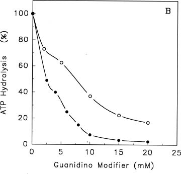
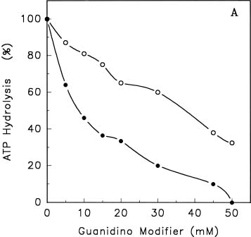
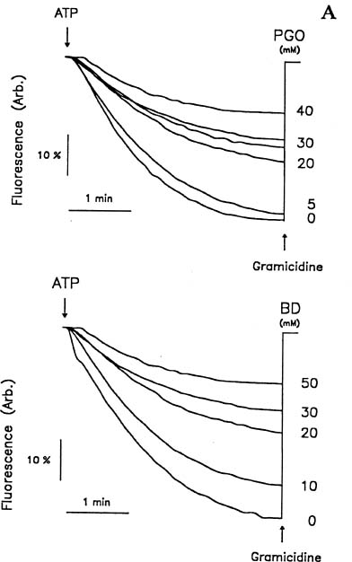
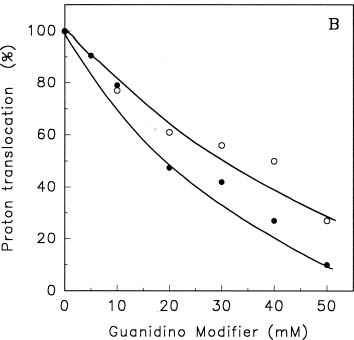
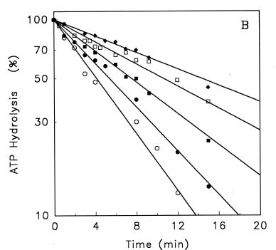
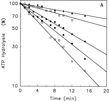
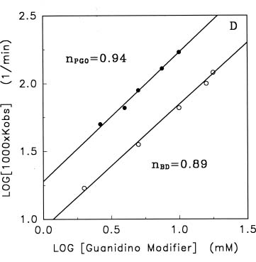
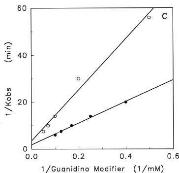
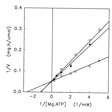
Abstract. Treatments of the tonoplast ATPase purified from mung bean seedlings (Vigna radiata L.) with guanidino modifiers, phenylglyoxal and 2,3-butanedione, caused a marked loss of the ATP hydrolysis activity and proton translocation in a concentration-dependent manner. Kinetic analysis yielded first order rate constants, k2, of 0.416 and 0.227 s-1 and steady-state dissociation constants, Ki, of 19.3 and 24.2 mM for phenylglyoxal- and butanedione-inhibition of vacuolar H+-ATPase, respectively. The reaction order of phenylglyoxal- and butanedione-inhibition was calculated to be 0.94 and 0.89, respectively, suggesting that at least one arginine residue of vacuolar H+-ATPase was modified by both reagents. Lineweaver-Burk plots showed that the mode of inhibition of vacuolar H+-ATPase by both modifiers is competitive. Mg-ATP, the physiological substrate of vacuolar H+-ATPase, but not its analogs, exerted preferentially partial protection against phenylglyoxal and butanedione, indicating that the arginine residue involved in the inhibition of enzymatic activity may be located at or near the active site and directly participate in the binding of the substrate.

Keywords: Chemical modification; H+-ATPase; Mung bean; Proton translocation; Tonoplast.

Abbreviations: BD, 2,3-butanedione; BZ-ATP, 3-O-(4-benzoyl) benzoyladenosine 5'-triphosphate; DCCD, N,N'-dicyclohexyl carbodiimide; DIDS, 4,4'-diisothiocyanostilbene-2,2'disulfonate; NBD-Cl, 7-chloro-4-nitrobenzo-2-oxa-1,3-diazole; NEM, N-ethylmaleimide; PGO, phenylglyoxal.

Introduction

Tonoplast H+-ATPase of higher plants plays a vital role in the secondary active transport of sugars, ions, and metabolites (Sze, 1985; Sze et al., 1992). Tonoplast H+-ATPase is discretely stimulated by Cl- and inhibited by NO3-, DIDS2, NEM, NBD-Cl, and DCCD (Sze, 1985; Sze et al., 1992). Molecular mass of tonoplast H+-ATPase from higher plants was estimated to be 400_600 kDa (Parry et al., 1989; Matsuura-Endo et al., 1990). Vacuolar H+-ATPase contains seven to nine polypeptides upon SDS-PAGE with two major bands of molecular masses of 65_80 kDa (A subunit) and 55_65 kDa (B subunit), and a 16-kDa protein (DCCD-binding proteolipid). The A subunit of vacuolar H+-ATPase is implicated as the catalytic site (Randall and Sze, 1987; Bowman et al., 1986), while the B subunit probably plays a regulatory role (Bowman et al., 1986; Manolson et al., 1985). The DCCD-binding proteolipid subunit might be involved in the proton translocation and is preferentially labeled by radioactive DCCD (Mandala and Taiz, 1986; Manolson et al., 1985; Randall and Sze, 1987).

The cDNA sequences and consequently their deduced amino acid sequences of A subunit from tonoplast H+-ATPases of carrot roots (Zimniak et al., 1988), mung bean (Chiu et al, 1995), cotton (Wilkins, 1993), and Neurospora crassa (Bowman et al., 1988) were recently obtained. Several essential amino acid residues involved in the catalytic activity of vacuolar H+-ATPases from coated vesicles and yeasts were investigated by substrate analogs (Vasilyeva and Forgac, 1996; Zhang et al., 1995; Feng and Forgac, 1994; Manolson et al., 1985; Mandala and Taiz, 1986) and site-directed mutagenesis (Liu et al., 1997; Liu et al., 1996). For instance, the sensitivities of vacuolar H+-ATPase to NEM and NBD-Cl imply the possible presence of cysteine and/or tyrosine residues at the active site (Manolson et al., 1985; Mandala and Taiz, 1986). Further mutation analysis reveals the roles of glycine and glutamate residues in the stability and the catalytic activity of vacuolar H+-ATPase (Liu et al., 1997). Our recent work also indicates, using the substrate analog dialdehyde derivative of ATP, the A subunit of vacuolar H+-ATPase contains a lysine residue essential to the enzymatic activity (Chow et al., 1992; Tzeng et al., 1992). In spite of these results, further efforts are required to identify other amino acid residues significant to the activity of vacuolar H+-ATPase and to elucidate their roles in the enzymatic mechanism.

3Corresponding author. Tel/Fax: (886)-(3)-5742688; E-mail: rlpan@RB.nthu.edu.tw


Botanical Bulletin of Academia Sinica, Vol. 40, 1999

Arginine residues were shown to participate in catalytic activities, substrate binding, and maintenance of enzyme conformation of F- and P-type H+-ATPases (Vallejo, 1973; Kasamo, 1988). However, no evidence directly reveals whether arginine residues are also involved in the catalytic activity of purified V-type H+-ATPase. Phenylgloxal (PGO) and 2,3-butanedione (BD) have been widely used to identify arginine residues and determine their roles in enzymatic activity of various enzymes (Riordan, 1973; Kuo and Pan, 1990). In this study, we demonstrated the presence of an arginine residue may be involved in the inhibition of ATP hydrolysis and H+-translocation by PGO and BD. Kinetic analysis indicates that modification of one mol arginine/mol ATPase is sufficient to inhibit the enzymatic activity of vacuolar H+-ATPase.

Materials and Methods

Plant Materials

After soaking in tapwater for 24 h, seeds of Vigna radiata L. (mung bean) were germinated at room temperature in the dark. Hypocotyls of 4-day old etiolated seedlings were excised, chilled, and then used as starting materials.

Isolation of Tonoplast Vesicles

Tonplast vesicles were prepared by a floating method of Matsuura-Endo et al. (1990) with minor modifications. Chilled hypocotyls were ground with a Waring blender in a homogenization medium containing 50 mM Mops/KOH (pH 7.6), 0.25 M sorbitol, 5 mM EGTA, 1 mM phenylmethylsulfonyl fluoride, 2.5 mM potassium metabisulfite, and 1.5% (w/v) polyvinylpyrolidone. The homogenate was filtered through four layers of cheesecloth and subjected to differential centrifugation at 3,600 g for 10 min and 150,000 g for 20 min. The pellet was suspended in a suspension solution containing 10 mM potassium phosphate (pH 7.8), 0.3 M sucrose, 1 mM EGTA, and 2 mM dithiothreitol. The suspension was overlaid in a medium containing 5 mM Mops/KOH (pH 7.3), 0.25 M sorbitol, 1 mM EGTA, and 2 mM dithiothreitol. After centrifugation at 120,000 g for 40 min, the interface portion was collected and diluted with 5 mM Mops/KOH (pH 7.3), 0.25 M sorbitol, 1 mM EGTA, and 2 mM dithiothreitol. The suspension was then centrifuged at 150,000 g for 20 min, and the resulting white pellet (vacuolar-membrane vesicles) was suspended in a medium containing 10 mM Tris/Mes (pH 7.5), 0.25 M sorbitol, 2 mM dithiothreitol, and the protein concentration of 3 mg/ml.

Purification of Vacuolar H+-ATPase

Vacuolar H+-ATPase was purified from resealed tonoplast vesicles according to the method of Matsuura-Endo et al. (1990) with minor modifications. Triton X-100 and NaCl were added dropwise to the membrane fraction to final concentrations of 5% (w/v) and 0.1 M, respectively.

The suspension was stirred at 4°C for 30 min and centrifuged at 150,000 g for 30 min. The pellet was suspended in Buffer A [20 mM Tris/acetate (pH 7.5), 1 mM EGTA, 1 mM dithiothreitol, 2 mM MgCl2, and 20% (v/v) glycerol]. Lysophosphatidylcholine was added to the suspension at a final concentration of 4 mg/ml. The mixture was stirred for 30 min at 25°C and centrifuged at 150,000 g for 40 min. The supernatant was applied onto a Mono Q HR5/5 anion-exchanger FPLC column (0.5 × 5 cm) pre-equilibrated with running buffer [Buffer A + 0.1% (w/v) Triton X-100] at room temperature. The sample was eluted at a flow rate of 0.5 ml/min with NaCl gradient (40 ml) from 0 to 0.5 M. Fractions (1 ml) were collected and aliquots (20 µl) were withdrawn for enzyme assay. Fractions with highest ATPase activity were pooled and stored at -70°C for further studies.

Enzyme Assay and Protein Determination

Activities of tonoplast H+-ATPase were measured in a 0.5 ml reaction solution. The mixture contained 30 mM Tris-HCl (pH 7), 3 mM MgSO4, 50 mM KCl, 3 mM ATP, 0.5 mM sodium azide, 0.1 mM sodium vanadate, 0.1 mM ammonium molybdate, 40 µg phosphatidylcholine, and 12_20 µg of protein. After 30 min incubation at 37°C, the reaction was stopped by adding a solution containing 1.7% (w/v) ammonium molybdate, 2% (w/v) SDS, and 0.02% (w/v) ANSA (1-amino-2-naphthol-4-sulfonic acid) at room temperature. The released Pi was measured spectrophotometrically (Wang et al., 1989).

Protein concentration was determined according to a modified Lowry method using bovine serum albumin as the standard (Larson et al., 1986).

Measurement of Proton Translocation

Proton translocation was measured as described previously using fluorescence quenching of Acridine Orange (Wang et al., 1989). The reaction mixture contained 5 mM Tris-HCl (pH 7), 250 mM sorbitol, 3 mM ATP, 3 mM MgSO4, 50 mM KCl, 0.5 mM NaN3, 0.1 mM Na-vanadate, 0.1 mM ammonium molybdate, 5 µM Acridine Orange, and 10_20 µg/ml membrane protein. The fluorescence quenching was initiated by adding 3 mM MgSO4. The ionophore gramicidin (2 µg/ml) was added at the end of each assay.

Modification of ATPase with Modifiers

Vacuolar H+-ATPase was modified with various concentrations of PGO and BD in 50 mM Mops-KOH (pH 7) for time periods indicated at room temperature. At the end of incubation the mixture was diluted 25 fold with assay solution as described above, and enzyme assay started immediately.

Kinetic Analysis

Lineweaver-Burk plots were obtained conventionally and values of Km and Vmax were thus determined directly from intercepts. The t1/2 values, time required for 50% inhibition of activity, at various concentration of modi


Kuo et al. — Arginine residue in V-ATPase

fiers were measured according to semi-logarithmic plots of percent residual activity versus time as described by Levy et al. (Levy et al., 1963). The first order rate constant, k2, and reaction order (n) with respect to the modifiers were determined from double-logarithmic plots of the apparent rate constant, Kobs, versus the concentration of modifiers as follows (Carlson, 1984; Rakitzis, 1978, 1984):

(1) log Kobs = n log [I] + log k2

where Kobs is the apparent reaction rate constant; k2 is the first order rate constant for inactivation; and [I] is concentration of modifers. The steady-state dissociation constant of inhibition, Ki, was estimated from the slope of the plot according to the equation 2 (Carlson, 1984):

(2)

Chemicals

PGO and BD were obtained from Sigma and ATP from Merck. All other chemicals were of the highest grade available from commercial sources.

Results and Discussion

Inactivation of ATPase by Phenylglyoxal and 2,3-Butanedione

Incubation of vacuolar membrane with BD and PGO resulted in a progressive loss of ATP hydrolysis in a concentration-dependent manner (Figure 1A). The half maximal inhibition of membrane-bound H+-ATPase by BD and PGO occurred approximately at 35 and 9 mM,

respectively. Similarly, I50 values (concentration of modifiers at which 50% activity of control is inhibited) of BD- and PGO-inhibition of purified vacuolar H+-ATPase were obtained (Figure 1B). In parallel experiments, BD- and PGO-inhibition of ATP-mediated H+-translocation into vacuole as measured by fluorescence quenching of Acridine Orange was also observed (Figure 2). The rate of proton-translocation was markedly decreased following the treatment of tonoplast vesicles with BD and PGO, respectively, at various concentrations. However, neither modifier likely exerts any significant effect on the membrane leakage, since the coupling efficiency (expressed as the ratio of proton translocation/enzymatic acitvity) of the vacuolar H+-ATPase (data not shown) was not changed after the addition of inhibitors. Concentration for half maximal inhibition of proton translocation by BD is similar to that for enzymatic activity of membrane-bound H+-ATPase. Moreover, a larger I50 value was obtained for inhibition of H+-translocation than ATP hydrolysis activity of vacuolar membrane by PGO. These results indicate that ATP-hydrolysis and H+-translocation of vacuolar H+-ATPase might be indirectly linked in situ. Similar phenomena were observed in F-type ATPase (Tu et al., 1990) and sulfhydryl and lysine modification of vacuolar H+-ATPase (Chow et al., 1992). Moreover, purified H+-ATPase is approximately fourfold more sensitive to guanidino modifiers than membrane-bound enzyme. The relatively higher sensitivity for purified H+-ATPase excludes the side effect of isolated membranes which might be a mixture of "right-side out" and "inside-out" orientated vesicles. It is suggested that membranes might play a significant role in blocking the access of inhibitors to the target amino acid residues in vacuolar H+-ATPase.

Figure 1. Inhibition of vacuolar H+-ATPase by guanidino modifiers. Membrane protein (1.0 mg/ml) and purified ATPase (0.6 mg/ml) were incubated with various concentrations of BD (¡) and PGO (l) in 50 mM Mops-KOH (pH 7) at room temperature. After 10 min, aliquots (20 µl) were removed and enzyme activity assayed as described under Materials and Methods. The control activities for membrane-bound (A) and purified ATPases (B) were 10.5 and 107.4 µmol Pi released/mg protein×h, respectively.


Botanical Bulletin of Academia Sinica, Vol. 40, 1999

Semi-logarithmic time courses of BD- and PGO-inhibition of vacuolar H+-ATPase follow a simple first order kinetics (Figure 3A and B). The first order rate constants, k2, for BD- and PGO-inhibition of vacuolar H+-ATPase were estimated as 0.227 and 0.416 s-1, respectively (Figure 3C). Furthermore, steady-state dissociation constants, Ki, of 19.3 and 24.2 mM for BD- and PGO-inhibition of vccuolar H+-ATPase were also calculated. The reaction order of vacuolar H+-ATPase with BD and PGO was determined as 0.89 and 0.94 from a double-logarithmic plot of Kobs against BD and PGO concentrations (Figure 3D), respectively, indicating that at least one arginine residue of the enzyme was involved in the inhibition by guanidino modifiers. A Lineweaver-Burk plot of the native vacuolar H+-ATPase yields Km and Vmax values of 0.39 mM and 122 µmol Pi released/mg protein×h, respectively (Figure 4). However, after incubation with guanidino modifiers, Vmax of vacuolar H+-ATPase remained unchanged while Km apparently increased twofold to approximately 0.83 mM. The mode of inhibition of vacuolar H+-ATPase by guanidino modifiers is competitive as determined by its effect on Km but not Vmax. It is thus confirmed kinetically that the an arginine residue is involved in the inhibition of enzymatic activity by BD and PGO and probably located at or near the catalytic site.

Protection by ATP Against BD and PGO Inhibition

To determine whether the labeling occurred at the catalytic site of vacuolar H+-ATPase, ligand protection was employed. Table 1 summarizes protection effects by its physiological substrate ATP, inorganic phosphate, inorganic pyrophosphate, p-nitrophenol phosphate, and several nucleotides. Substantial protection of the enzyme was provided by the physiological substrate ATP in the presence of 5 mM MgSO4. The addition of Mg2+ was crucial for the complete protection against guanidino modifiers. In its absence, the degree of protection of ligands was reduced. Other nucleotide triphosphates, which were poor substrates for ATPase, exerted widespread protection in the presence of Mg2+. Protection by ATP was compared to that by the nucleotides as listed in Table 1 with and without MgSO4. The order of protection of nucleotides against inhibitors was ADP > ATP > CTP > GTP > UTP > AMP. It is possible the difference in the protection effect of nucleotides might be due to the steric fitness of the ribose and diphosphate moieties of its physiological product ADP to the active pocket. Furthermore, a protective effect by phosphate and its derivatives such as pyrophosphate and p-nitrophenol phosphate was observed. Pyrophosphate provided substantial protection in the presence of Mg2+. However, phosphate and p-nitrophenol


Figure 2. Inactivation of H+-translocation by BD and PGO. Vacuolar membranes (1 mg/ml protein) were treated with various concentrations of modifiers in 50 mM Mops-KOH at room temperature for 10 min. Aliquots (20 µl) of vesicles were removed for assay of fluorescence quenching as described under Materials and Methods. The reaction was initiated by adding 3 mM MgSO4. The ionophore gramicidin (2 µg/ml) was added at the end of each assay. The initial rates of fluorescence quenching were determined from the changes in fluorescence in one min. (A), Reaction trace of fluorescence quenching of Acridine Orange with various concentrations of PGO (top panel) and BD (bottom panel); (B), Concentration curves for the inhibition of H+-translocation by modifiers. (¡), BD; (l), PGO.


Kuo et al. — Arginine residue in V-ATPase

Figure 3. Kinetics of inhibition of purified vacuolar H+-ATPase by guanidino modifiers. (A), (B) Time courses of inhibition of purified vacuolar H+-ATPase by various concentrations of guanidino modifiers. Vacuolar H+-ATPase (0.6 mg/ml) was incubated with various concentrations of BD and PGO in 50 mM Mops-KOH (pH 7) at room temperature. At appropriate time intervals, aliquots of solution (20 µl) were withdrawn, and the residual enzyme activity of modified vacuolar H+-ATPase was measured as described under Materials and Methods. The BD (A) concentrations were (¡) 2, (l) 5, (¦) 10, (o) 15, and (u) 20 mM, while those for PGO (B) were (¡) 2, (l), 2.5, (¦) 5, (o) 8, and (u) 10 mM, respectively. The control activity was approximately 124.6 µmol Pi released/mg protein×h. (C) Plots of Kobs versus the concentration of BD and PGO. First order rate constants (k2) and the steady-state dissociation constant of inhibitions, Ki, were each calculated directly from the slopes. (D) Double-logarithmic plots of pseudo-first order rate constants (Kobs) of the inhibition against concentrations of guanidino modifiers. Reaction order of 0.89 and 0.94 was determined for BD (nBD) and PGO (nPGO) inhibition.

phosphate exerted relatively poor protection. The results suggest that PGO and BD might modify the residue by charge-charge interaction of the guanidino group of arginine with the a and b Pi, at a site close to ribose moieties. Nevertheless, we can not exclude the possibility that PGO and BD modified the arginine residue in a domain other than the active site, inducing a conformational change and consequently the loss of enzymatic activity.

Recently, the cDNA sequence and its deduced amino acid sequence of the catalytic subunit (A) of higher plants, eubacteria, red alga, yeast, and bovine have been reported (Bowman et al., 1988; Chiu et al., 1995; Puopolo et al., 1991; Tsutsumi et al., 1991; Wilkins, 1993; Ziegler et al., 1995; Zimniak et al., 1988). The sequences of these A subunits show high homology. In addition, six regions within the sequences, designated A-F, are highly con


Botanical Bulletin of Academia Sinica, Vol. 40, 1999

Table 1. Protection of vacuolar H+-ATPase activity against inhibition by PGO and BD.

Protectors Activity as substrate Protection (%) against

(3 mM) (%) PGO BD

+Mg -Mg +Mg -Mg

ATP 100.0 100.0 14.5 100.0 54.9

ADP 18.2 100.0 2.6 100.0 4.0

AMP < 1.0 26.0 1.5 47.1 2.5

GTP 16.6 40.5 2.9 52.0 4.7

CTP 25.0 51.5 1.3 73.8 7.5

UTP 6.7 35.0 < 1.0 63.5 < 1.0

PPi 12.3 34.7 < 1.0 37.5 < 1.0

p-nitrophenyl phosphate 2.3 10.0 < 1.0 33.8 < 1.0

Pi _ <1.0 < 1.0 15.0 < 1.0

Vacuolar H+-ATPase (0.6 mg/ml) was preincubated with 3 mM ligand, if present, at room temperature in the presence (+Mg) or absence (-Mg) of 5 mM MgSO4. After incubation with 8 mM PGO and 10 mM BD for 10 min, aliquots (20 µl) were removed and assayed for ATPase activity. The percentage of protection was calculated according to the equation:

[SA(protected) - SA(unprotected)]

Percentage protection = ———————————— × 100

[SA(control) - SA(unprotected)]

where SA is specific activity. The control activity of vacuolar H+-ATPase was approximately 120_125 µmol Pi released/mg protein×h. Each value was the mean of three assays with standard deviation approximately less than 15%.


cated in the homologous region B (Chiu et al., 1995). We speculate that Arg-234 in the region B is the most likely candidate. If this is the case, this conserved region may fold within the active pocket in the vacuolar H+-ATPase and participate in the enzymatic activity as well as proton translocation. However, we can not exclude the possibility that modified arginine residue is located in a stretch other than the universal nucleotide binding region or that its modification may have a long range effect on the enzymatic activity. In addition, a recent report on the site-directed mutagenesis of yeast vacuolar H+-ATPase indicates that mutation at Arg-381 of B subunit contributes to the loss of catalytic activity (Liu et al., 1996). It is also possible that the essential arginine of plant vacuolar H+-ATPase modified by PGO and BD in this study is located at subunit B. Determinating the exact location of this essential arginine requires further investigation.

Acknowledgments. This work was supported by a grant from the National Science Council, Republic of China (NSC87-2311-B-007-009) to R.L.P.

Literature Cited

Bowman, E.J., S. Mandala, L. Taiz, and B.J. Bowman. 1986. Structural studies of the vacuolar membrane ATPase from Neurospora crassa and comparison with the tonoplast membrane ATPase from Zea mays. Pro. Natl. Acad. Sci. USA 83: 48_52.

Bowman, E.J., K. Tenney, and B.J. Bowman. 1988. Isolation of gene encoding the Neurospora crassa vacuolar ATPase: analysis of vma-1 encoding the 67 kDa subunit reveals homology to other ATPase. J. Biol. Chem. 263: 13994_14001.

Bowman, B.J., R. Allen, M.A. Wechser, and E.J. Bowman. 1988. Isolation of gene encoding the Neurospora crassa vacuolar ATPase: analysis of vma-2 encoding the 57 kDa polypeptide

Figure 4. Lineweaver-Burk plots of BD- and PGO-inhibition of vacuolar H+-ATPase. Purified vacuolar H+-ATPase (0.6 mg/ml) activity was measured in the prescence of 7.5 mM BD (¡), 2.5 mM PGO (l), or without modifer (o), in 50 mM Mops-KOH (pH 7) at room temperature, respectively. After 10 min, aliquots (20 µl) of enzyme were removed, and the ATPase activity was determined in an assay medium with various concentrations of ATP.

served as compared to the b (catalytic) subunit of F-type H+-ATPases (Zimniak et al., 1988). The region B between residues 231_260 of A subunit of mung bean H+-ATPase is homologous to residue 129_158 of the b subunit of E. coli F1-ATPase, which is to contain the universal nucleotide-binding site (Nelson and Taiz, 1989). The A subunit of mung bean vacuolar H+-ATPase contains 33 arginine residues, of which only one (Arg-234) is lo


Kuo et al. — Arginine residue in V-ATPase

and comparison to vma-1. J. Biol. Chem. 263: 14002_14007.

Carlson, G.M. 1984. Precautions when determining kinetically the order of inactivation of enzymes by functionally irreversible inhibitors. Biochim. Biophys. Acta 789: 347_350.

Chiu, S.J., S.H. Hung, L.Y. Lin, and R.L. Pan. 1995. A cDNA clone encoding the A subunit of the vacuolar H+-ATPase (Accession No. U26709) from etiolated mung bean seedlings. Plant Physiol. 109: 1125_1127.

Chow, W.M., C.M. Tzeng, C. S. Chen, S.Y. Kuo, M.Y. Wang, and R.L. Pan. 1992. Inhibition of tonoplast ATPase by 2',3'-dialdehyde derivative of ATP. Plant Physiol. 98: 44_52.

Feng, Y. and M. Forgac. 1994. Inhibition of vacuolar H+-ATPase by disulfide bond formation between cystein 254 and cystein 532 in subunit A. J. Biol. Chem. 269: 13224_13230.

Kasamo, K. 1988. Essential arginyl residues in the plasma membrane H+-ATPase from Vigna radiata L. (mung bean) roots. Plant Physiol. 87: 126_129.

Kuo, S.Y. and R.L. Pan. 1990. An essential arginine residue in the tonoplast pyrophosphatase from etiolated mung bean seedlings. Plant Physiol. 93: 1128_1133.

Larson, E., B. Howlett, and A.T. Jagendorf. 1986. Artifical reductant Enhancement of the Lowry method for protein determination. Anal. Biochem. 155: 243_248.

Levy, H.M., P.D. Leber, and E.M. Ryan. 1963. Inactivation of myosin by 2,4-dinitrophenol and protection by adenosine triphosphatase and other phosphate compounds. J. Biol. Chem. 238: 3654_3659.

Liu, Q., X.H. Leng, P.R. Newman, E. Vasilyeva, P.M. Kane, and M. Forgac. 1997. Site-directed mutagenesis of yeast V-ATPase A subunit. J. Biol. Chem. 272: 11750_11756.

Liu, Q., P.M. Kane, P.R. Newman, and M. Forgac. 1996. Site-directed mutagenesis of the yeast V-ATPase B subunit (Vma2p). J. Biol. Chem. 271: 2018_2022.

Mandala, S. and L. Taiz. 1986. Characterization of the subunit structure of the Maize tonoplast ATPase. J. Biol. Chem. 261: 12850_12855.

Manolson, M.H., P.A. Rea, and R.J. Poole. 1985. Identification of 3-O-(4-benzoyl)-benzoyladenosine-5'-triphosphate and N,N'-dicyclohexyl-carbodiimide-binding subunits of a higher plant H+-translocating tonoplast ATPase. J. Biol. Chem. 260: 12273_12279.

Matsuura-Endo, C., M. Maeshima, and S. Yoshida. 1990. Subunit composition of vacuolar membrane H+-ATPase from mung bean. Eur. J. Biochem. 187: 745_751.

Nelson, N. and L. Taiz. 1989. The evolution of H+-ATPase. Trends Biochem. Sci. 14: 113_116.

Parry, R.V., J.C. Tunner, and P.A. Rea. 1989. High purity preparations of higher plant vacuolar H+-ATPase reveal additional subunits: Revised subunit composition. J. Biol. Chem. 264: 20025_2032

Puopolo, K., C. Kumamoto, I. Adachi, and M. Forgac. 1991. A single gene encodes the catalytic A subunit of the bovine H+-ATPase. J. Biol. Chem. 266: 24564_24572.

Rakitzis, E.T. 1978. Kinetics of irreversible enzyme inhibition: the Tsou plot in irreversible binding cooperativity. J. Theor. Biol. 70: 461_465.

Rakitzis, E.T. 1984. Kinetics of protein modification reactions. Biochem. J. 217: 341_351.

Randall, S.K. and H. Sze. 1987. Probing the catalytic subunit of the tonoplast H+-ATPase from oat roots. J. Biol. Chem. 262: 7135_7141.

Riordan, J.F. 1973. Functional arginyl residues in carboxypeptidase A. Modification with butanedione. Biochemistry 12: 3915_3923.

Sze, H. 1985. H+-translocating ATPase: advances using membrane vesicles. Ann. Rev. Plant Physiol. 36: 175_208.

Sze, H., J. Ward, and S. Lai. 1992. Vacuolar H+-translocating ATPase from plants: structure, function, and isoforms. J. Bioenerg. Biomemb. 24: 371_381.

Tsutsumi, S., K. Denda, K. Yokoyama, T. Oshima, T. Daid, and M. Yoshida. 1991. Molecular cloning of genes encoding major two subunits of a eubacterial V-type ATPase from Thermus thermophilus. Biochim. Biophys. Acta 1098: 13_20.

Tu, S.I., D. Brauer, and E.C. Nungesser. 1990. Differential inhibition of tonoplast H+-ATPase activities by fluorescamine and its derivatives. Plant Physiol. 3: 1102_1109.

Tzeng., C.M., L.H. Hsu, and R.L. Pan. 1992. Inhibition of tonoplast ATPase from etiolated mung bean seedlings by fluorescein 5'-isothiocyanate. Biochem. J. 285: 737_743.

Vallejo, R.H. 1973. Chemical modification of chloroplast coupling factor 1. In B.R. Selman and S. Selman-Reimer (eds.), Energy Coupling in Photosynthesis. Elsevier-North Holland, Amsterdam, pp. 129_139.

Vasilyeva, E. and M. Forgac. 1996. 3'-O-(4-benzoyl)bezoyladenosine 5'-triphosphate inhibits activity of the vacuolar H+-ATPase from bovine brain clathrin-coated vesicles by modification of a rapidly exchangeable, noncatalytic nucleotide binding site on the B subunit. J. Biol. Chem. 271: 12755_12782.

Wang, M.Y., Y.H. Lin, W.M. Chow, T.P. Chung, and R.L. Pan. 1989. Characterization of tonoplast ATPase from etiolated mung bean seedlings. Plant Physiol. 90: 475_481.

Wilkins, T.A. 1993. Vacuolar H+-ATPase 69 kilodalton catalytic subunit cDNA from developing cotton (Grssypium hirsutum) ovules. Plant Physiol. 102: 679_680.

Zhang, J., E. Vasilyeva, Y. Feng, and M. Forgac. 1995. Inhibition and labeling of the coated vesicle V-ATPase by 2-azido-[32P]ATP. J. Biol. Chem. 270: 15494_15500.

Ziegler, K., G. Hauska, and N. Nelson. 1995. Cyanium caldarium genes encoding subunits A and B of V-ATPase. Biochim. Biophys. Acta 1230: 202_206.

Zimniak, L., P. Dittrich, J.P. Gogarten, H. Kibak, and L. Taiz. 1988. The cDNA sequence of the 69-kDa subunit of the carrot vacuolar ATPase: homology to the b-chain of FoF1-ATPase. J. Biol. Chem. 263: 9102_9112.


Botanical Bulletin of Academia Sinica, Vol. 40, 1999

¥Õ¤Æºñ¨§¥®­]²Gªw½è¤l¶Ç°e®Ö¥Ì¤TÁC»Ä¤ô¸Ñú@¤¤¤§°ò­nºë®ò»Ä

³¢¶¶¦t1 ´¿Æk©s1 ªL¤å³Ç2 ¦¿¤hª@1 ¬x²Q¼_1 ¼ïºa¶©1

1 °ê¥ß²MµØ¤j¾Ç¿ç®g¥Íª«¬ã¨s©Ò

2 ¤¸°öÂå¨Æ§Þ³N±M¬ì¾Ç®Õ©ñ®g§Þ³N¬ì

¥Ñ¥Õ¤Æºñ¨§¥®­]©â¥X¤§²Gªw½è¤l®Ö¥Ì¤TÁC»Ä¤ô¸Ñú@¡A¸g­f°ò¤A¤GîÇ (Phenylglyoxal) ©Î 2,3-¤B²mÂùଠ(2,3-Butanedione) ¤§³B²z¡A·|³y¦¨®Ö¥Ì¤TÁC»Ä¤ô¸Ñ§@¥Î»P½è¤l¶Ç°e§@¥Îªº§í¨î²{¶H¡C°Ê¤O¾Ç¤ÀªR±oª¾¡A­f °ò¤A¤Gîǹï²Gªw½è¤l®Ö¥Ì¤TÁC»Ä¤ô¸Ñ§í¨î¤§¤@¯Å¤ÏÀ³±`¼Æ (k2) »PíºA¸ÑÂ÷±`¼Æ (Ki) ¤À§O¬° 0.416 s-1 »P 19.3 mM¡F¦Ó 2,3-¤B²mÂùହï²Gªw½è¤l®Ö¥Ì¤TÁC»Ä¤ô¸Ñ§í¨î¤§¤@¯Å¤ÏÀ³±`¼Æ (k2) »PíºA¸ÑÂ÷±`¼Æ (Ki) «h¤À§O¬° 0.227 s-1 »P 24.2 mM¡C¦P®É¡A­f°ò¤A¤GîÇ»P¤B²mÂùହï²Gªw½è¤l®Ö¥Ì¤TÁC»Ä¤ô¸Ñ§í¨î¤§¯Å¼Æ¬° 0.94 »P 0.89¡A«üÃÒ¥X²Gªw½è¤l®Ö¥Ì¤TÁC»Ä¤ô¸Ñ×Q¤¤¦Ü¤Ö¦³¤@ºë®ò»Ä³Q¦¹¨â¤Æ¾Çª«©Ò­×¹¢¡C§õ-³Õ¤ó§@¹Ï (Lineweaver-Burk plot) Åã¥Ü¦¹¤G­×¹¢ª«¤§§í¨î§@¥Î¬°Ävª§©Ê¤ÏÀ³¡C²Gªw½è¤l®Ö¥Ì¤TÁC»Ä¤ô¸Ñú@ªº°ò½èÁâ2+-®Ö¥Ì¤TÁC»Ä¥i¥H¦³®Äªº«OÅ@­f°ò¤A¤GîÇ»P 2,3-¤B²mÂùହï²Gªw½è¤l®Ö¥Ì¤TÁC»Ä¤ô¸Ñ×Q¤§§í¨î§@¥Î¡C¦] ¦¹¡A§Ú­Ì±À½×´Óª«²Gªw½è¤l®Ö¥Ì¤TÁC»Ä¤ô¸Ñ×Q¤ÏÀ³¤¤¤ß¨ã¦³¤@°ò­nºë®ò»Ä¡A¦¹¤@ºë®ò»Ä°Ñ»P°ò½è¤§µ²¦X§@ ¥Î¡A¨Ã¥B«P¨Ï­f°ò¤A¤GîÇ»P 2,3-¤B²mÂùହï²Gªw½è¤l®Ö¥Ì¤TÁC»Ä¤ô¸Ñú@¤§§í¨î§@¥Î¡C

ÃöÁäµü¡J ²Gªw¡F½è¤l¶Ç°e¡F®Ö¥Ì¤TÁC»Ä¤ô¸Ñú@¡F¤Æ¾Ç­×¹¢¡Fºñ¨§¡C