Bot. Bull. Acad. Sin. (1999) 40: 237-242

Chen et al. — Aster gall

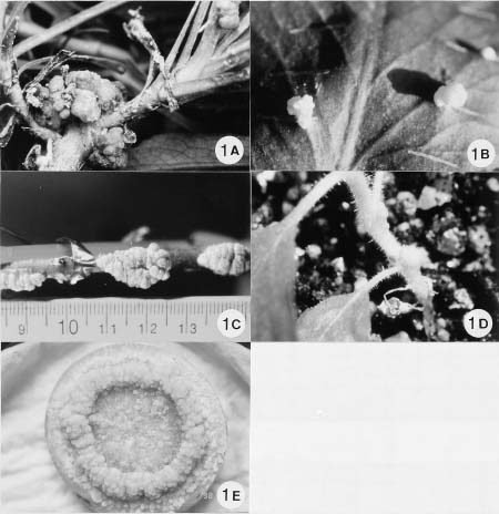
Leaf, stem and crown galls on perennial asters caused by Agrobacterium tumefaciens in Taiwan

Fure-Chyi Chen1,4, Shiow-Huey Hseu2, Shio-Tau Hung1, Ming-Chao Chen3, and Chien-Yih Lin2

1National Pingtung University of Science and Technology, Department of Plant Industry, Nei-pu, Pingtung 91207, Taiwan

2Taiwan Agricultural Research Institute, Department of Plant Pathology, Wu-feng, Taichung 413, Taiwan

3Kao-Hsiung District Agricultural Improvement Station, Pingtung City, Pingtung 900, Taiwan

(Received July 15, 1998; Accepted December 15, 1998)

Abstract. Leaf, stem, and crown galls induced by Agrobacterium tumefaciens were observed on field grown perennial asters (Aster spp.). Plants of purple flowers were more susceptible to infection than white flowers. The occurrence ranged from 25% in white flowered and 90% in purple flowered plants. Galls also occurred on leaves wounded by insect bites or mechanical shearing. Agrobacterium tumefaciens was isolated from crown and leaf galls with a selective medium NASA. The bacterial isolate was identified as A. tumefaciens using the Biolog GN system. Inoculation of selected A. tumefaciens strains on Kalanchoe pinnata leaves resulted in gall formation 8~10 days afterward. Several other A. tumefaciens strains from different gall samples also caused gall formation 6~8 days after inoculating on the stems of tobacco, and tomato seedlings. Re-inoculating virulent strains by scissors onto healthy aster leaves also induced galls 10 to 12 days after cut-inoculation. Biochemical tests of most Agrobacterium strains from aster galls showed that they belong to biovar 1.

Keywords: Agrobacterium tumefaciens; Aster sp.; Biovar 1; Crown gall; Selective medium.

Introduction

Agrobacterium tumefaciens is the causal agent of crown gall formation on many dicot plants, including ornamental species (De Cleene and De Ley, 1976). The bacteria transfers a segment of DNA (T-DNA) from Ti plasmid to a host cell, which then integrates itself into the host genome (Kado, 1991). As a result, the host develops a gall at the site of infection.

Perennial asters (Aster spp.) with pink, purple, or white florets on a long inflorescence stem are produced as cut flowers year round in central and southern Taiwan. We recently observed gall formation in the field grown purple variety in the southern part of Taiwan. Agrobacterium tumefaciens strain IL2 (possibly biovar 1) isolated from aster was briefly reported previously (Haas et al., 1995). However, a full characterization of aster Agrobacterium strains has not been done. Chrysanthemum, which also belongs to Compositae, was also reported to be susceptible to A. tumefaciens infection, which produced leaf, stem, crown and root galls (Miller, 1975; Bush and Pueppke, 1991). The Ti plasmid of Agrobacterium tumefaciens from chrysanthemum isolate, Chry5, was recently characterized molecularly (Bush and Pueppke, 1991). The isolation, de

tection and identification of A. tumefaciens depend on the use of several methods, including selective media, Biolog software characterization, and amplification of specific virulence and hormone biosynthesis regions by polymerase chain reaction (Haas et al., 1995; Moore et al., 1988; Ponsonnet and Nesme, 1994; Sawada et al., 1995; Serfontein and Staphorst, 1994).

In this study, we report the characterization of A. tumefaciens isolated from aster galls by a pathogenicity test, growth on selective medium, biochemical utilization pattern, and re-inoculation of healthy host asters.

Materials and Methods

Isolation and Maintenance of Agrobacterium tumefaciens

Leaf and crown galls of field grown, purple, perennial asters were collected from two locations in Pingtung County. The surface of the galls were removed by a handy blade and sterilized in 100 ml of 10% commercial bleach containing 4 drops of Tween-20 for 20 min. A 30 sec ultrasonic vibration (Branson 2200) was applied at the beginning of sterilization. After sterilization, the galls were washed three times with sterile water. They were then finely chopped and immersed in sterile water for 3 h or overnight. One loopful of the gall extract was streaked onto the Clark's selective medium as described in

4Corresponding author. Tel/Fax: 886-8-774-0371; E-mail: furechen@mail.npust.edu.tw


Botanical Bulletin of Academia Sinica, Vol. 40, 1999

Serfontein and Staphorst (1994). The medium contains nutrient broth, 50 mg/l sodium selenite, 250 mg/l cycloheximide, and 15 g/l Sigma agar, and was designated as NASA medium. The plates were incubated at 28°C for two days. Putative brick red colonies from NASA were streaked on the same medium to purify single colonies. The purified colonies were cultured on YM medium (0.04% yeast extract, 1% mannitol, 1.7 mM sodium chloride, 0.8 mM magnesium sulfate, and 2.2 mM dipotassium phosphate, pH 7.0, 1.5% Sigma agar) and stored at 4°C, or cultured in liquid YM medium overnight at 250 rpm and frozen at -80°C with 25% glycerol until use.

Diagnostic Tests and Carbon Utilization

Colonies confirmed to be Gram negative bacteria, according to the method of Suslow et al. (1982), were streaked on Tryptic soy agar (TSA medium: Bacto tryptone 15 g/l, Bacto Soytone 5 g/l, Sodium chloride 5 g/l, Difco-Bacto agar 15 g/l) and incubated at 28°C overnight. They were applied into a microtiter plate containing 95 substrates. The ability to catabolise different compounds was measured by an ELISA reader and compared by Biolog software (Biolog Inc., Hayward, CA, USA). Bacterial colonies were identified using the Biolog GN system according to manufacture's directions (Microlog 3.5, Biolog Inc., Hayward, CA, USA).

Diagnostic tests for physiological and biochemical characterizations were conducted according to Moore et al. (1988).

Pathogenicity Test

The bacteria slurry grown on the YM medium was scraped off with a sterile surgery scalpel and slash-inoculated on both sides of the upper leaf epidermis of kalanchoe, Kalanchoe pinnata (Minnemeyer et al., 1991). The inoculated plants were cultivated on a balcony outside the laboratory. A control slash was made without bacteria. Gall formation was scored two months after inoculation. Selected stains were also inoculated on the stems of tomato (Lycopersicum esculentum cv. Known-You 301) and tobacco seedlings (Nicotiana tabacum cv. Xanthi NC). Colonies were also applied onto the top surface of carrot slices after surface sterilization by immersion and shaking in diluted Clorox.

Re-inoculation of Agrobacterium to Aster Leaves

Healthy lateral shoots of purple aster without galls were collected from the field and rooted under mist conditions. Each leaf was cut twice at both sides from the margin to half of the blade. A pair of sterile scissors was dipped in overnight grown bacterial suspension and used to cut the leaves. Control leaves were cut with sterile scissors without bacteria. Challenged pot-grown asters were wrapped in a plastic bag for two days to retain high humidity. They were then grown in a greenhouse with ambient temperature during February 1997. Gall formation was scored after two weeks.

Results and Discussion

Perennial asters from two farmers' fields in southern Taiwan were found to have stem, crown, and leaf galls (Figure 1A) after the inflorescence stems were harvested during the spring of 1996. About 25~90% of the field grown plants were galled. The underground parts of some plants also had dark brown to gray large galls. Galls were more common during the spring and fall season in southern Taiwan. During the summer, galls became disintegraded. The source of the aster cuttings was probably cut flower stems imported long ago because the plant is not native to Taiwan. The bacteria likely was imported and spread by vegetative propagation of aster cuttings by farmers. Plants with pink and purple flowers are more susceptible while white flowered varieties are less susceptible and produce fewer and smaller galls. This indicates that the severity of gall formation in asters is cultivar dependent. When leaves of field or pot grown plants are attacked by insects, the injured areas also produce small galls.

We were able to isolate smooth, round colonies with dark red centers and light transparent rings in the margin on the NASA selective medium from different gall samples. Several colonies were further purified by streak-plating on solid YM medium. Analysis of several strains by the Biolog GN program suggested that most were A. tumefaciens. The similarity to A. tumefaciens ranges from 74.8~82.6%. Other biochemical tests using the standard protocol (Moore et al., 1988) confirmed that most isolated strains belong to biovar 1 (Table 1) when compared to the authentic strain of biovar 1 (Chry5), which was previously isolated from chrysanthemum (Bush and Pueppke, 1991). However, some tested strains did not fit typical metabolisms of biovar 1. These exceptional strains are currently under investigation and will be reported elsewhere.

When the purified strains were inoculated on the leaf surface of kalanchoe, signs of gall formation were observed after 8~10 days. This induced small galls on the wound sites after 2 to 3 weeks. The galls tended to show vertical, rather than horizontal, growth (Figure 1B). Another twenty-seven strains were further inoculated onto tobacco and tomato seedlings. Signs of gall formation were observed on tomato stems about four days after inoculation. The galls grew larger after two weeks (Figure 1C). Galls on tobacco (Figure 1D) were much smaller than those on tomato. Galls formed on tobacco stems about 8 days after inoculation. Tobacco seedlings infected with stem galls became very weak and died later. Small dense galls were visible on carrot slices about 5 days after inoculation (Figure 1E), indicating that Agrobacterium isolated from aster gall is more virulent than that from Ficus galls (Hseu et al., 1997).

Table 2 shows the results of inoculating 26 strains on tomato and tobacco stems. More than 50% of the strains isolated from NASA selective medium were able to induce galls on tobacco and tomato stems, suggesting that aster agrobacterium showed pathogenicity on both hosts. The


Chen et al. — Aster gall

Figure 1. A, Stem and leaf galls found on field grown asters after cutting the floral stalk; B, Galls produced on the leaf surface of Kalanchoe pinnata after slash-inoculating the Agrobacterium suspension; C, Stem galls on seedling of tomato cultivar Known-You 301 after inoculating the Agrobacterium suspension; D, Stem galls on seedling of tobacco cultivar Xanthi NC after inoculating the Agrobacterium suspension; E, Gall formation on the surface of a carrot slice after inoculation with aster Agrobacterium cells.

selective NASA medium is therefore suitable for Agrobacterium isolated from aster galls.

When leaves of healthy aster plants were inoculated with 10 Agrobacterium strains, they all produced visible small galls after 10 to 12 days on the wound sites (Figure 2). The control leaves, on the other hand, did not show any symptoms of galls. Galls on the treated leaves tended to emerge from the veins, an indication of Agrobacterium infection in the tissues active in cell division.

We previously reported the isolation of A. tumefaciens from ornamental Ficus microcarpa trees (Hseu et al., 1997) in central and southern Taiwan. The occurrence of Agrobacterium-induced galls in Taiwan in perennial asters has not been recorded previously. This study is perhaps the first report of galls from field grown asters in Taiwan. The morphology of aster Agrobacterium is simi

Figure 2. Gall formation 10 to 12 days after re-inoculation of aster Agrobacterium on aster leaf.


Botanical Bulletin of Academia Sinica, Vol. 40, 1999

Table 1. The characterization of biovar of Agrobacterium tumefaciens from aster and chrysanthemum.

Diagnostic test Biovar 1a Chrysanthemum Aster

(Chry5 strain)b (4 strains)

3-Ketolactose production + + +

Growth in 2% NaCl + + +

Growth at 35°C + + +

Action on litmus milk

Alkaline + + +

Acid _ _ _

Acid from:

Sucrose + + +

Erythritol _ _ _

Melezitose + + +

Alkali from:

Malonic acid _ _ _

L-tartaric acid _ _ _

Propionic acid V _ _

Mucic acid _ _ _

Ferric ammonium citrate + + +

L-tyrosine utilization _ _ _

Citrate utilization V + +

aData from Moore et al., 1988.

bChry5 was kindly provided by S.G. Pueppke as a reference for biovar 1.

lar to that of F. microcarpa when viewed under electron microscope (Hseu et al., 1997); both were rod-shaped peritrichous bacteria.

Since most asters are susceptible to Agrobacterium, it is desirable to produce healthy propagules free of the bacteria using meristem or leaf explant culture coupled with antibiotic treatment and other suitable control measures. One commercial aster supplier describes in its catalog its effort to eliminate the agrobacterium from stock plants in order to establish clean micropropagation materials (Danziger "Dan" Flower Farm Catalog, Israel, date unknown). Also, a method capable of detecting infected asters early, in the nursery or in the cut flowers, needs to be developed. Amplification of certain conserved regions of Ti plasmids by polymerase chain reaction has been suggested as a method of diagnosis (Haas et al., 1995; Ponsonnet and Nesme, 1994; Sachadyn and Kur, 1997; Sawada et al., 1995).

Since the isolated agrobacteria cells were able to induce galls on solanaceous crops such as tobacco and tomato, and on carrot slices, they supposedly contain tumor inducing genes for, e.g. auxin and cytokinin biosynthesis. They also cause the rapid growth of galls after inoculation, an indication of strong virulence on herbaceous plants. The ipt and rolC genes have been cloned from other agrobacteria strains/species and introduced into the genome of higher plants to modify their morphology (Brzobohaty et al., 1994; Michael and Spena, 1995). It is possible to isolate the same gene homologues from aster

Table 2. Gall formation in stems of tomato and tobacco inoculated with Agrobacterium tumefaciens strains isolated from aster.

Strain Gall on tomato stema Gall on tobacco stemb

Aa-1 + +

Aa-2 + +

Aa-3 + +

Aa-4 + +

Aa-5 + +

Aa-6 _ _

Aa-7 _ _

Aa-8 + +

Aa-9 _ _

Aa-10 _ _

Aa-11 _ _

Aa-12 _ _

Aa-13 + +

Aa-14 + +

Aa-15 + +

Aa-16 + +

Aa-17 + +

Aa-18 NDc ND

Aa-19 + +

Aa-20 _ _

Aa-21 + _

Aa-22 + +

Aa-23 ND +

Aa-24 ND +

Aa-25 _ ND

Aa-27 + ND

Control _ _

aKnown-You cultivar 301.

bNicotiana tabacum cv. Xanthi NC.

cND, not determined.


Chen et al. — Aster gall

agrobacteria that we reported here. Modified agrobacterium has also been used as a vector for plant genetic engineering, and we suggest that aster agrobacterium has potential as a tool to introduce foreign genes into important crop plants.

Acknowledgement. This study was supported by a grant from the Council of Agriculture, Executive Yuan, Republic of China, in part to F.-C. Chen under the contract number 85AST-1.1-FAD-49(10). We also thank anonymous reviewers for critically reading the manuscript.

Literature Cited

Brzobohaty, B., I. Moore, and K. Palme. 1994. Cytokinin metabolism: implications for regulation of plant growth and development. Plant Mol. Biol. 26: 1483-1497.

Bush, A.L. and S.G. Pueppke. 1991. Characterization of an unusual new Agrobacterium tumefaciens strain from Chrysanthemum morifolium Ram. Appl. Environ. Microbiol. 57: 2468-2472.

De Cleene, M. and J. De Ley. 1976. The host range of crown gall. Bot. Rev. 42: 389-466.

Haas, J.H., L.W. Moore, W. Ream, and S. Manulis. 1995. Universal PCR primers for detection of phytopathogenic Agrobacterium strains. Appl. Environ. Microbiol. 61: 2879-2884.

Hseu, S.H., C.Y. Lin, and F.C. Chen. 1997. The occurrence of crown gall caused by Agrobacterium tumefaciens on Ficus microcarpa in Taiwan. Plant Prot. Bull. 39: 195-205.

Kado, C.I. 1991. Molecular mechanisms of crown gall tumorigenesis. Crit. Rev. Plant Sci. 10: 1-32.

Michael, T. and A. Spena. 1995. The plant oncogenes rolA, B, and C from Agrobacterium rhizogenes. Effects on morphology, development, and hormone metabolism. Methods Mol. Biol. 44: 207-222.

Miller, I.N. 1975. Leaf, stem, crown, and root galls induced in Chrysanthemum by Agrobacterium tumefaciens. Phytopathology 65: 805-813.

Minnemeyer, S.L., R. Lightfoot, and A.G. Matthysse. 1991. A semiquantitative bioassay for relative virulence of Agrobacterium tumefaciens strains on Bryophyllum daigremontiana. J. Bacteriol. 173: 7723-7724.

Moore, L.W., C.I. Kado, and H. Bouzar. 1988. Agrobacterium. In N.W. Schaad (ed.), Laboratory Guide for Identification of Plant Pathogenic Bacteria. American Phytopathol. Soc., pp. 16-36.

Ponsonnet, C. and X. Nesme. 1994. Identification of Agrobacterium strains by PCR-RFLP analysis of pTi and chromosomal regions. Arch. Microbiol. 161: 300-309.

Sachadyn, P. and J. Kur. 1997. A new PCR system for Agrobacterium tumefaciens detection based on amplification of T-DNA fragment. Acta Microbiol. Pol. 46(2): 145-156.

Sawada, H., H. Ieki, and I. Matsuda. 1995. PCR detection of Ti and Ri plasmids from phytopathogenic Agrobacterium strains. Appl. Environ. Microbiol. 61: 828-831.

Serfontein, S. and J.L. Staphorst. 1994. Crown gall of hop caused by Agrobacterium tumefaciens biovar in South Africa. Plant Pathol. 43: 1028-1030.

Suslow, T.V., M.N. Schroth, and M. Isaka. 1982. Application of a rapid method for gram-differentiation of plant pathogenic and saprophytic bacteria without staining. Phytopathology 72: 917-918.


Botanical Bulletin of Academia Sinica, Vol. 40, 1999

¥xÆW¦a°Ï±J®Úµµ­b²ô³¡¤Î¸­¤ù¸~½F¬O¥Ñ²Óµß©ÊÀù¸~¯fµß (Agrobacterium tumefaciens) ©Ò¤Þ°_

³¯ºÖºX1 ³\¨q´f2 ¬x¨q®ç1 ³¯©ú¬L3 ªL«T¸q2

1 °ê¥ß«ÌªF¬ì§Þ¤j¾Ç¹A¶é¥Í²£¨t

2 ¥xÆW¬Ù¹A·~¸ÕÅç©Ò´Ó¯f¨t

3 °ª¶¯°Ï¹A·~§ï¨}³õ

±J®Úµµ­b¬°¤@·s¿³¤Áªá§@ª«¡A¦³¦hºØªá¦â¡A¨ä¤¤¨â­Ó«~ºØ `µµ¤Õ³¶' ¤Î `¥Õ¤Õ³¶' ¦b 85 ¦~¬K©ó«ÌªF¦a °Ïµo²{¦b­×°Å¹Lªº´Ó®è°ò³¡¡B®Ú«a¡B¸­¤ù¤Î¤Á¤f³B§¡ªø¥X¤j¤p¤£µ¥ªº¸~½F¡A¤jªÌ¬ù¥i¹Fª½®| 5 ¤½¤À¡A¦³¨Ç´Ó®è¨ü©øÂΫr­¹«á¤§¸­¤ù·|©µµÛ¶Ë¤fªø¥X¤p§Î¸~½F¡C¤£¦P«~ºØ¶¡µo¥Í¸~½Fªº¤ñ²v¦³®t²§¡Aµµ¤Õ³¶«~ºØ¦b ¥Ð¶¡¬ù¦³ 90% ¥H¤Wªº´Ó®è³£µo²{²£¥Í¸~½F¡A¥Õ¤Õ³¶¤§µo¥Í²v«h¸û§C¡A¬ù 25%¡A³Ìªñ¤SÆ[¹î¨ì¥t¤@­Ó·s¤Þ ¤Jªº¥Õªá«~ºØ³¡¥÷´Ó®è¤]¦b²ô³¡¤Î¸­¤ù¶Ë¤f²£¥Í¸û¤p¸~½F¡Cµµ¤Õ³¶¸~½F§Q¥ÎµLµß¤ôµÑ¨ú¨Ã¥H NASA °ö¾i°ò¤ÀÂ÷±o¨ì¿j¬õ¦â¤§¶ê§Îµß¸¨¡A¥H YM °ö¾i°ò¯Â¤Æ¤§µß¸¨¬°¥Õ¦â¡B¬ð°_¡B¥ú·Æ¥B¬°¶ê§Î¡C¸g¥H Biolog GN Microplate (Biolog, Inc., Hayward, CA, USA) °ö¾iŲ©w¬° Agrobacterium tumefaciens¡A·í¥H¥Í¤ÆÀË©w¤èªk»P¼Ð ·Ç biovar 1 µß¨t Chry5 ¤ñ¸û®É¡AÅã¥Ü¦h¼Æµµ­bµß¨t§¡ÄÝ©ó¦P¤@ biovar¡C±N²Óµß¥H YM ©Î²GÅ餧 YM °ö¾i°ò°ö¾i¹L©]«á¡A¥H°w¨ë©Î¸Ñ­å¤M±µºØ©ó¸¨¦a¥Í®Ú¸­¤ù¡B¹A¤Í 301 µf­X¤Î·Ï¯ó (Nicotiana tabacum cv. Xanthi NC) ¥®­]¤§¸­¤ù©Î²ô³¡ (µf­X¤Î·Ï¯ó)¡Aµf­X¤Î·Ï¯ó¦b±µºØ«á 6~8 ¤Ñ©ó±µºØ³B²£¥Í¥Õ¦â¸~½F¡A¨ä¼e«×¬ù 1~1.5 ¤½¤À¡A¦Ó¦b¸¨¦a¥Í®Ú¸­¤ù¤W¤§¸~½F¬ù¦b 8~10 ¤Ñ¤~¶}©l§Î¦¨¥B¸û¤p¡A¥B§e¬Wª¬©Î¤£³W«h¶ê²yª¬¡C±N ¤ÀÂ÷¤§µß¸¨°ö¾i¦¨Äa¯B²G¨Ã¥H°Å¤M¦^±µµµ­b¸­¤ù¡A¦b 10~12 ¤Ñ«á¡A©ó¤Á¤f¦ì¸mªø¥X¤p½F¡A¹ï·Ó²Õ«h¨S¦³ ÅܤơC¥Ñ¥H¤Wªº¸ÕÅçµ²ªG¡AÃÒ¹ê³y¦¨ `µµ¤Õ³¶' ¸~½F¯f¤§¯f­ìµß¬°¹A±ìÀù¸~¯fµß Agrobacterium tumefaciens biovar 1¡C

ÃöÁäµü¡G Àù¸~¯fµß¡Fµµ­bÄÝ¡F¥Íª««¬ 1¡F¸~½F¡F¿ï¾Ü©Ê°ö¾i°ò¡C